





| Research Article | ||
Open Vet. J.. 2026; 16(8): 5321-5326
Open Veterinary Journal, (2026), Vol. 16(8): 5321–5326 Research Article Anatomy and immunolocalization of androgen receptor in the adrenal glands of Javan mongoose (Urva javanica)Sekar Arum Krisna Putri1, Teguh Budipitojo2*, Irma Padeta2, Woro Danur Wendo2, Hery Wijayanto3, Farah Salma Fikriyah4 and Alifya Deraya Rahmawati41Doctoral Program in Veterinary Science, Faculty of Veterinary Medicine, Universitas Gadjah Mada, Yogyakarta, Indonesia 2Department of Anatomy, Faculty of Veterinary Medicine, Universitas Gadjah Mada, Yogyakarta, Indonesia 3Department of Anatomy, Wildlife and Zoonotic Disease Center, Faculty of Veterinary Medicine, Universitas Gadjah Mada, Yogyakarta, Indonesia 4Veterinary Science Study Program, Faculty of Veterinary Medicine, Universitas Gadjah Mada, Yogyakarta, Indonesia *Corresponding Author: Teguh Budipitojo. Veterinary Science Study Program, Faculty of Veterinary Medicine, Universitas Gadjah Mada, Yogyakarta, Indonesia. Email: budipitojo [at] ugm.ac.id Submitted: 03/02/2026 Revised: 06/06/2026 Accepted: 23/06/2026 Published: 08/08/2026 © 2025 Open Veterinary Journal
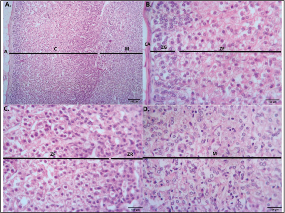
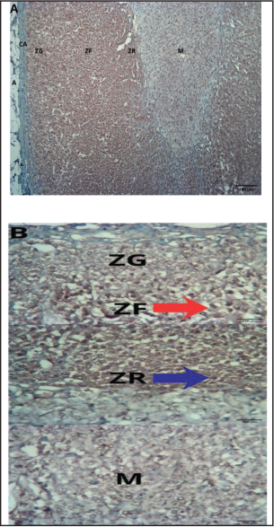
AbstractBackground: The adrenal gland is a key endocrine organ involved in hormonal regulation, stress response, and reproductive function across mammalian species. In wild carnivores such as the Javan mongoose (Urva javanica), information regarding adrenal gland morphology and androgen receptor (AR) distribution remains scarce. Aim: This research focuses on examining the anatomical and histological characteristics of the adrenal glands in U. javanica and investigating the distribution of AR–immunoreactive (AR-IR) cells within them. Methods: Six adrenal glands obtained from male and female Javan mongooses were examined using gross anatomical observation, routine histology, and immunohistochemical staining. Tissue samples were analysed to distinguish the cortical zones—zona glomerulosa (ZG), zona fasciculata (ZF), and zona reticularis (ZR)—as well as the medulla, and to determine the distribution of AR-IR cells. Results: Macroscopically, the adrenal glands were located near the cranial poles of the kidneys and exhibited a consistent morphology, with subtle asymmetry between the right and left glands. Histologically, the six pairs of adrenal glands showed a well-defined cortical and medullary organization, comparable to that of other mammals. Immunohistochemical examination showed that AR-IR cells were mainly concentrated in the ZF and ZR of the adrenal cortex, while no immunoreactivity was detected in the ZG or medulla. This distribution pattern indicates a distinct species-specific localization of ARs in the adrenal glands of the Javan mongoose. Conclusion: These findings demonstrate a distinct, species-specific pattern of AR distribution in the adrenal glands of the Javan mongoose. The restricted localization of AR-IR cells to specific cortical zones suggests a targeted role of androgen signalling in adrenal function and highlights interspecies variation in endocrine regulation among wild mammals. Furthermore, this study provides baseline anatomical and immunohistochemical data for a relatively understudied wild species, contributing to a better understanding of comparative adrenal endocrinology in wildlife. Keywords: Adrenal cortex, Adrenal gland, Androgen receptor, Immunohistochemistry, Javan mongoose. IntroductionThe Javan mongoose (Urva javanica) is a carnivorous mammal from the Herpestidae family, known for its opportunistic feeding habits and is widely distributed in Southeast Asia, including Indonesia (Chutipong et al., 2016; Putri et al., 2026). This species is considered to have a low risk of extinction (Susdiyanti et al., 2023; Putri et al., 2026). Despite its ecological importance, anatomical and endocrine studies of this species remain limited. In vertebrates, the adrenal glands are generally found as a pair of endocrine organs situated next to the kidneys. Their microscopic architecture reflects the typical structural–functional organization of endocrine organs, characterized by specialized secretory cells and rich vascularization that facilitate hormone production and systemic distribution. Endocrine organs lack excretory ducts, and their secretions (hormones) are directly released into the lymphatic and circulatory systems. Adrenal hormones are vital for the body’s essential functions, affecting tissue growth and differentiation, reproductive organ development, and sexual cycle progression. They also play crucial roles in maintaining ecosystem balance and sustainability (Kot et al., 2023; Budipitojo et al., 2025). The morphology and function of the adrenal glands can vary significantly among different animal classes (Budipitojo et al., 2025). In mongooses, the adrenal glands are generally elongated and lack the pronounced lobulation commonly observed in dogs. The left gland tends to be more elongated and flattened, while the right gland is usually thicker, blunter, and shows slightly angular borders on its lateral and ventral sides. Both glands are situated close to the dorsal body wall near the cranial poles of the kidneys (Tomich, 1965). The adrenal gland consists of two parts: the cortex and the medulla. The adrenal cortex consists of three distinct zones, namely the zona glomerulosa (ZG), zona fasciculata (ZF), and zona reticularis (ZR), which are involved in steroid hormone production, while the medulla forms the inner region of the gland (Budipitojo et al., 2025). Androgen receptors (ARs) are important regulators of endocrine activity and have been identified in the adrenal glands of several mammalian species. Budipitojo et al. (2025) reported that AR–immunoreactive (AR-IR) cells in the adrenal glands of Hystrix javanica were predominantly localized in the ZG and adrenal medulla, with fewer cells in the ZF and no immunoreactivity in the ZR. Similarly, Trejter et al. (2015) reported that AR expression was predominantly observed in the ZG and ZF, while little to no expression was noted in the ZR. In rodents, the adrenal medulla also showed evidence of AR expression. Previous studies on mongoose species have primarily focused on genetics, behavior, diet, and anatomical studies of the respiratory and digestive systems (Shil et al., 2012; Jerome et al., 2016; Mahmood and Adil, 2017; Hussein et al., 2018; Mahmood and Kadhim, 2018; Veron et al., 2022; Putri et al., 2026). Although Tomich (1965) used different mongoose species and employed approaches and objectives that did not specifically examine the histological characteristics or distribution of immunoreactive cells in the adrenal glands. This study was carried out to address the lack of data on the distribution pattern of AR-IR cells in the adrenal glands of the Javan mongoose. The findings provide a scientific foundation for understanding the role of the adrenal glands as endocrine organs and emphasize their importance in supporting animal adaptation to their environment. Materials and MethodsSample collection and tissue preparationThis research received approval from the Preclinical Research Ethics Committee of Universitas Gadjah Mada, Indonesia (Approval No. 18/EC-FKH/int./2025). The study was conducted at the Laboratory of Gross Anatomy and Histology, Faculty of Veterinary Medicine, Universitas Gadjah Mada, Indonesia. A total of six adrenal glands were collected from Javan mongooses (U. javanica), consisting of three males and three females, with age not taken into consideration. The number of specimens was considered adequate for a descriptive anatomical and immunohistochemical study, as this research focused on qualitative morphological observations rather than statistical inference. Before tissue collection, the animals were euthanized by intramuscular injection of a ketamine–xylazine combination consisting of ketamine (10 mg/kg body weight) and xylazine (2 mg/kg body weight). Following confirmation of deep anaesthesia and loss of reflexes, intracardiac exsanguination was performed before tissue collection. The adrenal gland tissues were fixed in formalin for 24 hours. After trimming, the samples were placed in tissue cassettes for processing into paraffin blocks. Routine histological staining was performed using haematoxylin and eosin (H&E) to observe the histological structure of the adrenal glands (Budipitojo et al., 2025). Immunohistochemistry procedureImmunohistochemical staining was carried out to identify the distribution of AR-IR cells within the adrenal glands. Paraffin sections were deparaffinized in xylene and rehydrated through a graded ethanol series. Antigen retrieval was performed using heat-induced epitope retrieval in citrate buffer (pH 6.0) before immunostaining. Endogenous peroxidase activity was subsequently blocked by incubating the tissue sections in 3% hydrogen peroxide diluted in methanol for 10 minutes at room temperature. To minimize nonspecific antibody binding, tissue sections were pre-incubated with a normal goat serum blocking solution for 30 minutes at 37°C. The slides were incubated overnight at 4°C with a polyclonal rabbit anti-AR primary antibody (catalogue number A16200, AB Clonal Technology, Wuhan, China) at a 1:200 dilution, which specifically recognizes the activated form of the AR. Following this step, the sections were rinsed with phosphate-buffered saline (PBS) and then incubated with the appropriate secondary antibody for 20 minutes at 37°C. Immunoreactive sites were visualized using a diaminobenzidine chromogen system. Counterstaining was done on tissue slides using Mayer’s haematoxylin, followed by stratified dehydration using ethanol, clearing with xylene, and ending with mounting. Each immunostaining session included both positive and negative control sections to ensure the specificity and reliability of the immunohistochemical reaction. Positive controls consisted of testicular tissue known to express AR immunoreactivity, whereas negative control sections were processed identically except that the primary antibody was omitted and replaced with PBS. No specific staining was observed in the negative control sections (Budipitojo et al., 2025). Microscopic observationHistological and immunohistochemical preparations were examined using a light microscope (BX60, Olympus, Tokyo, Japan). Representative images were captured with an OptiLab advance plus digital camera at various magnifications. The localization and distribution of AR-immunoreactive cells were described across the zones of the adrenal cortex and the medulla. The distribution of immunoreactive cells in the adrenal glands was analyzed descriptively (Putri et al., 2026). Ethical approvalThis research received approval from the Preclinical Research Ethics Committee of Universitas Gadjah Mada, Indonesia (Approval No. 18/EC-FKH/int./2025) ResultsAnatomical featuresThe adrenal glands of mongooses (Fig. 1) typically had a consistent shape without noticeable angular edges, deep grooves, or a distinctly bilobed appearance. Both glands were situated in the retroperitoneal fat near the front ends of the kidneys and displayed a yellowish color. The left adrenal gland was elongated and flat, whereas the right was thicker and more rounded, with slightly angular sides and bottom edges. Both adrenal glands taper toward the posterior end and lie close to the posterior abdominal wall, positioned medially to the anterior poles of the kidneys. The right gland is partly concealed by the postcaval vein and the caudate lobe of the liver, giving it a more restricted placement, while the left gland is more freely situated beside the postcaval vein and is gently compressed by the pancreas and stomach. An adrenolumbar vein runs across the upper and dorsolateral surfaces of the right gland, whereas on the left gland, a vein arching over its lower third often creates a slight medial notch as it enters the renal or postcaval vein.
Fig. 1. Topography of the adrenal glands of Javan mongoose (Urva javanica). The adrenal glands of Javan mongooses are typically consistent in shape, without prominent angular edges, deep grooves, or distinct bilobed structures. Both glands are in the retroperitoneal fat near the anterior end of the kidney and are yellowish. The left adrenal gland was elongated and flattened, whereas the right was thicker and more rounded, with slightly angular sides and lower edges. Blue arrows indicate the testes, green circles indicate the bulbourethral glands and penis, and red circles indicate the adrenal glands of the Javan mongoose. R=right region. Histological featuresIn Javan mongooses, the adrenal glands resemble those of other mammals and are composed of two main parts: the cortex and the medulla (Fig. 2A). The adrenal cortex is further divided into the ZG, ZF, and ZR (Fig. 2B and C), where in these zones, lipid-rich cells are arranged in cords or clusters surrounding the vascular sinusoids. The ZG is the superficial zone and is composed of polygonal cells with eosinophilic cytoplasm that are clustered; the cell nuclei appear euchromatic and relatively larger than those in the other zones. The ZF represents the central and largest zona of the adrenal cortex, located between the ZG and ZR; the cytoplasm often appears foamy due to numerous lipid vacuoles. The ZR forms the innermost zone of the adrenal cortex. It is composed of anastomosing cords and is surrounded by sinusoids; the cytoplasm is denser and darker. The adrenal medulla (Fig. 2D) consists of chromaffin cells that form interconnected clusters and cords separated by sinusoids. AR-IR cellsImmunohistochemical analysis identified AR-IR cells in the adrenal glands of Javan mongooses. The AR-IR cells were widely distributed within the ZG and ZR of the adrenal cortex, whereas no AR-IR cells were detected in the ZG and adrenal medulla (Fig. 3A and B). DiscussionThe present study demonstrated that the adrenal glands of the Javan mongoose exhibited a relatively regular and elongated shape without a distinctly bilobed appearance. Anatomically, both glands were positioned near the cranial poles of the kidneys and showed asymmetry between left and right sides, consistent with the previous description in another mongoose species (Tomich, 1965). Immunohistochemical analysis revealed that AR-IR cells were predominantly localized in the ZF and ZR of the adrenal cortex. In contrast, the ZG and adrenal medulla showed only weak, diffuse staining that was interpreted as non-specific background staining rather than true immunoreactivity. These findings indicate that AR distribution in the adrenal glands of the Javan mongoose is region-specific and may be associated primarily with the functional activity of the inner cortical zones.
Fig. 2. Histology of the adrenal gland in Javan mongoose (Urva javanica). The gland is enclosed by a dense connective tissue capsule (CA). The adrenal gland of the Javan mongoose consists of a cortex (C) and medulla (M). The adrenal cortex is divided into the ZG, composed of clustered polygonal cells with eosinophilic cytoplasm; the ZF, characterized by cells with foamy cytoplasm containing numerous lipid vacuoles arranged in cords; and the ZR, consisting of anastomosing cords with denser and darker cytoplasm. The medulla is composed of chromaffin cells arranged in interconnected clusters and cords separated by sinusoids. H&E staining, observed under light microscopy at 100× (A) and 400× (B–D) magnification. Scale bar=100 µm. Differences in AR distribution patterns have been reported among mammalian species (Abdulameer, 2025). In the Sunda porcupine (H. javanica), AR-IR cells were predominantly observed in the ZG and adrenal medulla, with weaker distribution in the ZF and absence in the ZR (Budipitojo et al., 2025). In rodents, AR expression has been reported in multiple cortical zones and medullary regions (Trejter et al., 2015; Gannon et al., 2019). Compared with these species, the Javan mongoose demonstrated a different localization pattern characterized by predominant immunoreactivity in the ZF and ZR. This variation may reflect species-specific differences in adrenal endocrine regulation and physiological adaptation among mammals. Previous studies have suggested that ARs contribute to adrenal cortical regulation, including steroidogenic activity and endocrine homeostasis (Cioni et al., 2020; Kot et al., 2023). Despite providing novel anatomical, histological, and immunohistochemical data on the adrenal glands of the Javan mongoose, this study has several limitations. First, the opportunistic collection of wild specimens precluded precise aging, thereby limiting our ability to assess age-related shifts in morphology and AR distribution. Second, as a descriptive qualitative study, sex-based differences in AR expression were not quantified using cell counts or staining intensity metrics. In addition, the relatively limited sample size may restrict broader biological interpretation. Furthermore, AR localization was evaluated only by immunohistochemistry without complementary molecular or protein validation techniques, such as immunofluorescence, Western blotting, or quantitative polymerase chain reaction. Nevertheless, this study provides baseline information on the adrenal glands of an understudied wild mammalian species and may serve as a foundation for future comparative and functional endocrine studies.
Fig. 3. Distribution of AR-IR cells in the adrenal glands of Javan mongoose (Urva javanica). The adrenal cortex consists of the ZG, ZF, and ZR, whereas the inner region is formed by the medulla (M). AR-IR cells were predominantly observed in the ZF and ZR. Red arrows indicate AR-IR cells in the ZG; whereas blue arrows indicate AR-IR cells in the ZR. IHC staining was performed using an anti-androgen receptor antibody at a dilution of 1:200, observed under light microscopy at 100× (A) and 400× (B) magnification. Scale bar=100 µm. A=adipose tissue; CA=capsule. ConclusionThe adrenal glands of the Javan mongoose (U. javanica) exhibit a relatively consistent morphology without sharp edges and are yellowish. They are located in the retroperitoneal fat tissue surrounding the anterior tip of the kidneys. Macroscopically, the left adrenal gland appeared more elongated and flattened than the right adrenal gland, which was relatively thicker and rounded, with slightly angled lateral and ventral borders. Both adrenal glands tend to narrow caudally and are located adjacent to the dorsal body wall and medial to the cranial pole of the kidney. Histologically, the adrenal glands of the Javan mongoose are composed of two main compartments: the cortex and medulla. The adrenal cortex was clearly distinguished into three zones: the ZG, ZF, and ZR. Immunohistochemical staining revealed that AR-IR cells were distributed throughout the ZF and ZR of the adrenal cortex, suggesting a potential role for androgens in regulating adrenal function in this species. AcknowledgmentThe authors would like to thank Universitas Gadjah Mada for providing research funding for the “Research and Development Grant of the Department of the Faculty of Veterinary Medicine, Universitas Gadjah Mada Year 2025”, based on the assignment letter Number: 1290/UN1/FKH/HK4/2025. Conflict of interestThe authors declare that no financial or commercial relationship existed during the course of this research that could be understood as a potential conflict of interest. FundingThe authors declare that this research received funding from the Department of Veterinary Medicine Research and Development Grant, Universitas Gadjah Mada in 2025 through BPPTN-BH FKH UGM, based on the Activity Implementation Agreement Letter Number 1290/UN1/FKH/HK4/2025. Authors’ contributionsSAKP contributed to the research implementation, including sample and data collection, data analysis, and the preparation and writing of the manuscript. TB, IP, WDW, and HW were involved in the study design, supervision of the research process, data analysis, and manuscript development. FSF and ADR contributed to sample and data collection and analysis. Data availabilityThe original data and contributions from this study are available in the article or supplementary materials. Furthermore, inquiries should be directed to the corresponding author. ReferencesAbdulameer, N. 2025. A review Anatomical and Histological Study of the Adrenal Gland in Domestic Animals. Kerbala J. Vet. Med. Sci. 1, 5–8. Budipitojo, T., Larasati, A.D., Putri, S.A.K., Wendo, W.D. and Fibrianto, Y.H. 2025. Distribution of androgen receptor immunoreactive cells in the adrenal glands of Hystrix javanica. Open. Vet. J. 15, 4090–4096; doi:10.5455/OVJ.2025.v15.i9.13 Chutipong, W., Duckworth, J.W., Timmins, R., Willcox, D.H.A. and Ario, A. 2016. Herpestes javanicus. The IUCN Red List of Threatened Species. 2016. Available via https://www.iucnredlist.org/ja/species/70204120/70204139 Cioni, B., Zaalberg, A., Van Beijnum, J.R., Melis, M.H.M., Van Burgsteden, J., Muraro, M.J., Hooijberg, E., Peters, D., Hofland, I., Lubeck, Y., De Jong, J., Sanders, J., Vivié, J., Van Der Poel, H.G., De Boer, J.P., Griffioen, A.W., Zwart, W. and Bergman, A.M. 2020. Androgen receptor signalling in macrophages promotes TREM-1-mediated prostate cancer cell line migration and invasion. Nat. Commun. 11(4498), 1–17; doi:10.1038/s41467-02018313-y Gannon, A.L., O’Hara, L., Mason, J.I., Jørgensen, A., Frederiksen, H., Milne, L., Smith, S., Mitchell, R.T. and Smith, L.B. 2019. Androgen receptor signalling in the male adrenal facilitates X-zone regression, cell turnover and protects against adrenal degeneration during ageing. Scientific Rep. 9(1), 1–16; doi:10.1038/s41598-019-46049-3 Hussein, A.J., Hussein, H.A. and Abdulzahra, H.K. 2018. A morphological and histological study of the liver and pancreas of the small Indian mongoose (Herpestes javanicus). Basrah J. Vet. Res. 17(3), 123–135. Jerome, A., Srivastava, S.K. and Sharma, R.K. 2016. Study on follicular characteristics, hormonal and biochemical profile in norgestomet+PMSG treated acyclic buffaloes. Iranian J. Vet. Res. 17(4), 247–252. Kot, T., Tkachuk, S., Usenko, S. and Prokopenko, V. 2023. Adrenal Gland of Poultry: anatomy, Microscopy, Morphometry, and Histochemistry. J. World’s. Poultry. Res. 13(1), 20–28. Mahmood, H.B. and Kadhim, K. 2018. Histomorphology and Histochemical Study of Duodenum and Pancreas in Gray Mongoose (Herpestes edwardsii) In Iraq. Indian. J. Natural. Sci. 9(51), 15368–15377. Mahmood, T. and Adil, A. 2017. Diet composition of small Indian mongoose (Herpestes javanicus) varies seasonally in its native range. Anim. Biol. 67(1), 69–80; doi:10.1163/15707563-00002516 Putri S, A.K., Budipitojo, T., Fibrianto, Y.H., Wendo, W.D. and Budiariati, V. 2026. Histological distribution of endocrine cells in the pancreas of Javan Mongoose (Urva javanica). Open Vet. J. 16(1), 669–678; doi:10.5455/OVJ.2026.v16.i1.62 Shil, S., Das, B., Uddin, M., Rahman, M. and Quasem, M. 2012. Anatomy of digestive and respiratory system of Indian grey mongoose (Herpestes edwardsii). Univ. J. Zool. Rajshahi. Univ. 31, 83–84; doi:10.3329/ujzru.v31i0.15438 Susdiyanti, T., Cita, K.D., Yuliani, N., Diantama, N. and Sasongko, D.A. 2023. Diversity of Mammals in 8 Years of Jati (Tectona grandis Linn. f) Unggul Nusantara (JUN). Jurnal Sains Natural 13(1), 21; doi:10.31938/jsn.v13i1.395 Tomich, P.Q. 1965. Weight Variation in Adrenal Glands of the Mongoose in Hawaii. Pacific Sci. 19(2), 238–243. Available via http://hdl.handle.net/10125/4415 Trejter, M., Jopek, K., Celichowski, P., Tyczewska, M., Malendowicz, L.K. and Rucinski, M. 2015. Expression of estrogen, estrogen related and androgen receptors in adrenal cortex of intact adult male and female rats. Folia Histochem. Cytobiol. 53(2), 133–144. Veron, G., Patou, M.L. and Jennings, A.P. 2022. Systematics and evolution of the mongooses (Herpestidae, Carnivora). In Small carnivores: evolution, ecology, behaviour and conservation. Eds., Rosalino, L.M. and Sillero-Zubiri, C. Chichester, United Kingdom: John Wiley & Sons Ltd, pp: 61–78; doi:10.1002/9781118943274.ch3 | ||

| How to Cite this Article |
| Pubmed Style Putri SAK, Budipitojo T, Padeta I, Wendo WD, Wijayanto H, Fikriyah FS, Rahmawati AD. Anatomy and immunolocalization of androgen receptor in the adrenal glands of Javan mongoose (Urva javanica). Open Vet. J.. 2026; 16(8): 5321-5326. doi:10.5455/OVJ.2026.v16.i8.26 Web Style Putri SAK, Budipitojo T, Padeta I, Wendo WD, Wijayanto H, Fikriyah FS, Rahmawati AD. Anatomy and immunolocalization of androgen receptor in the adrenal glands of Javan mongoose (Urva javanica). https://www.openveterinaryjournal.com/?mno=309076 [Access: August 08, 2026]. doi:10.5455/OVJ.2026.v16.i8.26 AMA (American Medical Association) Style Putri SAK, Budipitojo T, Padeta I, Wendo WD, Wijayanto H, Fikriyah FS, Rahmawati AD. Anatomy and immunolocalization of androgen receptor in the adrenal glands of Javan mongoose (Urva javanica). Open Vet. J.. 2026; 16(8): 5321-5326. doi:10.5455/OVJ.2026.v16.i8.26 Vancouver/ICMJE Style Putri SAK, Budipitojo T, Padeta I, Wendo WD, Wijayanto H, Fikriyah FS, Rahmawati AD. Anatomy and immunolocalization of androgen receptor in the adrenal glands of Javan mongoose (Urva javanica). Open Vet. J.. (2026), [cited August 08, 2026]; 16(8): 5321-5326. doi:10.5455/OVJ.2026.v16.i8.26 Harvard Style Putri, S. A. K., Budipitojo, . T., Padeta, . I., Wendo, . W. D., Wijayanto, . H., Fikriyah, . F. S. & Rahmawati, . A. D. (2026) Anatomy and immunolocalization of androgen receptor in the adrenal glands of Javan mongoose (Urva javanica). Open Vet. J., 16 (8), 5321-5326. doi:10.5455/OVJ.2026.v16.i8.26 Turabian Style Putri, Sekar Arum Krisna, Teguh Budipitojo, Irma Padeta, Woro Danur Wendo, Hery Wijayanto, Farah Salma Fikriyah, and Alifya Deraya Rahmawati. 2026. Anatomy and immunolocalization of androgen receptor in the adrenal glands of Javan mongoose (Urva javanica). Open Veterinary Journal, 16 (8), 5321-5326. doi:10.5455/OVJ.2026.v16.i8.26 Chicago Style Putri, Sekar Arum Krisna, Teguh Budipitojo, Irma Padeta, Woro Danur Wendo, Hery Wijayanto, Farah Salma Fikriyah, and Alifya Deraya Rahmawati. "Anatomy and immunolocalization of androgen receptor in the adrenal glands of Javan mongoose (Urva javanica)." Open Veterinary Journal 16 (2026), 5321-5326. doi:10.5455/OVJ.2026.v16.i8.26 MLA (The Modern Language Association) Style Putri, Sekar Arum Krisna, Teguh Budipitojo, Irma Padeta, Woro Danur Wendo, Hery Wijayanto, Farah Salma Fikriyah, and Alifya Deraya Rahmawati. "Anatomy and immunolocalization of androgen receptor in the adrenal glands of Javan mongoose (Urva javanica)." Open Veterinary Journal 16.8 (2026), 5321-5326. Print. doi:10.5455/OVJ.2026.v16.i8.26 APA (American Psychological Association) Style Putri, S. A. K., Budipitojo, . T., Padeta, . I., Wendo, . W. D., Wijayanto, . H., Fikriyah, . F. S. & Rahmawati, . A. D. (2026) Anatomy and immunolocalization of androgen receptor in the adrenal glands of Javan mongoose (Urva javanica). Open Veterinary Journal, 16 (8), 5321-5326. doi:10.5455/OVJ.2026.v16.i8.26 |


