





| Research Article | ||
Open Vet. J.. 2026; 16(5): 3039-3051 Open Veterinary Journal, (2026), Vol. 16(5): 3039-3051 Research Article Development of a suspension-based vero cell culture platform for the efficient propagation of lumpy skin disease virusHadeer M. Mossa1*, Soad Eid1, Amira A. El-Said1, Ausama A. Yousif2 ,and Ahmed A. El-Sanousi21Veterinary Serum and Vaccine Research Institute, Agriculture Research Center, Giza, Egypt 2Department of Virology, Faculty of Veterinary Medicine, Cairo University, Giza, Egypt *Corresponding Author: Hadeer M. Mossa. Veterinary Serum and Vaccine Research Institute, Agriculture Research Center, Giza, Egypt. Email: hadeervsvri [at] gmail.com Submitted: 01/02/2026 Revised: 20/04/2026 Accepted: 30/04/2026 Published: 31/05/2026 © 2025 Open Veterinary Journal
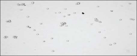
ABSTRACTBackground: Lumpy skin disease virus (LSDV) is an increasingly prevalent pathogen of significant global concern. It is indigenous to Africa and the Middle East and has recently spread to Europe and Asia. Vaccine-based control programs represent the most effective preventive strategy for disease management. The extraordinary worldwide dissemination of the virus necessitates establishing effective, scalable vaccine production systems to meet both local and global vaccine demand. Vaccine manufacturing has depended on LSDV multiplication in adherent cell cultures, thus constraining large-scale manufacturing. Aim: This study aimed to enhance and maximize the productivity of an LSDV on a suspension-adapted cell line. VERO cells are anchorage-dependent, and there are currently no commercially available suspension VERO cell lines. The creation of our customized cells constitutes a singular R&D investment. Vero cells were adapted for suspension growth to facilitate subcultivation, enhance process scalability, and reduce production costs. Methods: A robust small-scale suspension culture system of Vero cells was developed for the efficient propagation of LSDV. A preliminary step for industrial production in bioreactors, with optimized passage time and initial cell density, along with other factors such as cell density, medium composition, and agitation rates. Results: The modified cells exhibited remarkable viability and proliferation rates in suspension, reaching 1.94 × 10⁶ cells/ml. Suspension-adapted Vero cells efficiently replicated LSDV, as shown by cytopathic effect observation, quantitative polymerase chain reaction, and infectivity titration assays. After nine passages, suspension-adapted Vero cells reached 6.85 log10 tissue culture infectious dose (TCID50)/ml 96 hours post infection, compared to 5.5 log10in adherent cells. The study demonstrated that LSDV productivity was significantly influenced by a multiplicity of infection of 0.1, a viable cell density of 5.8 × 10⁵ cells/ml, and adequate nutritional supplementation. These findings confirm that suspension-adapted Vero cells offer a promising and potentially scalable platform for the production of LSDV vaccine. Although the obtained results suggest advantages in terms of process control and culture flexibility, further studies in bioreactors are needed to confirm performance at industrial scales. Conclusion: This study shows high-titer LSDV production in suspension-adapted Vero cell culture (up to 7 × 10⁶TCID10/ml), indicating its potential for future scale-up. These findings are intriguing, but bioreactor studies and process validation are needed to confirm industrial applicability and regulatory compliance. Keywords: Cell culture, Lumpy skin disease virus, New methods, Vero cells. IntroductionLumpy skin disease virus (LSDV) is a double-stranded DNA virus from the genus Capripoxvirus of the Poxviridae family. Cattle infected with this virus develop a systemic illness and telltale skin nodules (Tulman et al., 2001; Tuppurainen et al., 2015). Infected calves can experience fever, nodular skin lesions, enlarged lymph nodes, and a high morbidity rate with variable mortality rates. Transmission of the virus via insects is believed to be the primary cause of most illnesses. Therefore, holding animals in insect-proof pens prevents LSDV transmission (Leliso, 2021). Infection from direct contact is believed to occur at a low rate and is not a key component of transmission during epizootics, although it is still possible (Owada et al., 2024). In 1929, the clinical condition of lumpy skin disease (LSD) was initially observed in Zambia (Akther et al., 2023). In Egypt, animals imported from Africa in May 1988 were believed to be the source of LSD into the local quarantine station in Suez Governorate, Egypt (Ali et al., 1990). The disease seemed to have survived the winter with few or no clinical symptoms after spreading locally in the summer of 1988. Resurfacing in the summer of 1989, it expanded to 22 of Egypt’s 26 governorates in just 5 or 6 months after its first isolation (Salib and Osman 2011). Subsequently, further outbreaks were recorded in 2006, 2008, and 2012 in southeastern Europe, the Caucasus, Russia, Kazakhstan, India, and Bangladesh (Tuppurainen and Galon, 2016; Whittle et al., 2023). For effective LSD control, biosecurity measures, including animal quarantine and movement management, vector control, facility and equipment disinfection, and early detection and surveillance at the farm level, are necessary. Vaccination is the most common and effective control measure (Tuppurainen et al., 2021). LSD prevention in African cow populations has been achieved through the extensive and effective use of three separate vaccinations. The Romanian attenuated sheep-pox vaccine was used to protect cattle from LSDV. According to Khafagy et al. (2016)vaccination against LSDV elicits a strong immunogenic response, resulting in high antibody titers and long-lasting immunity. Historically, vaccination with the Kenyan sheep pox virus strain was used in Egypt as a heterologous vaccine to control LSD outbreaks. However, several studies have indicated that this approach did not provide complete protection in cattle, raising concerns regarding its safety and efficacy for use in bovine populations (Tuppurainen et al., 2014). The primary homologous vaccine, a live attenuated LSDV vaccine derived from the harmless and immunogenic Neethling strain of LSD, was propagated using either the VERO or MDBK cell lines (WOAH) (Bazid et al., 2023). The VERO cell line, a continuous cell line with the potential for an unlimited lifespan, was derived from the kidney of a healthy adult African green monkey (Cercopithecus aethiops) via spontaneous immortalization (Ammerman et al., 2008). The additional processing steps required for cell adhesion and detachment are one disadvantage of traditional static Vero cell culture, which increases production costs (Chun et al., 2019). Unfortunately, most of these systems rely on serum-containing media, which can be expensive and introduce a host of unknown contaminants that could damage cell cultures and disrupt subsequent processes (Randall Alfano et al., 2020). Geographical, seasonal, and production-related variables can also cause qualitative and quantitative variations in serum composition across batches, resulting in inconsistent cell growth and productivity. Biopharm International (2023)found that these differences can increase production delays and decrease volumetric viral productivity compared to serum-free systems, which are more regulated. Adapting Vero cells to suspension growth eliminates the dependence on anchorage-dependent culture systems and represents a promising strategy for simplifying large-scale vaccine manufacturing. Suspension cell culture platforms are widely used in the biopharmaceutical industry because of their compatibility with scalable bioreactor systems and good manufacturing practices. In principle, further process intensification strategies, such as fed-batch, perfusion, or continuous cultivation, may enhance productivity in cell culture–based viral vaccine production. However, the evaluation of these approaches was beyond the scope of the present study. Many recent studies have shown that suspension-adapted Vero cells cultured with microcarriers produced higher amounts of rabies virus than adherent Vero cells. However, some challenges associated with Vero cells, such as longer cell doubling time and low cell viability, remain to be addressed through formulation and appropriate adaptation processes (Rourou et al., 2019). Another study addressed the success of such a production method by using vesicular stomatitis virus (VSV) to evaluate viral productivity on suspension Vero cells. The suspension cell culture showed higher VSV productivity than the adherent Vero cell culture (Shen et al., 2019a,b). In addition, the suspension culture could be infected at higher cell densities by stirring, oxygenation, density of the cell inoculum, and type of growth surface, as well as chemical parameters, such as pH and medium composition (Shen et al., 2019a,b), which will all contribute to improving the volumetric virus productivity. Recently, VERO suspension cells have been tested for the propagation of the new emerging Zika virus (ZIKV), showing that the established perfusion process allows the provision of large amounts of ZIKV material, thereby facilitating further research and was the first step toward process development for manufacturing inactivated or live-attenuated ZIKV vaccines to overcome its spread in the Americas and the Pacific that reached warning levels (Nikolay et al., 2018). Generally, the results for the suspension-adapted culture systems usually give similar virus titers or even sometimes more compared to batch cultivations (Tapia et al., 2016; Shen et al., 2019a,b). The primary objective of this study is to develop and optimize a suspension culture system based on Vero cells as a foundational step toward scalable bioreactor-based production platforms. This study also aims to evaluate the growth performance of Vero cells under low-serum conditions and to assess the capacity of the system to support viral replication using LSDV as a Capripoxvirus model. Materials and MethodsCells and culture mediaGradual adaptation of candidate cells to grow in suspensionThe VERO adherent cell line, sourced from a serum-containing cell bank, was acquired from the Pox Vaccines Department at the Veterinary Serum and Vaccine Research Institute in Egypt. Seeds were cultivated in T-75 flasks at 37°C with 5% CO2 in Dulbecco’s Modified Eagle Medium (DMEM; Caisson, Grand, USA) supplemented with 5% fetal bovine serum (FBS) (Biowest, USA). After the cells were adapted to reduced-serum culture conditions, they were transferred from static T-75 flasks containing adherent Vero cells to a non-treated suspension culture system using a 500 ml three-neck spinner flask (Bellco, Cat. No. 1969-00500) to minimize cell attachment. Cells were inoculated into 200 ml of complete DMEM and maintained under continuous agitation in an incubator at 37°C using a digital magnetic stirrer, commencing at 80 rpm, with a seeding density of 0.5 × 10⁶ cells supplemented with 5% FBS. Media preparation and optimization of suspended VERO cellsChemically defined in-house DMEM with L-glutamine 10 gm/5 l, 100 IU/ml penicillin-streptomycin was purchased from SIGMA–ALDRICH. All solutions and equipment that come in contact with the cells were sterile. The media was supplemented regularly with additional components to enhance cell growth, such as essential amino acids (NEAA100X, Sigma Aldrich), vitamins (50×) (Sigma Aldrich), and growth factors (Quesney et al., 2003; Posung et al., 2021). Subculturing of the vero cell suspension cultureVero cells were adapted to suspension culture by passaging every 1–2 days with 10%–50% (v/v) conditioned medium from the previous culture (Rourou et al., 2019). The cells were considered adapted upon achieving reproducible exponential growth after 10 passages. During early adaptation, cultures were subcultured every 1–2 days by gentle centrifugation, and the spent medium was replaced with fresh medium to assess the impact on cell density and growth. To reduce shear stress and aggregation during this phase, the agitation speed was maintained at 80 rpm and then gradually increased to 120 rpm to prevent cell clumping. The pH was maintained at 7.4–7.8. Cell viability, growth, and morphologyCell viability was assessed daily using trypan blue staining with an automated cell counter (Bio-Rad, TC20). In brief, a sample of 10 μl of suspended cells was mixed with 0.4% trypan blue stain and 0.85% NaCl (Lonza®) in an equal volume. Then, 10 μl of the stained suspended cells was added to a disposable glass slide for automated cell counting (Strober, 1997), and the average cell count of two samples was recorded at each passage. The sample was thoroughly mixed before pipetting to ensure uniform cell distribution and then split into two technical replicates for representative counting. Cell growth [population doubling time (PDT)] was calculated according to a series of cell counts, and samples were measured twice at 0, 18, 36, 48, and 72 hours. The sampling times for each viable cell density (VCD) per ml were recorded. Cells were cultured at 5.81 × 10⁵ cells/ml (initial density). PDT=(t × log(2)) / log(Nt / N0) PDT: Population Doubling Time, t: time between measurements (in hours), N0: initial cell number, Nt: final cell number. Cell growth data collected over 12 serial passages during the adaptation of Vero cells to suspension culture were analyzed using Python (v3.10) with the Matplotlib and NumPy libraries (Harris et al., 2020). Parameters, including total cell concentration, VCD, viability percentage, dead cell density (DCD), and death percentage, were plotted across passages (Piccinini et al., 2017). Cell morphologyVERO cells in cultured media were morphologically examined daily in terms of shape and arrangement of cells by an inverted microscope. Visual monitoring of aggregation behavior, morphology, and suspension stability. Sterile cell lineDirect inoculation on TG mediaA volume of 0.5 ml of the collected culture medium with VERO-S cells was transferred into a 10 ml; sterile tube containing thioglycolate broth (Sagar Aryal, 2022; Sigma-Aldrich, St. Louis, MO), which supports mycoplasma growth while suppressing the development of other microorganisms. The tube was then sealed and incubated at 37°C for 7–14 days under aerobic conditions for each passage. Sabouraud dextrose agar (SDA) is commonly used for the detection of fungal contamination in cell cultures. A 0.1 ml of the collected VERO S cells with culture medium was streaked onto SDA plates (Sigma-Aldrich, St. Louis, MO). The agar plates were then incubated at 30°C for 3–7 days for each passage. Tryptone Soya Broth (TSB) (Oxoid, UK) is a general-purpose enrichment medium suitable for detecting aerobic and facultative anaerobic microorganisms. A 0.5 ml of VERO-S sample in culture media was inoculated into broth, which was incubated at 37°C for 14 days under aerobic conditions and visually inspected daily for signs of microbial growth, including turbidity, sediment formation, or color change. The parallel negative controls (uninoculated broth) were included for each passage. Establishing a master cell seed bankThe fully suspension-adapted Vero line at passage 12 was cryopreserved using prepared cell-freezing media. A total of 10 ml of adapted VERO-S cells was added to a 15-ml Falcon tube (Greiner®). The cryopreservation medium was prepared by mixing 80% FBS with 5% dimethyl sulfoxide (DMSO) (Sigma-Aldrich, USA) and 15% MEM. Then, the cell suspension was gently mixed with an equal volume of the cryopreservation medium at 4°C to achieve a final concentration of 5 × 10⁵ cells/ml. Cryovials were placed in a refrigerator and pre-cooled to −80°C for a controlled-rate freezing process. After 24 hours, the cryovials were transferred to a liquid nitrogen tank for long-term storage at −196°C after 24 hours (Ammerman et al., 2008). The cryopreserved cells were revived after 10 days and 1 month. Upon thawing—typically performed swiftly in a 37°C bath—the cells were revived, transferred into spinner flasks containing pre-warmed growth medium, incubated at 37°C for 24 hours, and then assessed for viability. Infection parametersVirus: Neethling strain LSDV vaccine and serum were provided by the Veterinary Research Institute. Suspended cell inoculationVERO-S cells were inoculated in a spinner flask at a density of 1.73 × 10⁶ cells/ml (passage 11) for 4 days of incubation, then they were counted. The cells were directly infected during the mid- to late-exponential growth phase, when the VCD reached 75%. Cells were infected at an MOI of 0.1 without proceeding to medium exchange. The temperature was switched to 34°C after cell infection to improve infectivity and yield. The pH was maintained at 7.6 by the daily addition of NaHCO3 at 80 g/l. Samples were taken (0, 18, 36, 48, 72, and 96) hours post-infection (hpi), after three cycles of freezing and thawing, to detect the effect of virus propagation on cell viability and compare it to control uninfected cells. Adherent cell inoculationFor adherent cells that grew to 90% confluency, the previous media were discarded, and then the cells were infected at a multiplicity of infection (MOI=0.1). After 2 hours from virus adsorption, fresh MEM with Earle’s salts and 10% fetal calf serum was added, and cells were incubated for up to 4–5 days post infection (PI), until cytopathic effects (CPE) was observed. Kinetics of LSDV genome synthesis [real-time polymerase chain reaction (PCR)] in adherent and suspension VERO cellsThe virus harvested from the inoculated VERO-sus cells (P11) and the inoculated VERO adherent cells was investigated for viral replication by real-time PCR. A quantitative PCR (q-PCR) was performed using the Genetic PCR Solutions™ dtec-qPCR SHORT PROTOCOL. The PCR primers and probes were explicitly designed to detect the LSDV genome in harvested samples, including LSDV in adherent and suspension VERO cells in triplicate reactions. The assay was conducted using the following program: 95°C for 2 minutes, followed by 40 cycles of 95°C for 5 seconds and 60°C for 20 seconds. Each 20-μl PCR reaction comprised 5 μl of DNA, 9 μl of DNase/RNase-free water (genetic PCR solutions™), 5 μl of GPS mix, 1 μl of Target Species dtec-qPCR-mix, 15 μl of Reaction pre-mix volume dispensed, and 5 μl of samples. The final reaction volume is 20 μl. The cycle threshold (Ct) values were analyzed using the software of the instrument. Based on the manufacturer’s kit guidelines. According to the kit guidelines, samples with Ct values <38 were considered positive for LSDV DNA. Titration of the LSDV virus on adherent and suspension VERO cellsVirus titers were determined by measuring the Median Tissue Culture Infectious Dose (TCID50). Adherent Vero cells were seeded in 96-well plates following the method described by Stelzner et al. (1994). Samples collected from both adherent and suspension cultures were titrated using the standard end-point dilution assay, performed in two technical replicates for each sample. Viral titers were calculated using the Reed–Muench method and expressed as log10 TCID50/ml (Reed and Muench, 1938; Ramakrishnan et al., 2018). Virus detection in VERO sus using transmission electron microscopy (TEM) and imagingAt 48 hpi, LSDV-infected VERO-S cells were collected by gentle centrifugation for 5 minutes. Cell pellets were then fixed in a primary fixative solution containing 2.5% glutaraldehyde in 0.1 M phosphate buffer (pH 7.4) for at least 2 hours at 4°C. The cell pellets were washed three times in 0.1 M phosphate buffer (pH 7.4) for 10 minutes each after primary fixation. Post-fixation was performed using 1% osmium tetroxide in 0.1 M phosphate buffer (pH 7.4) for 1 hour at room temperature (Hyun, 2022). After washing three times in distilled water, the samples were dehydrated for 10 minutes through a graded series of ethanol solutions. The dehydrated samples were then resin-infiltrated. The resin-infiltrated cell pellets were embedded in fresh resin in embedding molds and polymerized by heating at 60°C for 48 hours. Ultrathin sections of 70–90-nm thickness were cut using an ultramicrotome (Leica EM UC7). Sections were collected on copper grids with a mesh size of 200. The ultrathin sections were then stained to enhance the contrast. The stained grids were examined using a transmission electron microscope (JEOL JEM-1400) operating at an accelerating voltage. Images were acquired using a digital camera at various magnifications. Measurements of viral particle dimensions were performed using the imaging software of the microscope. Ethical approvalThis study was conducted in accordance with the ethical guidelines of Cairo University, Egypt, with the number CU/IACUC/2/G/210/2025. ResultsGradual adaptation of candidate cells to growth in suspensionThe cells were adapted to suspension culture and cultivated in DMEM with 5% FBS in spinner flasks, exhibiting consistent cell growth after 10 passages. Cultures were grown at 37°C, and 80–120 rpm on a magnetic stirrer. The highest cell density level reached 2.4 × 10⁶ cells/ml. VERO-S cells grew mainly as individual cells in media, although small to medium aggregates (approximately 5–20 cells) started to appear in the first two passages. Optimizations were performed to minimize the aggregates. By increasing the stirring speed to 120 rpm, nutrients such as glucose, l-glutamine, amino acids, vitamins, and tryptose are provided to enhance media nutrition, thereby balancing cell growth and cell aggregation. In addition to mechanical dissociation, efficient shaking enabled Vero-S to form small aggregates (<10 cells). Cell viability, growth, and morphology (growth curve analysis)Cell viability: The morphology and viability of the suspension-adapted Vero cells were assessed using trypan blue exclusion. As shown in Figure 1, quantitative analysis using an automated cell counter confirmed that the cell viability remained consistently high throughout the passages, and cells maintained a uniform distribution in suspension without significant clumping. These observations demonstrated the successful adaptation of Vero cells to growth in suspension while retaining viability and normal morphological levels.
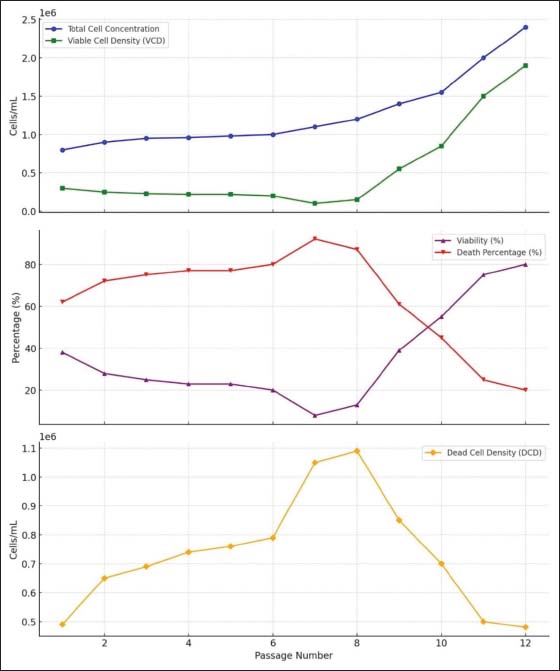
Fig. 1. Assessment of the viability of Vero suspension-adapted cells stained with trypan blue using an automated hemocytometer. Cell growth curve: To monitor adaptation, the total cell concentration, VCD, viability percentage, DCD, and death percentage were recorded at each passage. The data in Figure 2 show a gradual improvement in cell viability and density, indicating successful adaptation of Vero cells to suspension growth conditions. VERO-sus cells achieve concentrations of 2.43 × 10⁶ in spinner flasks. Growth profile of VERO-S adapted in spinner flasks in 12 successive passages with reduced fixed serum concentrations, starting with a total cell concentration of 7.86 × 10⁵ cells/ml. An average cell density of 1.11 × 10⁶ cells/ml was obtained after passage 7, when the total cell concentration began to increase, with minimal viability and high cell death due to new culture conditions such as agitation, altered oxygen transfer, and shear stress, which can transiently reduce cell viability (Sieck et al., 2014). The medium was refreshed every 1–2 days in most passages to improve cell health.
Fig. 2. Analysis of growth curve across the 12 passages. An initial phase of slow adaptation and stress (especially in Passage 7) followed by a local minimum viability after stress induction, followed by viability recovery, due to the selection and expansion of a more adapted cell population capable of growing in suspension. By Passages 10–12, the cells exhibited the characteristics of a successfully adapted suspension culture, with high viability and proliferation, which are crucial for optimizing the adaptation protocol and understanding the cellular response to the transition from adherent to suspension growth. Doubling time and time-dependent growth rate: The growth kinetics of VERO-S cells during adaptation were monitored over 72 hours when cells showed consistent growth and an increase in number, were monitored over 72 hours. The seeding density was 5.8 × 10⁵cells/ml at (passage 9), reaching 1.7 × 10⁶ cells/ml in 72 hours. Cells grew exponentially, with a PDT of 46 hours, indicating that they are dividing slowly but actively proliferating. This suggests that healthy, good-adapting conditions are present during the early adaptation process. Doubling time formula=Td=(t × log(2)) ÷ log(N / N0)
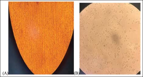
t × log(2)=72 × 0.6931 Log(N /N0)=1.0767 Td=(72 × 0.6931) / 1.0767 Doubling time=46 hours Specific growth rate : μ=specific growth rate (per hour) In, Xn - 1=VCD at time in and tn − (tn − 1) tn – (tn - 1)=time interval in hours The cell morphology of VERO-S: Microscopic observation of VERO-sus cells grown in spinner flasks during cell adaptation. Cells were observed under an inverted light microscope at 20× without staining. The suspension culture primarily consisted of cell aggregates, and some adherent cells were also observed during the 1st two passages, which disappeared as the agitation rate in the spinner flask gradually increased and the serum content decreased. All cells successfully remained in suspension from the third passage onward (Fig. 3).
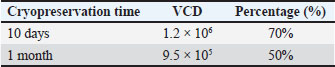
Fig. 3. Microscopic observation of non-stained cells using an inverted light microscope at 40×. (A) Morphologies of Vero cells grown as adherent culture and (B) suspension culture. Cell line sterility: Microbial contamination was absent across all passages of the cell line. No growth was detected in thioglycolate broth, SDA, or tryptic soy broth cultures inoculated with cell culture supernatant from any passage. Establishment of a master cell seed bankCryopreservation of VERO-S cells from passage 12 was achieved using a medium consisting of 80% FBS, 5% DMSO, and 15% MEM. Following cryopreservation, the cells were thawed and incubated in growth medium for 24 hours in a spinner flask at 37°C. Cell viability, assessed by Trypan Blue exclusion, was high post-thaw (Table 1), indicating a high recovery rate following cryopreservation. No significant morphological changes were observed in the suspension-adapted cells upon revival, and they displayed a typical rounded morphology indicative of healthy, proliferating cells. In addition, the cells adapted to suspension culture maintained high viability throughout the 48-hour incubation period, with no signs of significant cell death or aggregation observed in the spinner flask. Table 1. Cell viability after cryopreservation.
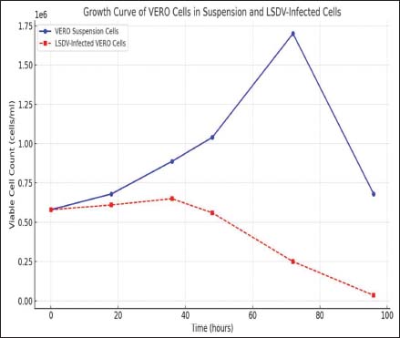
Infection parametersSuspended cell inoculation: Fig. 4 shows the growth kinetics of both uninfected and LSDV-infected VERO-S cells. LSDV-infected cells showed a marked decline in viability at 36 hpi. The rapid reduction to fewer than 104 viable cells/ml by 72 hours and the near-complete collapse of the culture by 96 hours reflect extensive cell death associated with LSDV replication and propagation in suspension-adapted Vero cells.
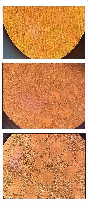
Fig. 4. Growth curves of VERO cells in suspension and LSDV-infected vero cells over time. The curves demonstrate the different proliferative patterns of VERO sus cells compared with VERO cells infected with LSDV over 96 hours. Adherent cell inoculationMorphological changes in adherent cells infected with the virus can provide insight into infection progression and severity. Infected VERO cells typically show distinct CPEs: cell rounding, cell detachment, syncytium formation, and increased cell lysis (Fig. 5).
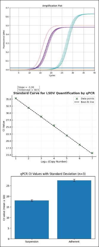
Fig. 5. It shows the cytopathic progression during a 96-hour PI. The uninfected control cells displayed typical adherent epithelial morphology; in contrast, the infected cultures showed a clear CPE. Progressive disruption of the monolayer and an increased number of infected cells. Kinetics of LSDV genome synthesis (real-time PCR) in adherent and suspension VERO cellsReal-time polymerase chain reaction (PCR) amplification curves for LSDV for two samples using the Genetic PCR Solutions™ (dtec-qPCR SHORT PROTOCOL) kit. As shown in Fig. 6a, no amplification was observed in the negative control samples and in the no-template control, confirming the assay specificity.
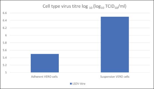
Fig. 6. a. Amplification plots of LSDV by real-time PCR in adherent and suspension Vero cells. Each curve represents a technical replicate. Suspension culture showed earlier amplification with lower CT values than that of adherent cultures. b. Standard curve for LSDV quantification using real-time PCR. c. Comparison of qPCR CT values for virus propagation in adherent and suspension cell cultures. Bars represent the mean CT values ± SD calculated from three technical replicates (n=3). LSDV obtained from suspension cultures showed earlier amplification with Ct values of approximately 18 (n=3), whereas adherent cultures exhibited later amplification with Ct values ranging from approximately 27 to 28 (n=3). The positive control was amplified at Ct ≈16, confirming proper assay performance, whereas the negative control showed no detectable amplification throughout the reaction cycles. Quantification using the standard curve (Fig. 6b) confirmed this observation. The standard curve generated from serial dilutions of LSDV DNA followed a linear relationship with a slope of 3.30 and a Y-intercept of 38.5. Based on these values, the viral genome copy number was calculated for each sample. The suspension culture showed a substantially higher copy number than the adherent cell culture, indicating more efficient virus production in the suspension-adapted system. A linear relationship was observed between Ct values and the viral genome copy number logarithm. The curve was used to estimate the viral load in test samples from their Ct values. To estimate the number of LSDV genome copies in each sample, the Ct values obtained from real-time PCR were converted into copy numbers using the standard curve equation provided with the kits: Ct=Y intercept+slope×log(copy number) Copy number=10 (ct Y intercept)/slope Using the following standard curve parameters: Slope=3.30 Y-intercept=38.5 Adherent cells with a mean CT=27.5 27.5–38.5/−3.30=3.3 copy number=2 × 10³ for suspension cells with a mean CT=18 18–38.5/−3.30=6.2 copy number=1.6 × 10⁶ This calculation clearly shows a markedly higher viral genome load in suspension-adapted Vero cells than in adherent cells under the same experimental conditions (Fig. 6c). Titration of the LSDV virus on adherent and suspension VERO cellsThe virus titer was calculated by inoculating at a cell count of 2 ± 0.07 × 10⁶ at an MOI of 0.1 for both samples (LSDV on adherent cells and suspension VERO cells). The virus was serially diluted tenfold from both cell types and inoculated into 96-well plates seeded with confluent VERO cell monolayers. After a 4-day incubation period at 37°C, the CPE was monitored daily, and the number of positive wells per dilution was recorded, showing 5.5 log10 TCID50/ml and 6.85 log10 TCID50/ml, respectively (Fig. 7). CPE was detected as granular, rounded cells, leaving irregular, empty batches, and vacuolation was completed after 96 hours PI.
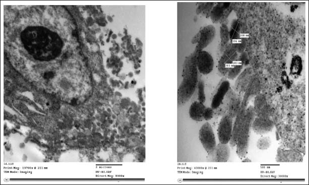
Fig. 7. Titration of the LSDV virus on adherent and suspension VERO cells. Virus detection in VERO sus using TEM and Imaging: Virus particles typically have a dumbbell or oval shape and measure approximately 200–300 nm in diameter. Signs of cellular damage include swollen organelles, distorted cell membranes, and blebbing of the cell surface (Fig. 8). The cells appear slightly irregular in shape, a common feature of suspended cells, with prominent nuclei and visible nucleoli (Shao et al., 2002).
Fig. 8. Cell death (typically necrosis) due to the overwhelming replication of the virus. Virions bud from the cell membrane and form large aggregates of viral particles near the plasma membrane. DiscussionAdherent VERO cells, a well-established cell line derived from African green monkey kidney tissue, have been widely used to propagate many viruses (Rourou et al., 2019), including LSDV. The adaptation procedure lasted 35 days to achieve growth in a low-serum, homemade medium using a stepwise method to obtain VERO-S cells growing as individual cells in suspension. This study explores the challenges and successes encountered during the adaptation process, focusing on key factors such as media optimization, agitation conditions, and the impact of suspension growth on LSD viral titers and yield. During the early stages of adaptation, gradual modifications to the culture conditions were implemented. The serum concentration in the medium was progressively reduced to 5% during the transition from Earl’s salts to high-glucose DMEM. This strategic shift facilitated a smoother transition by allowing cells to adjust to reduced adherence and lower serum dependency, both of which are prerequisites for successful adaptation to suspension growth. This phase laid the foundation for subsequent suspension adaptation, increasing tolerance to low-serum environments—an essential key characteristic for cells propagated in bioreactors for vaccine production (Quesney et al., 2001). In the next phase, the conditions were selected to balance adequate oxygenation and nutrient distribution while minimizing shear stress—a critical factor for maintaining cell viability and supporting progressive adaptation (Tapia et al., 2016). Cells were subcultured every 1–2 days over eight or more passages using 10%–50% of the spent medium from the previous passage. This carryover approach allowed cells to benefit from the conditioned medium and support growth in suspension environments. Successful adaptation was determined by the ability of the cells to maintain exponential growth, achieve a uniform suspension without excessive clumping, and exhibit consistent viability across passages. Cell viability and growth parameters across passages were monitored using the trypan blue exclusion assay coupled with an automated cell counter. Most cells appeared as bright, unstained spherical structures, indicating that the intact plasma membranes could exclude the dye. Only a limited number of dark-stained cells were observed, representing nonviable cells with compromised membrane integrity. This observation suggests that the cell population maintained acceptable viability under the experimental conditions(Strober, 1997). The key metrics—including total cell concentration, VCD, and viability percentage—were consistently recorded during each passage, offering a comprehensive view of the cellular response throughout the adaptation process. Initial passages (1–6) showed moderate increases in total cell number but remained characterized by low viability (20%–38%) and high levels of DCD, indicating that a significant portion of the population was not yet acclimated to suspension conditions. Also, this decrease in cell viability is usually associated with the substrate depletion or accumulation of acid or other metabolic products (Freshney, 2010). By passage 7, although the total cell concentration increased to 1.11 × 10⁶ cells/ml, viability dropped sharply to 6%, suggesting intense selection pressure and a cellular stress response during this transition. The marked decrease in cell viability observed during the early adaptation passages may be associated with metabolic stress experienced by the cells during the transition from adherent to suspension growth. Previous studies have reported that adaptation to suspension culture conditions can lead to a reduction of approximately 20% in glycolytic activity, which correlates with a decrease in cell growth of about 25%–30% and a reduction in maximal metabolic performance (Pech et al., 2021). Such metabolic alterations may impair cellular energy production and proliferation capacity, contributing to transient reductions in viable cell numbers during the adaptation phase. The data indicated that the cell growth in the suspension culture might not be limited only by the presence of nutrients or inhibited by the common metabolites, lactate and ammonia, but also by the accumulation of unknown metabolites in the cellular microenvironment that is probably responsible for the inhibition of cell growth observed under the optimized environmental condition (Shen et al., 2019a,b). This bottleneck appears to represent a critical turning point in adaptation, where initial growth is accompanied by high mortality until subpopulation cells emerge. From passage 8 onwards, a gradual improvement in the viability and VCD was observed. By passage 12, Vero suspension cultures reached a peak VCD of 1.94 × 10⁶ cells/ml with 80% viability—clear evidence of successful adaptation. Cultures were maintained at a pH of 7.6 throughout the adaptation period, and the medium was refreshed every 1–2 days to remove toxic metabolites and improve nutrient availability—practices aligned with standard protocols to maintain cell health during adaptation (Shen et al., 2019). The doubling time, although slower than that of classical suspension lines, reflects active proliferation and suggests that the cells progressed through the adaptation process without significant metabolic impairment (Shen et al., 2019). The acceleration in the cell doubling time of suspension-adapted Vero cells in cultures was due to the partial removal of serum, as the decrease or removal of serum usually increased the cell doubling time, reaching 46 hours. These data validate the phased nature of Vero cell adaptation to suspension. The initial stress-induced decline in viability was followed by a stable and reproducible increase in VCD and viability, indicating that a robust subpopulation was successfully selected. This stable phenotype is critical for large-scale viral vaccine production because it ensures the reliability and scalability of the cell line in suspension bioreactor systems. Establishing a master cell seed bank was critical to ensure the long-term availability and consistency of suspension-adapted Vero cells. A master cell seed bank was established from the 12 passage—the point at which the cells demonstrated optimal viability and stable suspension growth. Cultures were inoculated with LSDV to evaluate the susceptibility and infection kinetics of suspension-adapted Vero cells, and cell viability was tracked post-infection. A pronounced decline in viability was observed following viral exposure (Fig. 4). At 36 hpi, the VCD dropped to 6.5 × 10⁵ cells/ml, and by 96 hours, the population had decreased to 3.5 × 10⁴ cells/ml, with extensive cell death and visible CPEs. These findings confirm the cytolytic nature of LSDV in Vero cells and highlight the utility of suspension-adapted lines for infection modeling. Our results align with previous observations that Vero cells remain highly permissive for Capripoxvirus replication and provide a robust platform for virus production (Pervin et al., 2023). Quantitative PCR analysis was performed to compare viral genome levels in suspension and adherent Vero cell cultures. The amplification curves demonstrated earlier detection in suspension cultures than in adherent cultures. The CT values obtained from three technical replicates showed consistent amplification profiles with low variability between replicates. The mean Ct value for suspension samples was approximately 18.0 ± 0.4, whereas that for adherent cultures was approximately 27.5 ± 0.5. These results indicate substantially higher viral genome levels in suspension cultures than in adherent cells. This finding aligns with the increased viability and growth kinetics observed in later passages of suspension-adapted cells, attributed to the homogeneous distribution of cells and nutrients in suspension and to reduced spatial constraints that can limit virus spread in monolayer cultures. The higher viral titers obtained in suspension-adapted Vero cells suggest that this system may provide advantages over traditional adherent cultures. Suspension cell cultures are generally considered more suitable for scale-up and industrial bioprocessing (St.Amand and Gitschier, 2021). Therefore, the system described here may offer potential for scalable production of the LSD virus. TEM was used to visualize ultrastructural changes in suspension-adapted Vero cells after infection with LSDV. A clear depiction of the nuclear region, surrounded by densely packed cytoplasm, is observed. The presence of multiple electron-dense particles—morphologically and size-wise consistent with poxvirus virions—suggests active viral replication within the cytoplasmic compartment (Hyun, 2022). This observation aligns with the known replication strategy of capripoxviruses, which complete their life cycle entirely in the cytoplasm. Collectively, these observations suggest that suspension-adapted Vero cells not only survive and proliferate well in a dynamic culture system but also support more robust LSDV replication, thus presenting a scalable and efficient platform for future vaccine production. This study is of great significance for LSDV vaccines’ upcoming industrial production. It provides a reference point for the rapid transformation of LSDV adherent cell culture technologies to an intensive level of output. This work will also pave the way for the adaptation of other existing adherent cell lines used for biological production to suspension culture under chemically defined conditions. ConclusionThe successful adaptation of Vero cells to suspension culture represents a promising advancement for the production of high-density LSDV. Although scalability in bioreactors remains to be experimentally verified, these results suggest the potential for improving process control and yield in future vaccine manufacturing. Suspension-adapted Vero cells maintained high viability and enhanced viral replication. Real-time PCR analysis revealed significantly higher LSDV genome yields in suspension cultures than in traditional adherent cells, confirming the suitability of this system for efficient virus amplification. Overall, this approach provides a cost-effective and reproducible method for LSDV propagation and could facilitate future scale-up in controlled bioreactor systems for vaccine production and research. AcknowledgmentsNot applicable. Conflict of interestNone. FundingNot applicable. Authors’ contributionAll authors contributed equally to this study. Data availabilityAll data are included in the manuscript. ReferencesAkther, M., Akter, S.H., Sarker, S., Aleri, J.W., Annandale, H., Abraham, S. and Uddin, J.M. 2023. Global burden of lumpy skin disease, outbreaks, and future challenges. Viruses 15, 1861. Alfano R, Pennybaker A, Halfmann P, Huang, C.Y.H. 2020. Formulation and production of a blood-free and chemically defined virus production media for VERO cells. Biotechnol. Bioeng. 117, 3277–3285; doi: 10.1002/bit.27486 Ali, A.A., Esmat, M., Attia, H., Selim, A. and Abdel-Hamid, Y.M. 1990. Clinical and pathological studies on lumpy skin disease in Egypt. Vet. Rec. 127(22), 549–550. Ammerman, N.C., Beier-Sexton, M. and Azad, A.F. 2008. Growth and maintenance of Vero cell lines. Curr. Protoc. Microbiol. 4, 11. Bazid, A.H., Wasfy, M., Fawzy, M., Nayel, M., Abdelmegeid, M., Thabet, R.Y., Yong, H.S., El-Sayed, M.M., Magouz, A. and Badr, Y. 2023. Emergency vaccination of cattle against lumpy skin disease: evaluation of safety, efficacy, and potency of MEVAC® LSD vaccine containing Neethling strain. Vet. Res. Commun. 47, 767–777. Biopharm International. 2023. The role of media development in process optimization: a historical perspective. Available via https://www.biopharminternational.com/view/role-media-development-process-optimization-historical-perspective Freshney, R.I. 2010 Culture of animal cells: a manual of basic technique and specialized applications. 6th Edition, Hoboken, NJ: John Wiley & Sons Ltd; doi: 10.1002/9780470649367 Harris, C.R., Millman, K.J., Van Der Walt, S.J., Gommers, R., Virtanen, P., Cournapeau, D. and Oliphant, T.E. 2020. Array programming with NumPy. Nature 585, 357–362; doi: 10.1038/s41586-020-2649-2 Hyun, J. 2022. Poxvirus under the eyes of the electron microscope. Appl. Microsc. 52(1), 11. Khafagy, H.A., Abdelwahab, M.G., Mustafa, A.M. and Saad, M.A. 2016. Preparation and field evaluation of live attenuated sheep pox vaccine for protection of calves against lumpy skin disease. Benha. Vet. Med. J. 31, 1–7. Leliso, S.A. 2021. Overview of diagnostic techniques and economic importance of lumpy skin disease. J. Vet. Med. Res. 8(4), 1218. Nikolay, A., Léon, A., Schwamborn, K. Genzel, Y. and Reichl, U. 2018. Process intensification of EB66® cell cultivations leads to high-yield yellow fever and Zika virus production. Appl. Microbiol. Biotechnol. 102, 8725–8737; doi: 10.1007/s00253-018-9275-z Owada, K., Mahony, T.J., Ambrose, R.K., Hayes, B.J. and Soares Magalhães, R.J. 2024. Epidemiological risk factors and modelling approaches for risk assessment of lumpy skin disease virus introduction and spread: methodological review and implications for risk-based surveillance in Australia, Transboundary Emerg. Dis. 3090226, 16; doi: 10.1155/2024/3090226 Pech, S., Rehberg, M., Janke, R., Benndorf, D., Genzel, Y., Muth, T., Sickmann, A., Rapp, E. and Reichl, U. 2021. Tracking changes in adaptation to suspension growth for MDCK cells: cell growth correlates with levels of metabolites, enzymes, and proteins. Appl. Microbiol. Biotechnol. 105(5), 1861–1874. Pervin, S., Ahamed, M., Chouhan, C., Jahan, M., Ahmed, R., Nazir, K., Siddique, M., Rahman, M., Kafi, M. and Rahman, M. 2023. Isolation, adaptation, and characterization of lumpy skin disease virus from cattle in Bangladesh. J. Adv. Vet. Anim. Res. 10(3), 563–569. Piccinini, F., Tesei, A. and Arienti, C. 2017. Cell counting and viability assessment of 2D and 3D cell cultures: expected reliability of the trypan blue assay. Biol. Proced. 19, 8. Posung, M., Promkhatkaew, D., Borg, J. and Tongta, A. 2021. Development of a modified serum-free medium for Vero cell cultures: effects of protein hydrolysates, l-glutamine, and SITE liquid media supplement on cell growth. Cytotechnology 73(5), 683–695. Quesney, S., Marc, A., Gerdil, C., Gimenez, C., Marvel, J., Richard, Y. and Meignier, B. 2003. Kinetics and metabolic specificities of Vero cells in bioreactor cultures with serum-free medium. Cytotechnology 42, 1–11; doi: 10.1023/A:1026185615650 Reed, L.J. and Muench, H. 1938. A simple method of estimating the 50% endpoints. Am. J. Epidemiol. 27, 493–497. Rourou, S., Ben Zakkour, M. and Kallel, H. 2019. Adaptation of Vero cells to suspension growth for rabies virus production in different serum-free media. Vaccine 37(47), 6987–6995. Salib, F.A and Osman, A.H. 2011. Incidence of lumpy skin disease among Egyptian cattle in Giza Governorate, Egypt. Vet. World. 4(4), 162–167. Shao, M., Jiang, L., Cong, W. and Ouyang, F. 2002. Analysis of Vero cell growth behavior on microcarrier by means of environmental scanning electron microscopy. Sci. China Ser. C.-Life Sci. 45, 149–158; doi: 10.1360/02yc9017 Owada, K., Mahony, T.J., Ambrose, R.K., Hayes, B.J. and Soares Magalhães, R.J. 2024. Epidemiological risk factors and modelling approaches for risk assessment of lumpy skin disease virus introduction and spread: methodological review and implications for risk-based surveillance in Australia, Transboundary Emerg. Dis. 3090226, 16; doi: 10.1155/2024/3090226 Sieck, J.B., Budach, W.E., Suemeghy, Z., Leist, C., Villiger, T.K., Morbidelli, M. and Soos, M. 2014. Adaptation for survival: phenotype and transcriptome response of CHO cells to elevated stress induced by agitation and sparging. J. Biotechnol. 189, 94–103; doi:10.1016/j.jbiotec.2014.08.002 St.Amand, T. and Gitschier, H. 2021. Success with adherent or suspension cell cultures: corning Life Sciences suggests ways to balance factors such as scalability, control, and cost. Gen. Engin. Biotechnol. News 41(12), 42–43. Strober, W. 1997. Trypan blue exclusion test of cell viability. Curr. Protocols Immunol. 21, A.3B.1–A.3B.2; doi: 10.1002/0471142735.ima03bs21 Tapia, F., Vázquez-Ramírez, D., Genzel, Y. and Reichl, U. 2016. Bioreactors for high cell density and continuous multi-stage cultivations: options for process intensification in cell culture-based viral vaccine production. Appl. Microbiol. Biotechnol. 100(5), 2121–2132. Tulman, E.R., Afonso, C.L., Lu, Z., Zsak, L., Kutish, G.F. and Rock, D.L. 2001. Genome of lumpy skin disease virus. J. Virol. 75(15), 7122–7130. Tuppurainen, E. and Galon, N. 2016. Technical item II lumpy skin disease: current situation in Europe and neighboring regions and necessary control measures to halt the spread in South-East Europe. Technical Report. OIE Regional Commission, Europe Tuppurainen, E., Dietze, K., Wolff, J., Bergmann, H., Beltran-Alcrudo, D., Fahrion, A., Lamien, C.E., Busch, F., Sauter-Louis, C., Conraths, F.J., De Clercq, K., Hoffmann, B. and Knauf, S. 2021. Review: vaccines and vaccination against lumpy skin disease. Vaccines 9, 1136. Tuppurainen, E.S., Pearson, C.R., Bachanek-Bankowska, K., Knowles, N.J., Amareen, S., Frost, L., Henstock, M.R., Lamien, C.E., Diallo, A. and Mertens, P.P. 2014. Characterization of sheep pox virus vaccine for cattle against lumpy skin disease virus. Antiviral. Res. 109, 1–6. Tuppurainen, E.S.M., Stoltsz, W.H., Troskie, M., Wallace, D.B., Oura, C.A.L., Mellor, P.S., Coetzer, J.A.W. and Venter, E.H. 2011. A potential role for ixodid (Hard) tick vectors in the transmission of lumpy skin disease virus in cattle. Transbound. Emerg. Dis. 58, 93–104. Tuppurainen, E.S.M., Venter, E.H., Shisler, J.L., Gari, G., Mekonnen, G.A., Juleff, N., Lyons, N.A., De Clercq, K., Upton, C., Bowden, T.R., Babiuk, S. and Babiuk, L.A. 2017. Review: capripoxvirus diseases: current status and opportunities for control. Transbound Emerg Dis. 64(3), 729–745; doi: 10.1111/tbed.12444 Whittle, L., Chapman, R. and Williamson, A.L. 2023. Lumpy skin disease: an emerging cattle disease in Europe and Asia. Vaccines (Basel) 11(3), 578. | ||
| How to Cite this Article |
| Pubmed Style Mossa HM, Eid S, El-said AA, Yousif AA, El-sanousi AA. Development of a suspension-based vero cell culture platform for the efficient propagation of lumpy skin disease virus. Open Vet. J.. 2026; 16(5): 3039-3051. doi:10.5455/OVJ.2026.v16.i5.46 Web Style Mossa HM, Eid S, El-said AA, Yousif AA, El-sanousi AA. Development of a suspension-based vero cell culture platform for the efficient propagation of lumpy skin disease virus. https://www.openveterinaryjournal.com/?mno=308793 [Access: June 26, 2026]. doi:10.5455/OVJ.2026.v16.i5.46 AMA (American Medical Association) Style Mossa HM, Eid S, El-said AA, Yousif AA, El-sanousi AA. Development of a suspension-based vero cell culture platform for the efficient propagation of lumpy skin disease virus. Open Vet. J.. 2026; 16(5): 3039-3051. doi:10.5455/OVJ.2026.v16.i5.46 Vancouver/ICMJE Style Mossa HM, Eid S, El-said AA, Yousif AA, El-sanousi AA. Development of a suspension-based vero cell culture platform for the efficient propagation of lumpy skin disease virus. Open Vet. J.. (2026), [cited June 26, 2026]; 16(5): 3039-3051. doi:10.5455/OVJ.2026.v16.i5.46 Harvard Style Mossa, H. M., Eid, . S., El-said, . A. A., Yousif, . A. A. & El-sanousi, . A. A. (2026) Development of a suspension-based vero cell culture platform for the efficient propagation of lumpy skin disease virus. Open Vet. J., 16 (5), 3039-3051. doi:10.5455/OVJ.2026.v16.i5.46 Turabian Style Mossa, Hadeer M., Soad Eid, Amira A. El-said, Ausama A. Yousif, and Ahmed A. El-sanousi. 2026. Development of a suspension-based vero cell culture platform for the efficient propagation of lumpy skin disease virus. Open Veterinary Journal, 16 (5), 3039-3051. doi:10.5455/OVJ.2026.v16.i5.46 Chicago Style Mossa, Hadeer M., Soad Eid, Amira A. El-said, Ausama A. Yousif, and Ahmed A. El-sanousi. "Development of a suspension-based vero cell culture platform for the efficient propagation of lumpy skin disease virus." Open Veterinary Journal 16 (2026), 3039-3051. doi:10.5455/OVJ.2026.v16.i5.46 MLA (The Modern Language Association) Style Mossa, Hadeer M., Soad Eid, Amira A. El-said, Ausama A. Yousif, and Ahmed A. El-sanousi. "Development of a suspension-based vero cell culture platform for the efficient propagation of lumpy skin disease virus." Open Veterinary Journal 16.5 (2026), 3039-3051. Print. doi:10.5455/OVJ.2026.v16.i5.46 APA (American Psychological Association) Style Mossa, H. M., Eid, . S., El-said, . A. A., Yousif, . A. A. & El-sanousi, . A. A. (2026) Development of a suspension-based vero cell culture platform for the efficient propagation of lumpy skin disease virus. Open Veterinary Journal, 16 (5), 3039-3051. doi:10.5455/OVJ.2026.v16.i5.46 |


