





| Case Report | ||
Open Vet. J.. 2026; 16(8): 5544-5554 !
Open Veterinary Journal, (2026), Vol. 16(8): 5544–5554 Case Report Concurrent idiopathic vestibular syndrome and idiopathic facial nerve paralysis in cats: A case reportSiyuan Shi1†, Yuwei Lin2†, Lingsen Lou3, Zhuohua Ao4 and Ming Kang1*1College of Agriculture and Animal Husbandry, Qinghai University, Qinghai, China 2Department of Neurology, Blue Wisdom Veterinary Medical Center, Shanghai, China 3Department of Neurology, Meilianzhonghe Veterinary Hospital, Hangzhou, China 4Department of Neurology, Ainuo Blessing Animal Hospital, Guangzhou, China †Siyuan Shi and Yuwei Lin contributed equally to this work and co-first authors *Corresponding Author: Ming Kang. College of Agriculture and Animal Husbandry, Qinghai University, Qinghai, China.Email: qhukang [at] 163.com Submitted: 27/01/2026 Revised: 11/06/2026 Accepted: 20/06/2026 Published: 08/08/2026 © 2025 Open Veterinary Journal
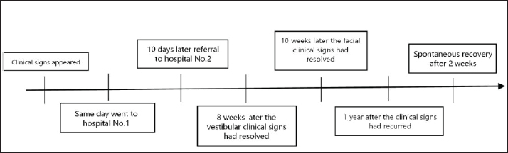
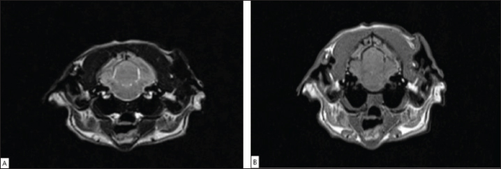
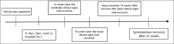
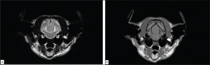
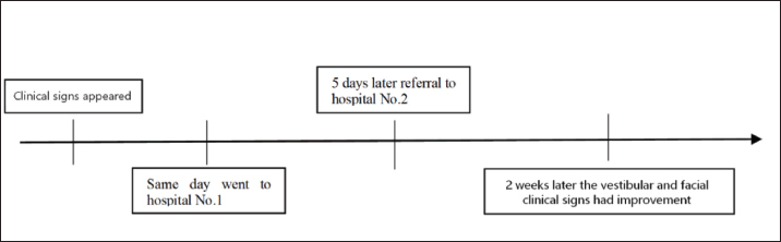
AbstractBackgrounds: Concurrent idiopathic vestibular syndrome (IVS) and idiopathic facial paralysis (IFP) are rare, benign, and self-limiting conditions with a favorable prognosis in cats. Given the rarity of this condition, only a single case report has described its clinical profile. Case Description: This report describes five feline cases with concurrent IVS and IFP. All five cats exhibited clinical signs, including head tilt, vestibular ataxia, and palpebral reflex absence. The diagnosis was established through a comprehensive minimum database, neurological examination, and magnetic resonance imaging, which revealed no significant abnormalities. Notably, all cats gradually improved within 14 weeks, with vestibular signs resolving first. However, two cats experienced recurrence after initial recovery, with spontaneously resolved clinical signs without intervention. Conclusion: Concurrent IVS and IFP in cats are self-limiting, benign conditions with a favorable prognosis. Breed predisposition can be suspected in China. Keywords: Facial nerve paralysis, Feline, Idiopathic vestibular syndrome, MRI, Neurology. IntroductionThe vestibular system receptors are located in the inner ear. The sympathetic trunk to the eyes, including its synapses and postganglionic fibers, is located close to the middle ear cavity. The facial nerve traverses the internal auditory canal alongside the vestibulocochlear nerve into the cranial vault. Consequently, vestibular dysfunction, facial nerve paralysis, and Horner’s syndrome commonly occur in cats with ear disease (Cook, 2003). However, vestibular dysfunction and facial nerve paralysis may also stem from non-otogenic causes, such as head trauma, peripheral nerve damage, intracranial tumors (e.g., meningiomas), systemic polyneuropathies, or idiopathic etiologies. Although hypothyroidism has been linked to these conditions in dogs, there is currently no evidence supporting this association in cats. In the literature, approximately 25% of cats with facial nerve paralysis and 22% with vestibular dysfunction are reported as idiopathic (Braund et al., 1979; Kern et al., 1987; Grapes et al., 2021; Moretto et al., 2021). Although the simultaneous occurrence of idiopathic vestibular syndrome (IVS) and idiopathic facial nerve paralysis (IFP) has been frequently reported in dogs, such cases are exceedingly rare in cats, with only one previous case report (Fraser et al., 2015). This report aims to present five cases of cats experiencing concurrent IVS and IFP, elucidate their clinical signs and prognosis, and explore possible underlying causes. Case DetailsCase 1A 5-year-old male gray American Shorthair cat presented to Shanghai Naughty Family Animal Hospital with a sudden right head tilt. The patient initially exhibited a right head tilt with reduced activity and hiding in the dark. Before referral, the cat was treated with prednisone (unknown dosage), vitamin B, and mirtazapine for 10 days without any improvement. The specific timeline is shown in Fig. 1. Neurological examination identified the following abnormalities, with normal findings omitted for brevity: Gait: Vestibular ataxia with right-sided circling tendency, although the patient could turn to the left. Posture: Right-sided head tilt. Cranial nerves: Absence of the right palpebral reflex and right menace response. These findings suggest neuroanatomical localization to the right peripheral vestibular system and right facial nerve without mentation or proprioceptive deficits. Routine blood tests, including hematological and serum biochemical analyses, and T4 staining, revealed no significant abnormalities. MRI was performed using an MR system (VET0.4, ChuanShanJia Electrical and Mechanical Co., Ltd., Ningbo, China) 9 days later at the owner’s request. MRI findings were within normal limits. Brain MRI is shown in Figure 2. Based on the clinical signs and examinations, IVS and IFP were diagnosed. The cat was treated with a 7-day course of prednisolone (1 mg/kg for the first 4 days and then 0.5 mg/kg for the following 3 days). The head tilt gradually resolved within approximately 8 weeks, followed by facial paralysis resolution within 10 weeks. Approximately 1 year after recovery, similar vestibular signs recurred. Based on the videos provided by the owner, the left palpebral reflex was weakened but not completely absent, and head tilt was observed on the same side. No intervention was performed, and the symptoms resolved spontaneously within 2 weeks. Case 2A 2-year-old male bicolor (yellow and white) British Shorthair cat was referred to Shanghai Naughty Family Animal Hospital due to a sudden inability to close the left eyelid for 6 days and a left head tilt that emerged 4 days earlier. The specific timeline is shown in Figure 3. Neurological examination revealed the following abnormalities, with normal findings omitted for brevity:Gait: Mild vestibular ataxia, especially evident when jumping down onto specific surfaces, with a tendency to drift to the left. Posture: Left-sided head tilt. Cranial nerves: Absence of the left palpebral reflex and left menace response.
Fig. 1. Timeline of case 1.
Fig. 2. Brain MRI of case 1 transverse T2-weighted (A) and T1-weighted (B) images obtained at the level of the ears show no bilateral tympanic bullae or labyrinth abnormalities. The eighth cranial nerves (CN VIII) are also unremarkable.
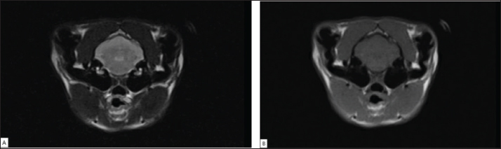
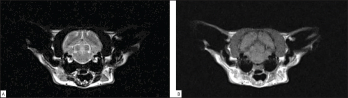
Fig. 3. Timeline of case 2. These findings suggest neuroanatomical localization to the left peripheral vestibular system and left facial nerve without mentation or proprioceptive deficits. Hematological and serum biochemical analyses, serum amyloid A (SAA), and cardiac ultrasound had been performed before referral, all of which yielded normal results. The cat also underwent computed tomography (CT), magnetic resonance imaging (MRI), and cerebrospinal fluid (CSF) analysis. The CT scan did not reveal any abnormalities. Brain MRI was performed using a low-field magnetic resonance system (VET0.35T, Superpet, Kampo Medical System Co., Ltd., Liaoning, China), revealing normal intracranial and osseous structures. The CSF analysis was also normal. Based on the clinical signs and examinations, IVS and IFP were diagnosed. Brain MRI is shown in Figure 4. The owner was advised to replace the tear and monitor the cat’s condition. A follow-up phone call 4 days later revealed that the head tilt had decreased, and the cat could blink slightly when the eye drops were administered. Two weeks later, the owner reported that the cat had fully recovered. Case 3A 2-year-old male British Shorthair cat presented to Hangzhou Meilianzhonghe Veterinary Hospital due to sudden head tilt, vomiting, impaired balance, and ataxia that emerged 4 days earlier. No prior treatment had been administered before the visit. Figure 5 shows the specific timeline. Neurological examination revealed the following abnormalities, with normal findings omitted for brevity:Gait: Mild vestibular ataxia with a tendency to drift to the right without circling. Posture: Right-sided head tilt. Cranial nerves: Absence of the right palpebral reflex and right menace response. Horizontal nystagmus with the fast phase directed to the left. These findings suggest neuroanatomical localization of the right peripheral vestibular system and right facial nerve. Routine blood tests (hematological and serum biochemistry, and T4 level) along with cardiac ultrasonography showed no abnormalities. Blood pressure was 160 mmHg with a heart rate of 180 bpm. MRI using a low-field system (VET0.3, ChuanShanJia Electrical and Mechanical Co., Ltd., Ningbo, China) was performed immediately, revealing normal intracranial structures and vestibular system morphology. Brain MRI is shown in Figure 6. The clinical presentation and diagnostic findings supported the diagnosis of IVS and IFP. The treatment consisted of oral administration of prednisolone (1 mg/kg daily for 2 weeks) combined with acupuncture, traditional Chinese herbal therapy, and physical rehabilitation over 3 weeks. Despite this multimodal approach, no significant improvement was observed. Notably, the head tilt resolved spontaneously within 12 weeks, followed by complete resolution of facial paralysis by week 14. Remarkably, the cat exhibited identical clinical signs ipsilateral to the initial presentation at 16 weeks post-recovery. Repeat MRI demonstrated no structural abnormalities, with spontaneous resolution of symptoms within 12 weeks without therapeutic intervention. Case 4A 3-year-old spayed female silver-gray British Shorthair cat was presented to Guangzhou Ainuo Blessing Animal Hospital due to the sudden onset of vestibular signs and right-sided facial paralysis. Before referral, the cat had been treated with an unknown dose of cephalosporin antibiotics, prednisolone, and vitamin B12, which resulted in a slight improvement in gait and a weak palpebral reflex in the right eyelid. The specific timeline is shown in Figure 7. Neurological examination revealed the following abnormalities, with normal findings omitted for brevity:Gait: Mild vestibular ataxia with a tendency to drift to the right without circling. Posture: Right-sided head tilt. Cranial nerves: Absence of the right palpebral reflex and right menace response. Horizontal nystagmus with the fast phase directed to the left. These findings suggest neuroanatomical localization of the right peripheral vestibular system and right facial nerve. Routine blood tests, including hematological and serum biochemical analyses, SAA, T4 testing, cardiac ultrasonography, and ear examinations, revealed no abnormalities. Brain MRI was scheduled for the following day using a low-field MR system (MAGNETOM C!, Siemens Healthineers, Erlangen, Germany). MRI revealed normal intracranial and osseous structures. CSF analysis was not performed because of the owner’s consideration. A preliminary diagnosis of right-sided IVS and IFP was made. Brain MRI is shown in Figure 8. The cat was discharged with oral administration of prednisolone (1.6 mg/kg) daily for 2 weeks, a 4-week course of mecobalamin (0.16 mg/kg for the first 2 weeks, then 0.08 mg/kg for the following 2 weeks), and doxycycline (8.3 mg/kg) daily for 2 weeks. After 2 weeks, a follow-up examination revealed resolution of the head tilt, and the cat could close its eyes.
Fig. 4. Brain MRI of case 2: Transverse T2-weighted (A) and T1-weighted (B) images showing normal bilateral tympanic bullae, labyrinths, and CN VIII.
Fig. 5. Timeline of case 3.
Fig. 6. Brain magnetic resonance imaging of Case 3: Transverse T2-weighted (A) and T1-weighted (B) images show normal bilateral tympanic bullae and labyrinths, with no abnormalities detected in CN VIII. Case 5A 1-year-old intact male white British Shorthair cat was presented to Guangzhou Ainuo Blessing Animal Hospital with an acute onset of right-sided head tilt and inability to close the right eye, which was noted 2 days prior. No medications were administered before presentation. The specific timeline is shown in Figure 9. Neurological examination revealed the following abnormalities, with normal findings omitted for brevity:Gait: Mild vestibular ataxia with a tendency to drift to the right without circling. Posture: Right-sided head tilt. Cranial nerves: Absence of the right palpebral reflex and right menace response. Horizontal nystagmus with the fast phase directed to the left. These findings suggest neuroanatomical localization of the right peripheral vestibular system and right facial nerve.
Fig. 7. Timeline of case 4.
Fig. 8. Brain MRI of case 4. Transverse T2-weighted (A) and T1-weighted (B) images reveal normal bilateral tympanic bullae and labyrinths, with CN VIII appearing normal.
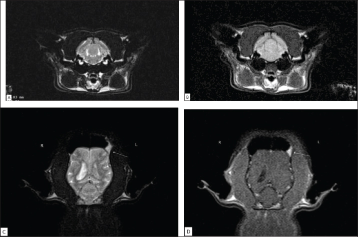
Fig. 9. Timeline of Case 5. The patient underwent a comprehensive diagnostic workup, including hematological and serum biochemical analyses, SAA, T4, cardiac and abdominal ultrasonography, otoscopic examination, and fecal analysis, which revealed no abnormalities. MRI was performed using a low-field MR system (MAGNETOM C!, Siemens Healthineers, Erlangen, Germany) and revealed a small, unknown T2-weighted and T1-weighted hyperintense lesion in the left frontal sinus, mild dilation of the right ventricle, and no abnormal signal areas in the brain parenchyma or in the osseous vestibular structures. CSF analysis was not performed due to the owner’s decision. Based on the clinical presentation and examination, we made a preliminary diagnosis of right-sided IVS and IFP. Brain MRI is shown in Fig. 10. The cat was discharged with an 8-day course of oral administration of prednisolone (0.5 mg/kg) once daily and maropitant (1.8 mg/kg) once daily for 2 days. Initially, no improvement was observed; however, the head tilt and facial paralysis resolved within 20 days and 11 weeks, respectively.
Fig. 10. Figure 5.2. Brain MRI of case 5: Transverse T2-weighted (A) and T1-weighted (B) images showing normal bilateral tympanic bullae, labyrinths, and eighth cranial nerves (CN VIII). A focal hyperintense lesion is identified within the left frontal sinus on the dorsal T2-weighted (C) and T1-weighted (D) images (white arrow). R=right; L=left.
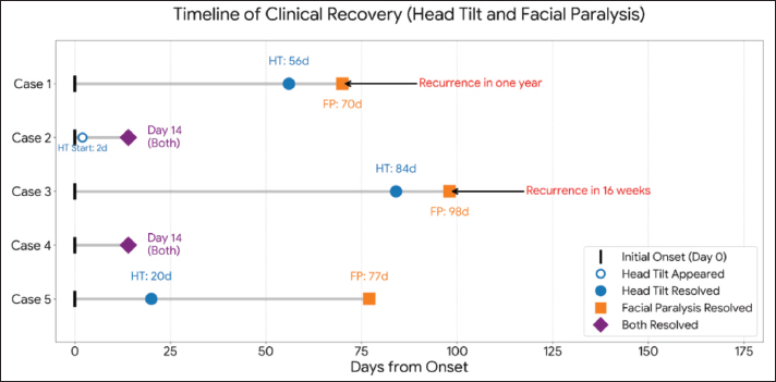
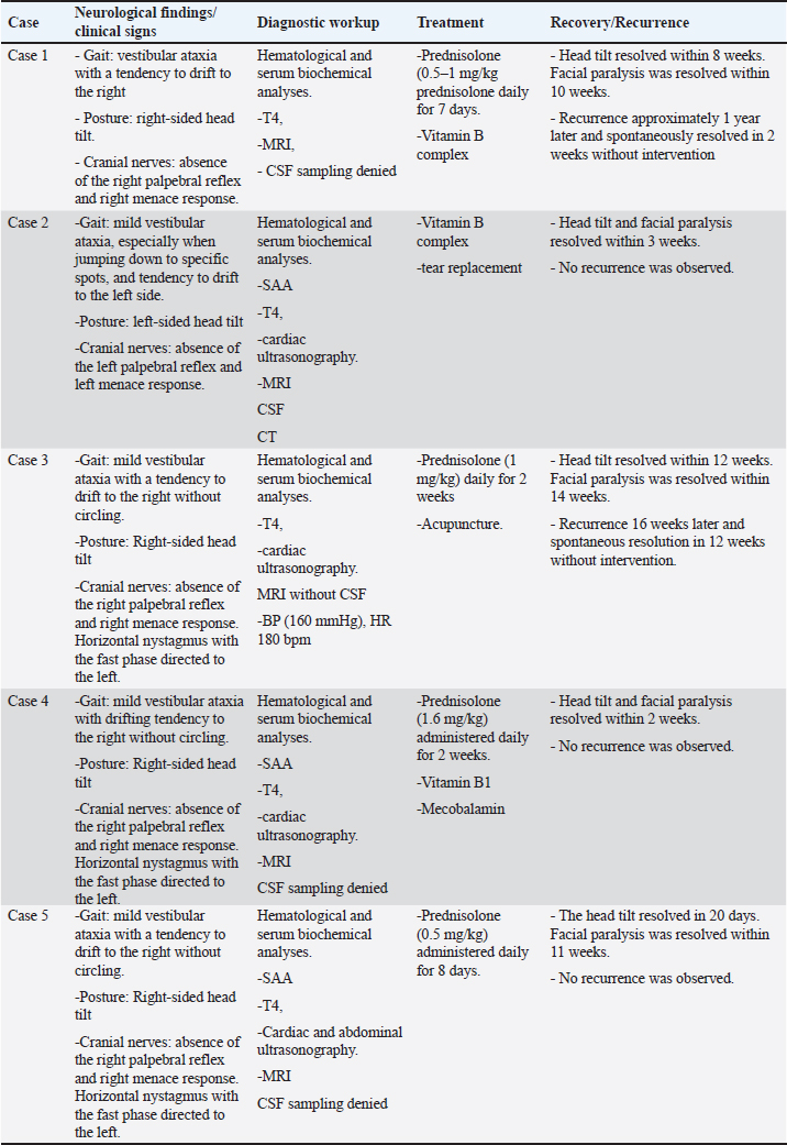
Fig. 11. Timeline of five felines cases clinical recovery. A timeline diagram (Fig. 11) was constructed to facilitate comparison of disease course and clinical outcome among the five cases to summarize the onset and resolution of head tilt and facial paralysis. This figure illustrates the temporal relationships among initial clinical signs, symptom duration, and recurrence, enabling visualization of inter-individual variability in clinical progression and resolution. Ethical approvalThe five cases were reported with informed owners’ consent. According to national regulations, no further ethical statement is necessary. DiscussionThis case series identified several consistent clinical patterns in cats with concurrent IVS and IFP. First, the clinical progression of this dual neuropathy appears to follow a benign and self-limiting course, mirroring the known natural history of isolated feline IVS (PNegrin et al., 2010). All five cats in our cohort achieved full clinical resolution of both vestibular and facial nerve deficits within a 2–14-week window. A distinctive temporal pattern was observed: vestibular signs (e.g., ataxia and head tilt) typically showed earlier improvement than the more protracted recovery of facial nerve function. Most cats with IVS exhibit spontaneous clinical improvement within weeks, with no definitive treatment proven effective (Mertens et al., 2023). The current treatment approach for cats with IVS is similar to that used in dogs, primarily involving supportive care such as fluid therapy and antiemetic medications. Glucocorticosteroid use has been reported in some veterinarians in North America, although its benefit in feline IVS remains unclear (Kent et al., 2009; PNegrin et al., 2010; Mertens et al., 2023). Our observations suggest that recovery occurs independently of specific pharmacological intervention. While five cats received varying doses of glucocorticoids, one cat that was managed solely with vitamin B complex recovered spontaneously within 3 weeks. Most notably, during the second episode, the 2 cases of recurrence (Cases 3 and 5) achieved complete resolution without any medical treatment. This spontaneous recovery in the absence of therapy provides strong evidence for this syndrome’s self-limiting nature and suggests that aggressive corticosteroid protocols may not significantly alter the long-term prognosis. However, studies in human medicine have shown that prednisone may facilitate early recovery in vestibular neuritis, although it does not appear to influence long-term prognosis (Shupak et al., 2008). In addition, evidence from clinical trials supports the use of early glucocorticosteroids in IFP (Sullivan et al., 2007; Madhok et al., 2016). In our case, one cat who was administered a relatively higher dose of prednisolone (1.6 mg/kg once daily) recovered within 2 weeks. However, given the limited number of cases in which we used high-dose glucocorticoids, further investigation is required to determine whether high-dose glucocorticoids can accelerate symptomatic recovery. Our findings deviate from the typical geriatric profile associated with canine IVS in terms of signaling (Mertens et al., 2023). All cats in this series were 6 years of age. Although 80% (4/5) of the affected cats were British Shorthairs, we interpret this as a reflection of regional breed popularity rather than a definitive genetic predisposition. Given that British Shorthairs represent approximately 20% of the local feline population, their overrepresentation in this small cohort remains descriptive rather than statistically predictive (Daxue, 2025). Collectively, these findings suggest that concurrent IVS and IFP in young to middle-aged cats carries a highly favorable prognosis regardless of the treatment regimen employed. In this case series, the majority (3/5) of patients showed significant improvement or complete recovery of vestibular symptoms within 2–3 weeks. This finding is consistent with previous reports indicating that IVS in cats typically improves within 2–4 weeks, and the prognosis for IVS in cats is generally good to excellent (PNegrin et al., 2010; Fraser et al., 2015). The remaining 2 cats showed resolution of vestibular symptoms within 8 and 12 weeks, respectively. Furthermore, in all five cases, the vestibular symptoms resolved before the resolution of facial paralysis. This finding is consistent with previous canine studies, which indicate that recovery from idiopathic facial nerve paralysis typically involves longer periods in dogs than in cats (Varejão et al., 2006; Fraser et al., 2015). Furthermore, the prognosis for dogs with concurrent IVS and IFP appears to be poorer than that for affected cats. One report documented complete recovery in only 4/16 of the dogs, with 9/16 exhibiting residual neurological deficits (Sjögren et al., 2019). A second study found persistent clinical signs in 47/94 dogs diagnosed with this comorbidity (Orlandi et al., 2020). In dogs, approximately 2 of 16 cases of concurrent IVS and IFP experienced recurrence, a phenomenon also observed in humans with BP (Jeandel et al., 2016). Two cats (2/5) exhibited recurrence, with clinical signs similar to the initial episode. Notably, one cat experienced a significantly shorter symptom duration during the second episode. A summary of the five reported cases is presented in Table 1. However, the small sample size in our study limited the ability to draw definitive conclusions about the prevalence of recurrence in cats with concurrent IVS and IFP. This study aimed to enhance the understanding of this rare condition and offer guidance in its diagnosis and management. Despite these findings’ clinical significance, several limitations must be acknowledged. First, the diagnosis of “idiopathic” condition in this series remains presumptive rather than definitive. Owing to the retrospective nature of the study and clinical constraints, a standardized diagnostic protocol was not universally applied. CSF analysis and comprehensive infectious disease screening (specifically for feline herpesvirus-1 titers) were not performed in all cases. Concurrently, studies have detected canine herpesvirus-1 in the vestibular labyrinth and ganglia of some normal dogs, and the presence of feline herpesvirus-1 has been demonstrated in the vestibular ganglia of domestic cats. These studies suggest that reactivated herpesvirus infection may result in anterograde transmission from the trigeminal nerve to the vestibular nucleus in cats and dogs (Parzefall et al., 2010, 2011). Recurrent vestibulopathy is associated with the reactivation of herpes simplex virus type 1 in humans (Richard et al., 2008). Therefore, although the association of herpesvirus infection with feline idiopathic vestibular disease has not been clearly demonstrated by any reports, screening for herpesvirus is recommended in the diagnosis of feline IVS, and this also constitutes a limitation of our study. Table 1. Summary of five cases with IVS and concurrent IFP.
In many instances, peripheral neuroanatomical localization combined with rapid early clinical improvement led owners to decline invasive procedures. As veterinary neurology is a burgeoning specialty in China, the owner’s perceived risk-benefit ratio frequently influences clinical decisions, which may result in a less exhaustive diagnostic workup than that seen in academic teaching hospitals. Second, potential systemic contributors, specifically for hypertension, were not consistently ruled out (Mertens et al., 2023). In clinical practice, routine otoscopy and advanced techniques, such as MRI and CSF analysis, are used to diagnose IVS. However, the diagnostic approaches differ between cats and dogs. Thyroid levels are prioritized in dogs, whereas blood pressure measurement is more critical in cats (Mertens et al., 2023). Studies have suggested that IFP in dogs resembles Bell’s palsy in humans, which can be either idiopathic or associated with hypertension (Varejão et al., 2006; Moretto et al., 2021). MRI scans in cats with IFP have identified changes in the midbrain, which are consistent with hypertension-induced hemorrhage, but no changes in the facial nerve nucleus region (Moretto et al., 2021). Systemic hypertension is a known cause of cranial neuropathies in cats; however, blood pressure was routinely measured in only 1 case in this cohort. Although that individual showed a systolic reading of 160 mmHg, the lack of repeated blood pressure measurements over a 4–8 week period prevents us from definitively excluding hypertensive neuropathy as a differential diagnosis (Acierno et al., 2018). Inherent limitations exist in the diagnostic imaging employed. While MRI is the gold standard for distinguishing central from peripheral vestibular disease, conventional sequences may lack the sensitivity to detect subtle inflammatory or degenerative changes in small cranial nerves (Bischoff et al., 2004; Fraser et al., 2015). The absence of advanced high-resolution sequences, such as volumetric interpolated breath-hold examination, which has shown superior sensitivity in canine facial neuropathy, may have resulted in the oversight of subtle structural abnormalities in our feline patients (Couturier et al., 2005; Smith et al., 2012). Finally, the small sample size (n=5) limits our ability to draw broad conclusions regarding breed predispositions or the definitive efficacy of glucocorticoid therapy. Although this series provides a much-needed expansion of the existing literature on this rare comorbidity, future prospective studies with standardized diagnostic criteria—including universal CSF analysis and blood pressure screening—are necessary to further elucidate the underlying pathophysiology of concurrent IVS and IFP in the feline population. ConclusionAll cats in this study were diagnosed with concurrent IVS and IFP, and all patients demonstrated spontaneous recovery within a range of 2–14 weeks without requiring antibiotic or corticosteroid therapy. In our case, although the majority (n=4) received pharmacotherapy, no improvement was observed during the treatment period. Concurrent IVS and IFP in cats is a self-limiting, benign condition with a favorable prognosis. Breed predisposition can be suspected in China. Given the limited number of reported cases of feline patients presenting with both symptoms simultaneously, this study aims to provide valuable references and considerations for clinical reasoning. By presenting these cases, we seek to enhance the understanding of this rare condition and offer guidance in its diagnosis and management. AcknowledgmentsThe authors would like to thank the staff of Shanghai Naughty Family Animal Hospital, Hangzhou Meilianzhonghe Veterinary Hospital, and Guangzhou Ainuo Blessing Animal Hospital for their cooperation with the clinical cases. FundingThis research was funded by the National Natural Science Foundation of China (Grant No. 31660698 and No. 32600806). Authors’ contributionsAll authors met the criteria for authorship. Shi S.Y., Lin Y.W., Lou L.S., Ao Z.H., and Kang M. participated in clinical case management. J Clin Investig Shi S.Y. drafted the manuscript and prepared Figures and Table 1. Lin Y.W. reviewed the neuroimaging studies, Shi S.Y., Lin Y.W., and Kang M. reviewed and edited the manuscript, Lin Y.W., Lou L.S., and Ao Z.H. provided the medical case information, and Kang M. provided funding. All authors have read and approved the published version of the manuscript. Conflicts of interestThe authors have no conflicts of interest to declare. Data availabilityAll data were provided in this case report. ReferencesAcierno, M.J., Brown, S., Coleman, A.E., Jepson, R.E., Papich, M., Stepien, R.L. and Syme, H.M. 2018. ACVIM consensus statement: guidelines for the identification, evaluation, and management of systemic hypertension in dogs and cats. J. Vet. Intern. Med. 32(6), 1803–1822; doi:10.1111/jvim.15331 Bischoff, M.G. and Kneller, S.K. 2004. Diagnostic imaging of the canine and feline ear. Vet. Clin. North. Am. Small. Anim. Pract. 34(2), 437–458; doi:10.1016/j.cvsm.2003.10.013 Braund, K., Luttgen, P., Sorjonen, D. and Redding, R. 1979. Idiopathic facial paralysis in the dog. Vet. Rec. 105(13), 297–299; doi:10.1136/vr.105.13.297 Cook, L.B. 2004. Neurologic evaluation of the ear. Vet. Clin. North. Am. Small. Anim. Pract. 34(2), 425–435; doi:10.1016/j.cvsm.2003.12.001 Couturier, L., Degueurce, C., Ruel, Y., Dennis, R. and Begon, D. 2005. Anatomical study of cranial nerve emergence and skull foramina in the dog using magnetic resonance imaging and computed tomography. Vet. Radiol. Ultrasound 46(5), 375–383; doi:10.1111/j.1740-8261.2005.00068.x Daxue Consulting. 2025. 2025 China’s pet economy report. Available at https://daxueconsulting.com/wp-content/uploads/2025/06/2025-Chinas-Pet-Economy-Report-Daxue-Consulting.pdf Fraser, A., Long, S. and Le Chevoir, M. 2015. Concurrent idiopathic vestibular syndrome and facial nerve paralysis in a cat. Aust. Vet. J. 93(7), 252–254; doi:10.1111/avj.12338 Gacek, R.R. 2008. Evidence for a viral neuropathy in recurrent vertigo. ORL 70(1), 6–15; doi:10.1159/000111042 Grapes, N.J., Taylor-Brown, F.E., Volk, H.A. and De Decker, S. 2021. Clinical reasoning in feline vestibular syndrome: which presenting features are the most important?. J. Feline Med. Surg. 23(8), 669–678; doi:10.1177/1098612X20970869 Jeandel, A., Thibaud, J.L. and Blot, S. 2016. Facial and vestibular neuropathy of unknown origin in 16 dogs. J. Small. Anim. Pract. 57(2), 74–78; doi:10.1111/jsap.12428 Kent, M., Platt, S.R. and Schatzberg, S.J. 2010. The neurology of balance: function and dysfunction of the vestibular system in dogs and cats. Vet. J. 185(3), 247–258; doi:10.1016/j.tvjl.2009.10.029 Kern, T.J. and Erb, H.N. 1987. Facial neuropathy in dogs and cats: 95 cases (1975-1985). J. Am. Vet. Med. Assoc. 191(12), 1604–1609. Mertens, A.M., Schenk, H.C. and Volk, H.A. 2023. Current definition, diagnosis, and treatment of canine and feline idiopathic vestibular syndrome. Front. Vet. Sci. 10, 1263976; doi:10.3389/fvets.2023.1263976 Moretto, L., Herzig, R., Beckmann, K., Wolfer, N., Dennler, M. and Glaus, T.M. 2021. Reversible facial nerve paralysis in a cat suspected to be associated with systemic hypertension. JFMS. Open Rep. 7(2), 20551169211063454; doi:10.1177/20551169211063454 Orlandi, R., Gutierrez-Quintana, R., Carletti, B., Cooper, C., Brocal, J., Silva, S. and Gonçalves, R. 2020. Clinical signs, MRI findings and outcome in dogs with peripheral vestibular disease: a retrospective study. BMC. Vet. Res. 16(1), 159; doi:10.1186/s12917-020-02366-8 Parzefall, B., Fischer, A., Blutke, A., Schmahl, W. and Matiasek, K. 2011. Naturally-occurring canine herpesvirus-1 infection of the vestibular labyrinth and ganglion of dogs. Vet. J. 189(1), 100–102; doi:10.1016/j.tvjl.2010.06.014 Parzefall, B., Schmahl, W., Fischer, A., Blutke, A., Truyen, U. and Matiasek, K. 2010. Evidence of feline herpesvirus-1 DNA in the vestibular ganglion of domestic cats. Vet. J. 184(3), 371–372; doi:10.1016/j.tvjl.2009.03.030 PNegrin, A., Cherubini, G.B., Lamb, C., Benigni, L., Adams, V. and Platt, S. 2010. Clinical signs, magnetic resonance imaging findings and outcome in 77 cats with vestibular disease: a retrospective study. J. Feline Med. Surg. 12(4), 291–299; doi:10.1016/j.jfms.2009.10.001 Salinas, R.A., Alvarez, G. and Ferreira, J. 2016. Corticosteroids for Bell‘s palsy (idiopathic facial paralysis). Cochrane Database Syst. Rev. 7(7), CD001942; doi:10.1002/14651858.CD001942.pub5 Shupak, A., Issa, A., Golz, A., Kaminer, M. and Braverman, I. 2008. Prednisone treatment for vestibular neuritis. Otol. Neurotol. 29(3), 368–374; doi:10.1097/MAO.0b013e3181692804 Sjögren, J., Magnusson, M., Tjernström, F. and Karlberg, M. 2019. Steroids for acute vestibular neuronitis-the earlier the treatment, the better the outcome?. Otol. Neurotol. 40(3), 372–374; doi:10.1097/MAO.0000000000002106 Smith, P.M., Gonçalves, R. and Mcconnell, J.F. 2012. Sensitivity and specificity of MRI for detecting facial nerve abnormalities in dogs with facial neuropathy. Vet. Rec. 171(14), 349; doi:10.1136/vr.100877 Sullivan, F.M., Swan, I.R.C., Donnan, P.T., Morrison, J.M., Smith, B.H., Mckinstry, B., Davenport, R.J., Vale, L.D., Clarkson, J.E., Hammersley, V., Hayavi, S., Mcateer, A., Stewart, K. and Daly, F. 2007. Early treatment with prednisolone or acyclovir in Bell‘s palsy. N. Engl. J. Med. 357(16), 1598–1607; doi:10.1056/NEJMoa072006 Varejão, A.S.P., Muñoz, A. and Lorenzo, V. 2006. Magnetic resonance imaging of the intratemporal facial nerve in idiopathic facial paralysis in the dog. Vet. Radiol. Ultrasound. 47(4), 328–333; doi:10.1111/j.1740-8261.2006.00148.x | ||


| How to Cite this Article |
| Pubmed Style Shi S, Lin Y, Lou L, Ao Z, Kang M. Concurrent idiopathic vestibular syndrome and idiopathic facial nerve paralysis in cats: A case report. Open Vet. J.. 2026; 16(8): 5544-5554. doi:10.5455/OVJ.2026.v16.i8.48 Web Style Shi S, Lin Y, Lou L, Ao Z, Kang M. Concurrent idiopathic vestibular syndrome and idiopathic facial nerve paralysis in cats: A case report. https://www.openveterinaryjournal.com/?mno=308185 [Access: August 08, 2026]. doi:10.5455/OVJ.2026.v16.i8.48 AMA (American Medical Association) Style Shi S, Lin Y, Lou L, Ao Z, Kang M. Concurrent idiopathic vestibular syndrome and idiopathic facial nerve paralysis in cats: A case report. Open Vet. J.. 2026; 16(8): 5544-5554. doi:10.5455/OVJ.2026.v16.i8.48 Vancouver/ICMJE Style Shi S, Lin Y, Lou L, Ao Z, Kang M. Concurrent idiopathic vestibular syndrome and idiopathic facial nerve paralysis in cats: A case report. Open Vet. J.. (2026), [cited August 08, 2026]; 16(8): 5544-5554. doi:10.5455/OVJ.2026.v16.i8.48 Harvard Style Shi, S., Lin, . Y., Lou, . L., Ao, . Z. & Kang, . M. (2026) Concurrent idiopathic vestibular syndrome and idiopathic facial nerve paralysis in cats: A case report. Open Vet. J., 16 (8), 5544-5554. doi:10.5455/OVJ.2026.v16.i8.48 Turabian Style Shi, Siyuan, Yuwei Lin, Lingsen Lou, Zhuohua Ao, and Ming Kang. 2026. Concurrent idiopathic vestibular syndrome and idiopathic facial nerve paralysis in cats: A case report. Open Veterinary Journal, 16 (8), 5544-5554. doi:10.5455/OVJ.2026.v16.i8.48 Chicago Style Shi, Siyuan, Yuwei Lin, Lingsen Lou, Zhuohua Ao, and Ming Kang. "Concurrent idiopathic vestibular syndrome and idiopathic facial nerve paralysis in cats: A case report." Open Veterinary Journal 16 (2026), 5544-5554. doi:10.5455/OVJ.2026.v16.i8.48 MLA (The Modern Language Association) Style Shi, Siyuan, Yuwei Lin, Lingsen Lou, Zhuohua Ao, and Ming Kang. "Concurrent idiopathic vestibular syndrome and idiopathic facial nerve paralysis in cats: A case report." Open Veterinary Journal 16.8 (2026), 5544-5554. Print. doi:10.5455/OVJ.2026.v16.i8.48 APA (American Psychological Association) Style Shi, S., Lin, . Y., Lou, . L., Ao, . Z. & Kang, . M. (2026) Concurrent idiopathic vestibular syndrome and idiopathic facial nerve paralysis in cats: A case report. Open Veterinary Journal, 16 (8), 5544-5554. doi:10.5455/OVJ.2026.v16.i8.48 |


