





| Research Article | ||
Open Vet. J.. 2026; 16(5): 2939-2947 Open Veterinary Journal, (2026), Vol. 16(5): 2939-2947 Research Article Genetic diversity and molecular identification of sheep infected with Theileria ovis in Central IraqMonyer Abdulameir Abd Alfatlawi* and Mohammed Qasim HadiDepartment of Veterinary Microbiology, College of Veterinary Medicine, University of Al-Qadisiyah, Al Diwaniyah, Iraq *Corresponding Author: Monyer Abdulameir Abd Alfatlawi. Department of Veterinary Microbiology, College of Veterinary Medicine, University of Al-Qadisiyah, Al Diwaniyah, Iraq. Email: monyerr.abd [at] qu.edu.iq Submitted: 18/01/2026 Revised: 15/03/2026 Accepted: 25/03/2026 Published: 31/05/2026 © 2025 Open Veterinary Journal
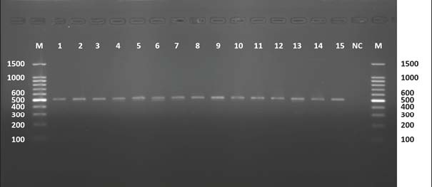
AbstractBackground: The parasitic disease Theileria ovis (Ovine Theileriosis) affects the health and productivity of infected sheep and sheep-based industries, especially in areas impacted by endemic tick-borne disease. Aim: This study aimed to achieve molecular characterization of T. ovis in sheep in Central Iraq and analyze the genetic diversity and phylogenetics of the local isolates through partial 18S ribosomal RNA (rRNA) gene sequencing. Methods: A complete blood sample was collected from the sheep, from which genomic DNA was extracted. For the molecular characterization of T. ovis, a nested conventional polymerase chain reaction (PCR) was performed targeting the 18S rRNA gene. Positive PCR amplicons were selected and subjected to Sanger sequencing, after which the sequencing data were edited and compared to GenBank and other countries’ data using NCBI-BLAST. The phylogenetic analysis was based on the Maximum Likelihood method and the Tamura-Nei model. Sequence alignment was performed to determine the variation and similarity in nucleotides among the different isolates. Results: The expected 520-bp fragment of the 18S rRNA gene in the sheep samples was successfully amplified using nested PCR, confirming the presence of T. ovis infections. Ten representative isolates were sequenced, and the resulting sequences were submitted to GenBank with the following accession numbers: PX755062 to PX755071. A BLAST analysis of the submitted sequences showed a high nucleotide identity percentage to the previously characterized T. ovis isolates from different countries, ranging from 98.81% to 100%. The isolates from Egypt and Türkiye exhibited the greatest similarity, whereas the isolates from Pakistan, China, Japan, and Iraq had slightly lower identity values than the isolates from other countries. Phylogenetic analysis indicated that the T. ovis clade also contained the Iraqi sheep isolates, and in conjunction with the global reference strains, the identity of the T. ovis species was confirmed, indicating a low level of genetic diversity. Conclusion: This study offers clear molecular data regarding the circulation of T. ovis in sheep in central Iraq. Iraqi isolates and global strains have high genetic similarity. This proves that a conserved 18S rRNA gene with minor regional variation exists. The data hydra provides to the epidemiology of ovine theileriosis in Iraq is significant and exemplifies the need for complex molecular surveillance to monitor and control the disease. Keywords: Theileria ovis, Sheep, Nested PCR, 18S rRNA gene, Genetic diversity, Phylogenetic analysis, Iraq. IntroductionOvine theileriosis is a small ruminant disease caused by the tick-borne hemoprotozoan parasite of the genus Theileria, with Theileria ovis being one of the most common species reported to infect sheep (Tanveer et al., 2022; Almahallawi et al., 2024). Theileria ovis is considered to have the lowest pathogenicity of the species. However, this parasite is present in a large geographic range. It has a high infection rate of ovis of small ruminant infections, resulting in high rates of subclinical infections and contributing to economic losses to sheep breeding systems. Theileria ovis infections have become a significant problem in unsophisticated sheep breeding in tick-infested areas. Theileria ovis infection and its clinical symptoms are significant from an epidemiological standpoint in T. ovis-infested areas. Microscopic examination of blood smears forms the basis of ovine theileriosis diagnosis. This method has poor sensitivity in subclinical, chronic, or carrier state infections and is the basis of the poor diagnostic accuracy in subclinical, chronic, or carrier state infections. The need for advanced diagnostic methods has led to the development of a new diagnostic approach. Theileria species infections are currently being diagnosed and characterized at the species level (Nangru et al., 2022). Due to its structure and variability, the small subunit ribosomal RNA (18S rRNA) gene has been used for the distinctive detection, genetic characterization, and phylogenetic analysis of Theileria species (Prajapati et al., 2023). Recent research on the molecular level of T. ovis has been conducted in different parts of the globe, and a large degree of genetic conservation of the organism and a few nucleotides that differ may indicate a geographical separation, host adaptation, or localized transmission of the organism (Arif et al., 2023; Irfan et al., 2023). Phylogenetic studies focusing on 18S rRNA gene sequencing have shown that T. ovis isolates cluster by geography. This proves that there is a strong molecular value, and it is also a contributing factor toward understanding the dispersal of the parasites and their evolutionary patterns. This also imposes the need to develop certain local sequenced data, which is imperative to guarantee the placement of certain mixture isolates within a global phylogenetic context. Iraq, like other nations in the Middle East, has huge sheep production volumes, which are significant to food security and the sustenance of rural livelihoods. There is also an obvious shortage of molecular data on T. ovis in sheep, which is either scattered, too fragmented, or limited to a certain region. There has also been a shortage of studies dealing with the comprehensive genetic characterization of sheep isolates from Iraq. Some recent studies from countries that border Iraq, such as Saudi Arabia, Türkiye, Pakistan, and Iran, have reported the molecular detection and genetic analysis of T. ovis in small ruminants (Tanveer et al., 2022; AlFaleh, 2025). This scenario also causes a definite gap that limits the study of regional genetic diversity, the epidemiology of the parasites, and the Iraqi strains with regard to the global T. ovis populations. Nested polymerase chain reaction (PCR) integrated with Sanger sequencing and phylogenetic examination offers a solid method for verifying the species and analyzing the genetic divergence of T. ovis isolates. Additionally, the submission of locally obtained sequences into common databases, such as GenBank, enhances longitudinal surveillance and assists with comparative analyses in different endemic areas (Aziz and Hamadamin, 2025). Therefore, increasing molecular data from Iraq is essential for enhancing diagnostic precision, establishing appropriate control measures, and facilitating future epidemiological and evolutionary analyses. The objectives of the present study were to molecularly identify T. ovis in sheep in Central Iraq using nested PCR of the 18S rRNA gene and to analyze the genetic diversity and phylogenetic relationships of local isolates of T. ovis through sequence analysis in comparison with global reference strains. Materials and MethodsStudy area and collection of samplesDuring the study period (Jan–-Dec 2025), blood samples were collected aseptically from 146 sheep raised in Central Iraq. Blood was collected through the jugular vein using sterile disposable syringes and venipuncture, and then transferred into ethylenediaminetetraacetic acid tubes to prevent blood coagulation. The samples were labeled, placed in cooling boxes, and transported to the laboratory. Sprat blood samples were processed immediately and stored at 4°C, then kept at −20°C until genomic DNA was obtained. Extraction of genomic DNA from blood samplesA DNA extraction kit from AddBio (South Korea) was used to extract genomic DNA from blood samples. The manufacturer’s instructions were followed. As an overview, 200 µl of blood was aliquoted into Eppendorf tubes, and the kit protocol steps were undertaken. Using the manufacturer’s instructions, DNA concentrations were measured using a Quantus™ Fluorometer (Promega, USA). The genomic DNA samples were quantified for concentration and purity using a Quantus™ Fluorometer (Promega, USA) as per the manufacturer’s guidelines, and the DNA samples that were deemed to have an appropriate quantity were kept at −20°C for further molecular analysis. Nested PCR amplificationTo molecularly detect T. ovis, the target small 18S rRNA gene was used, and nested conventional PCR was performed. Two sets of primers were used as described in the literature. Primers Used: First round PCR primers: For the Forward primer, it is AAGCCATGCATGTCTAAGTATAAGCTTTT, and the Reverse primer is CTTCTCCTTCCTTTAAGTGATAAGGTTCAC. The target gene was the 18S small subunit ribosomal RNA, and AY533144 was the GenBank accession reference. The anticipated product size was ~1,600 bp. Second round (nested) PCR primers: TCGAGACCTTCGGGT (for) and AAAGACTCGTAAAGGAGCAA (rev) that amplify the 18S rRNA gene (GenBank accession AY533144). The expected product size is 520 bp. The PCR reaction was performed at a total volume of 20 µl. The first round of PCR reactions contained 10 µl of PCR master mix (AddBio, South Korea), 1 µl of the forward primer (0.5 pmol/20 µl), 1 µl of the reverse primer (0.5 pmol/20 µl), 6 µl of PCR-grade water, and 2 µl of the genomic DNA template (approximately 100 ng). The second round of PCR included the following reaction mixture: 10 µl of PCR master mix, 1 µl of the forward primer, 1 µl of the reverse primer, 7 µl of PCR-grade water, and 1 µl of the amplified product from the first round of PCR as a template. The T100™ Thermal Cycler (Bio-Rad, USA) was used for PCR amplification. The thermocycling conditions consisted of a denaturation step at 95°C for 3 minutes, followed by 39 cycles of denaturation at 95°C for 35 seconds, annealing at 60°C for 35 seconds, and elongation at 72°C for 35 seconds. A final elongation step was performed at 72°C for 5 minutes. The same conditions were used for both PCR rounds. Using the agarose gel electrophoresis method, the PCR products were examined. In a 100 ml 1× TBE solution, 1.5 g of agarose powder was dissolved and then microwaved to dissolve. Once the agarose was stabilized at approximately 60°C, 30 μl of ethidium bromide was added and mixed. The samples were run at 100 V and 80 mA for 1 hour. A gel documentation system was used to capture and record amplified DNA fragments. DNA sequencing and phylogenetic analysisOnly samples confirming the presence of T. ovis were selected for sequencing. Sanger sequencing for the 10 T. ovis isolates retrieved from sheep was performed by Macrogen Inc. (South Korea). Nucleotide sequences were acquired and edited to remove sequences of poor quality. The edited sequences were retrieved and submitted to the GenBank database for the assignment of accession numbers. Sequences were compared for similarity using the NCBI-BLAST. Relationships were established using MEGA version 11. Phylogenetic trees were generated using the Maximum Likelihood method and the Tamura-Nei model with 1,000 bootstrap iterations. The ClustalW alignment algorithm was used to assess nucleotide relatedness and the divergence and similarity of local and global T. ovis isolates. Ethical approvalThe procedures that involved the animals in this study were performed in compliance with the ethical guidelines of the College of Veterinary Medicine, University of Al-Qadisiyah, Iraq, and were approved by the institutional animal ethics committee (214-Sept-2025). ResultsDetection of T. ovis in sheep using nested PCRTheileria ovis DNA was detected in sheep blood samples using nested PCR targeting the 18S rRNA gene. After the first round of PCR amplification, the positive samples showed the predicted large amplicon for the outer primer set. The second nested PCR round confirmed the presence of T. ovis with the amplification of a 520-bp specific DNA fragment from the tested sheep samples. On agarose gel electrophoresis (Fig. 1), the negative control did not show any amplification, confirming that the gel had no contamination, while amplifications were observed with the expected molecular size. Theileria ovis DNA was found in 10 of the 146 sheep samples that were tested using nested PCR targeting the 18S rRNA gene, which was 6.85% of the samples.
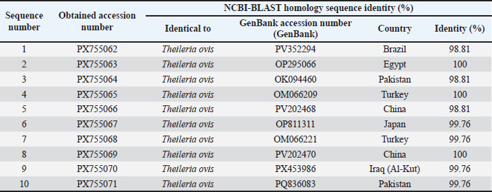
Fig. 1. Agarose gel electrophoresis (1.5 %) shows the amplicons (1–15) of T. ovis in sheep by inner primers of nested PCR (size=520 bp). NC is the negative control in which H2O was added instead of the template DNA. M is a molecular marker (Science Direx, South Korea). The band intensity and clarity confirmed successful amplification and that the DNA was of sufficient quality for subsequent processes, including sequencing. The molecular marker (100–1,500 bp ladder) verified that the fragments were of the expected size. These results validated the specificity and sensitivity of the nested PCR assay for detecting T. ovis infections in sheep. Sequencing output and GenBank submissionOut of the 10 sheep samples with positive PCR, we chose samples based on the clearness of PCR bands for sequencing, and all selected samples had successful partial sequencing of the 18S rRNA gene. Each isolate was processed and received trimmed, high-quality sequences of 419 BP. These sequences were submitted to GenBank for molecular data accessibility and traceability, where they can be accessed using accession numbers PX755062 to PX755071 (Table 1). Table 1. NCBI-BLAST homology sequence identity (%) in local T. ovis in sheep.
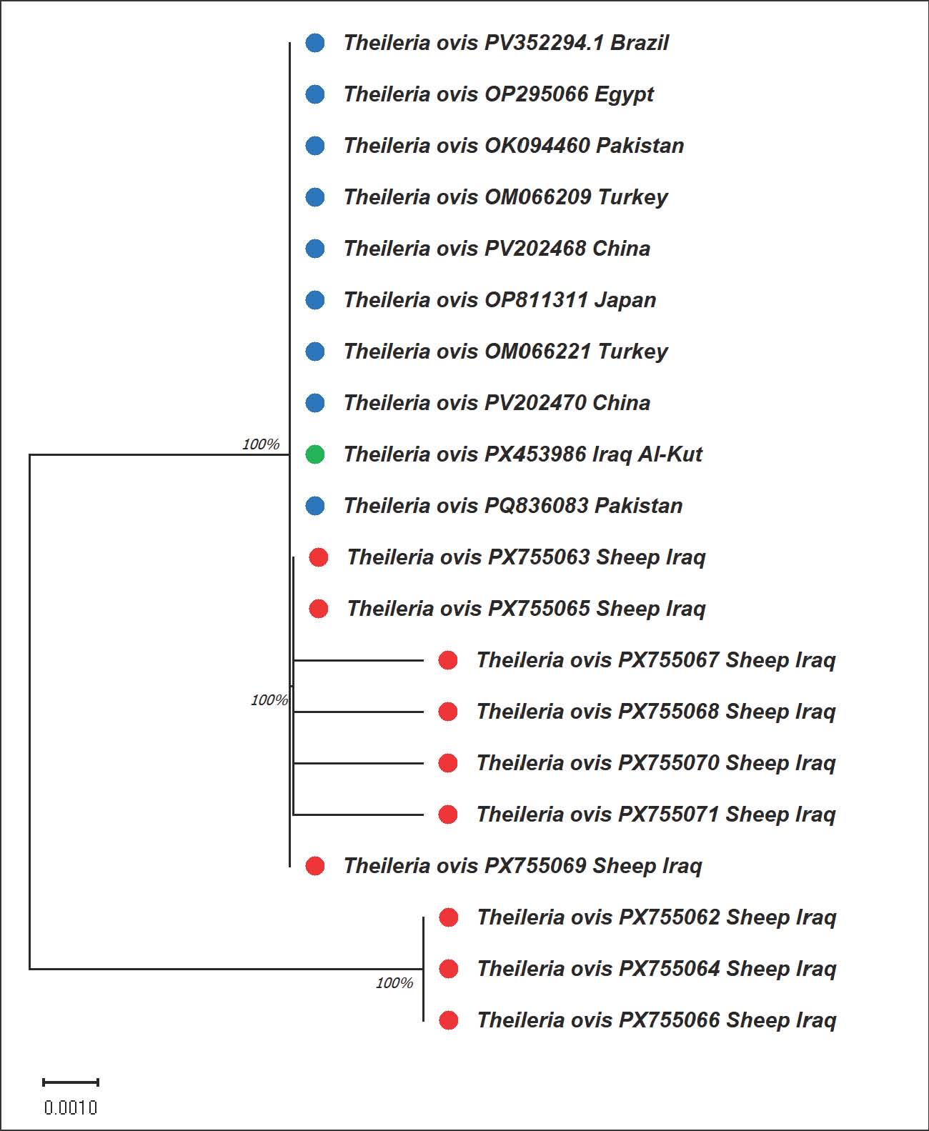
The submitted sequences contained complete metadata, including the host species (sheep), isolation source (blood), collection date (year 2021), and collection location (Iraq). The successful deposition of these sequences represents the first molecular record of the species T. ovis infecting sheep from Central Iraq based on 18S rRNA gene sequencing. BLAST homology and genetic identity analysisThe sequences obtained from NCBI-BLAST were all determined to be T. ovis. The result sequences contained previously reported T. ovis sequences from various areas, and with the result records, NCBI-BLAST reports show results with percentage matches of 98.81%–100%, indicating that the 18s rRNA gene region was highly conserved (Table 1). Iraqi sheep isolates showed 100% identity to reference sequences from Egypt and Türkiye and slightly lower identity values (98.61%–99.57%) to sequences from Pakistan, China, Japan, and Al-Kut, Iraq. These differences may reflect a geographical divergence that may not change the species designation due to a lack of sufficient nucleotide polymorphism. Theileria ovis phylogenetics of sheep isolatesUsing the Maximum Likelihood method and the Tamura-Nei model, we estimated the phylogenetic relationship among global reference sequences and local sheep isolates. A total of 20 sequences with 419 aligned sequence positions were used to build the phylogenetic tree (Fig. 2).
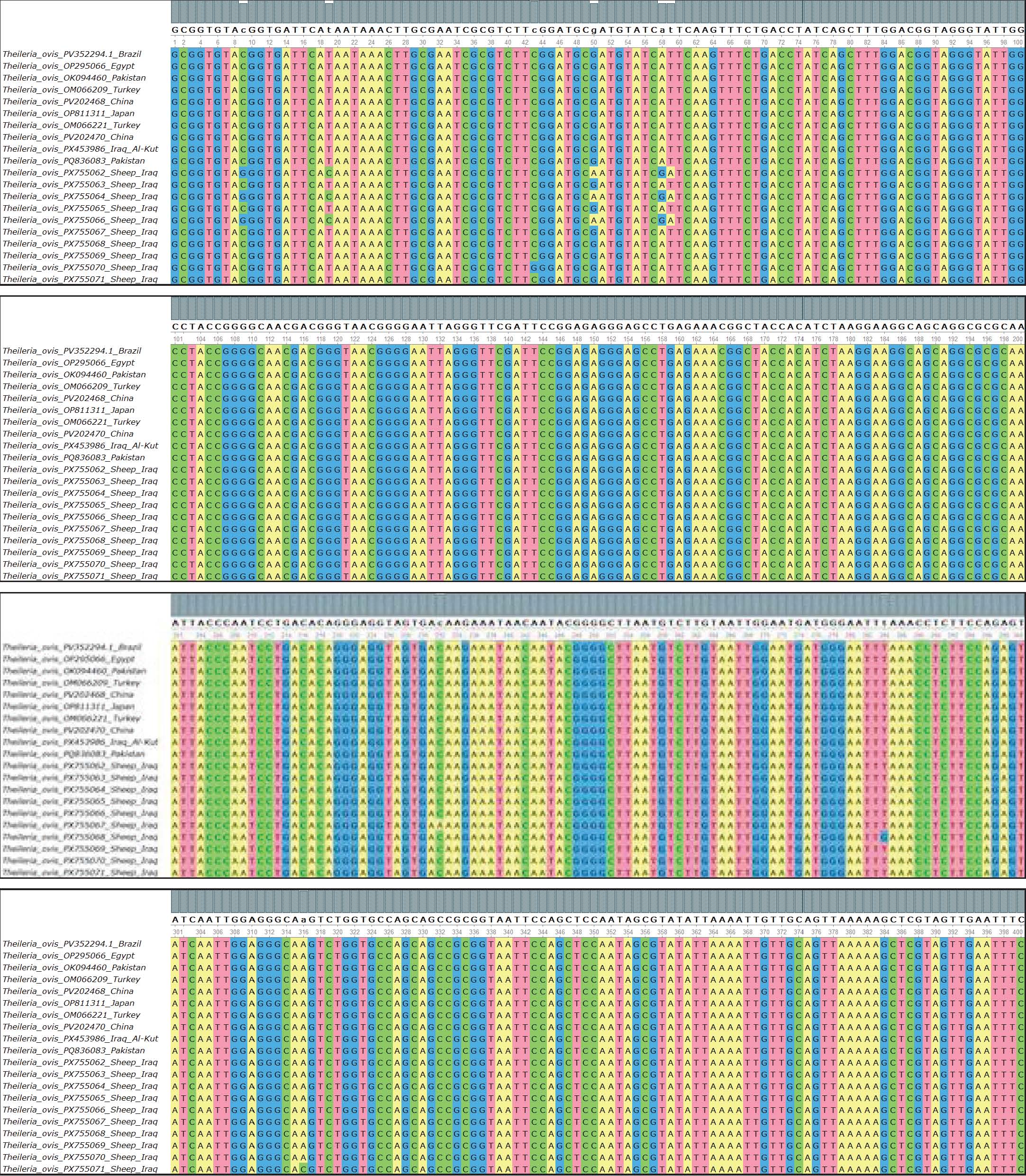
Fig. 2. Evolutionary tree analysis of T. ovis in sheep using the maximum likelihood method. This was inferred by using the Maximum Likelihood method and Tamura-Nei model. The tree is drawn to scale, with branch lengths measured in the number of substitutions per site. All Iraqi sheep isolates were within a single, robust clade of T. ovis, distinct from other Theileria species. The local isolates had closely positioned subclades with reference strains from Türkiye, Pakistan, Egypt, China, and Japan, illustrating a common ancestor. High bootstrap values strengthened the inferred relationships. The phylogenetic structure verified the identity of the parasites and corroborated the close genetic relationship of the Iraqi isolates with T. ovis strains found in other geographic regions. Analysis of multiple sequence alignmentThe analysis of the multiple sequence alignment of the partial 18S rRNA gene sequences showed high conservation of nucleotide sequences among the T. ovis isolates derived from sheep and GenBank reference sequences (Fig. 3). For the aligned sequences, most of the positions of the nucleotides are the same, indicating the strong genetic stability of the captured gene region.
Fig. 3. Multiple sequence alignment of a partial genetic region of T. ovis in sheep compared with other global sequences. This shows the similarities and differences between the sequences. From a few nucleotides, only some isolates had substitutions. These variations were ungrouped, and no motifs that had affiliation to a particular host or country were found. No insertions, deletions, and frameshift mutations were found. The results from the alignment further prove the 18S rRNA gene to be a significant molecular marker for T. ovis identification and ascertain the genetic homogeneity of the isolates derived from sheep in Central Iraq. DiscussionBased on the amplification and partial sequencing of the 18S rRNA gene via nested PCR, the current study documents the molecular and genetic characterization of T. ovis infecting sheep for the first time in Central Iraq. The current field isolates have high sequence identity compared with the molecular evidence available for T. ovis, confirming the widespread distribution of T. ovis in sheep worldwide. Recent studies from Asia, the Middle East, and Europe confirmed the utility of similar molecular techniques directed toward the 18S rRNA gene for low-cost and reliable species-level identification (Zhou et al., 2025; Celik et al., 2025). The present study’s BLAST analysis shows that Iraqi sheep isolates have a high nucleotide identity (98.81%–100%) with T. ovis sequences from Turkey, Egypt, Pakistan, China, and Japan. This finding corresponds with Na et al. (2025)who noted that T. ovis isolates from Xinjiang, China, were genetically highly conserved, although there were detectable local polymorphisms. The genetic identity of the Iraqi and Chinese isolates suggests that T. ovis populations in disparate geographies may have the same evolutionary roots due to livestock movement, tick dispersal, and cycles of long-lasting endemic transmissions. The present study’s phylogenetic analysis shows that all Iraqi sheep isolates unambiguously established their position in the T. ovis clade and were very closely related to most of the global reference strains. The clustering pattern corroborates the phylogenetic reconstructions from Turkey and Pakistan, in which T. ovis isolates from sheep exhibited well-supported monophyletic groups that were distinct from Theileria lestoquardi and other Theileria species (Riaz et al., 2024; Celik et al., 2025). Once again, the lack of host-specific subclades within the T. ovis lineage in the present study suggests the extreme conservation of the 18S rRNA gene relative to the sheep population (s) and geographical area(s). A comparative analysis alongside studies originating from Egypt shows remarkable consistency while considering the genetic identity levels. Studies conducted by Mahmoud et al. (2024)reported high sequence similarity among T. ovis isolates from sheep and goats in Luxor and low nucleotide divergence. The identity percentages in the current Iraqi isolates suggest that T. ovis in the Middle East has a low genetic diversity for the 18S rRNA. The genetic stability of T. ovis infections in various regions may be attributed to the subclinical nature of the infections. The current findings also support previous studies on Theileria DNA in tick vectors. The molecular presence of T. ovis in hard and soft ticks from Iran, China, and Turkey has been documented, confirming the vector capacity of ticks as reservoirs and dissemination agents (Norouzi et al., 2023; Ma et al., 2023; Liu et al., 2024). Although tick samples are not addressed in this research, the close phylogenetic connection of Iraqi sheep isolates to those found in ticks from other places implies the likely presence of active, vector-mediated transmission cycles in Central Iraq. Given the high rate of tick parasitism in small ruminants, this is further supported by regional research. Epidemiological studies have indicated that the presence of T. ovis in livestock is usually concomitant with the presence of other tick-borne pathogens, such as Anaplasma ovis and T. lestoquardi, which may further complicate clinical presentation and disease outcomes (Abdelsalam et al., 2023; Mahmoud et al., 2024). Although this study is limited to T. ovis, the molecular evidence of T. ovis in sheep calls for the need to establish national surveillance and monitoring strategies for ovine hemoparasitism that could encapsulate a multitude of pathogens, which would provide a comprehensive understanding of the associated burden of tick-borne disease in animals in Iraq. More recent evidence suggests that Theileria infections may be associated with issues other than subclinical parasitemia. Jurković Žilić et al. (2025) identified hemotropic pathogens in aborted ruminant fetuses, which raises the question of whether such parasitic infections might cause adverse reproductive outcomes. Although the present study did not consider reproductive outcomes, the confirmed presence of T. ovis in sheep populations raises the need to examine whether T. ovis may be a causative agent of reproductive loss in some unexplored ecological or immunological contexts. Research on Mediterranean ecosystems has shown the variety of tick-borne pathogens in ruminant ticks and the role of environmental changes in the distribution of these pathogens (Masià‐Castillo et al., 2025). In this situation, the present study provides important molecular information from Iraq, which has a similar climate and ecology, and aids in better understanding the distribution of T. ovis in the region and the world. Most of the findings of this study agree with the recent molecular studies in Asia, the Middle East, and Europe, which also point out that T. ovis, which infects sheep, has a high degree of genetic conservation and does not vary much from region to region. When added to the existing global phylogenetic literature, the Iraqi sequence data provide additional support to the claims of the wide geographical distribution and stability of T. ovis. It also emphasizes the need for continued molecular surveillance to ensure that control and prevention measures in small ruminant production systems are more effective. The molecular characterization of the parasite confirmed that it was T. ovis. The amplified 18S rRNA gene fragments from Iraqi sheep isolates had high nucleotide identity (between 98.75% and 98.81%) with reference sequences from Turkey, Pakistan, Egypt, China, Japan, and Brazil. Given the high sequence similarity, it may be concluded that the 18S rRNA gene is highly conserved globally among different T. ovis populations. Such a high level of gene conservation is a favorable indication of the use of this gene for detection and phylogenetic studies. The grouping of Iraqi isolates alongside strains from other regions confirmed that there is considerable genetic homogeneity among small ruminant T. ovis parasites. We performed multiple sequence alignment of 18S rRNA gene sequences with the GenBank reference sequences. The majority of nucleotide positions were conserved across the Iraqi sheep isolates and global strains. Minor sequence variation was found in a limited number of nucleotide substitutions in some isolates. Substitutions in the sequences were restricted to the studied gene region and included no insertions or deletions. Consequently, T. ovis was the only valid phylogenetic isolate. The high sequence conservation of the 18S rRNA gene confirms its utility for the detection and phylogenetic study of this parasite. ConclusionThe present study demonstrates the presence of T. ovis in sheep in central Iraq by nested PCR amplification and sequencing of the 18S rRNA gene. The nucleotide identities and lengths of the phylogenetic branches of the 18 isolates and the references support the presence of a genetically homogeneous parasite population with little global variation. AcknowledgmentsThe authors would like to express their sincere appreciation to the College of Veterinary Medicine, University of Al-Qadisiyah, for providing laboratory support and guidance throughout this study. Conflict of interestThe authors have no conflicts of interest to declare. FundingThe authors have self-funded the study. No external funding source is available. Authors’ contributionsAll authors have participated in the study. Data availabilityData are available when requested by the corresponding author. ReferencesAbdelsalam, M.A., Felefel, W., Fadl, S. and Bessat, M. 2023. Molecular prevalence and associated infection risk factors of tick-borne protozoan and rickettsial blood pathogens in small ruminants. BMC. Vet. Res. 19(1), 138; doi:10.1186/s12917-023-03702-4 Alfaleh, F.A. 2025. Molecular prevalence, associated risk factors, and genetic characterization of Theileria ovis in sheep and goats. Acta. Parasitol. 70(4), 178; doi:10.1007/s11686-025-01110-2 Almahallawi , R.S., Omer, S.A., Al-Shaebi, E.M., Al-Hoshani, N., Al-Malki, E.S., Abdel-Gaber, R. and Mohammed, O.B. 2024. Prevalence of Theileria ovis in sheep and goats in northwestern Saudi Arabia with notes on potential vectors. PeerJ 24, 12; doi:10.7717/peerj.18687 Arif, M., Saeed, S., Bashir, A., Farooq, M., Nasreen, N., Khan, A., Asif, M., Khalil, M.A., Ijaz, M., Muqaddas, H., Mehmood, N., Iqbal, F. and Chen, C.C. 2023. Molecular prevalence and phylogeny of Anaplasma marginale, Anaplasma ovis and Theileria ovis in goats and sheep enrolled from a hill station in Punjab, Pakistan. PLoS One 18(11), e0291302; doi:10.1371/journal.pone.0291302 Aziz, K.J. and Hamadamin, B.Q. 2025. Epidemiological and molecular study of Theileria spp. in sheep and goats in Erbil, Iraq. Trop. Anim. Health Prod. 57, 80; doi:10.1007/s11250-025-04330-w Celik, B.A., Kara, M., Ayan, A., Selcuk, M.A. and Celik, O.Y. 2025. Molecular prevalence and phylogenetic analysis of Theileria ovis and Theileria lestoquardi in small ruminants in Siirt Province, Türkiye. Vet. Med. Sci. 11(5), e70522; doi:10.1002/vms3.70522 Irfan, M., Chang, S.C., Iqbal, R.K., Tanveer, M., Asif, M., Khan, A., Nasreen, N., Atif, F.A., Shaikh, R.S., Aktas, M., Ben Said, M., Iqbal, F. and Chen, C.C. 2023. Seasonality, epidemiology and phylogeny of Theileria ovis with a note on hematological and biochemical changes in asymptomatic infected goats from Pakistan. PLoS One 18(8), 290620; doi:10.1371/journal.pone.0290620 Liu, D., Wang, J., Liu, Y., Wang, S., Zhu, H., Jiang, B., Li, Y., Zhang, Y., Chahan, B. and Zhang, W. 2024. Molecular analysis of Anaplasma ovis, Theileria ovis and Brucella abortus in adult Ornithodoros lahorensis soft ticks (Acari: ixodida: Argasidae) isolated from the Xinjiang Uygur Autonomous Region, China. J. Vet. Res. 68(3), 355–361; doi:10.2478/jvetres-2024-0049 Ma, Z., Ceylan, O., Galon, E.M., Mohanta, U.K., Ji, S., Li, H., Do, T.T., Umemiya-Shirafuji, R., El-Sayed, S.A.E.S., Zafar, I., Liu, M., Sevinc, F. and Xuan, X. 2023. Molecular identification of piroplasmids in ticks from infested small ruminants in Konya Province, Turkey. Pathogens 12(9), 1123; doi:10.3390/pathogens12091123 Mahmoud, H.Y.A.H., Tanaka, T., Ali, A.O. and Emeish, W.F.A. 2024. Molecular detection and characterization of Anaplasma ovis, Theileria ovis, and Theileria lestoquardi in sheep and goats in Luxor, Egypt. BMC. Vet. Res. 20(1), 260; doi:10.1186/s12917-024-04109-5 Masià‐Castillo, M.T., Maiques‐Rodriguez, E., Martínez‐Alfaro, A., García‐Bacete, I., Ingresa‐Capaccioni, S., Ahuir‐Baraja, A.E., Alarcón‐Elbal, P.M., Garijo‐Toledo, M.M. and Sansano‐Maestre, J. 2025. Tick-borne pathogens from ruminant ticks in a Mediterranean ecosystem from Eastern Spain. Med. Vet. Entomol. 39(2), 301–314; doi:10.1111/mve.12781 Nangru, A., Maharana, B.R., Vohra, S. and Kumar, B. 2022. Molecular identification of Theileria species in naturally infected sheep using nested PCR-RFLP. Parasitol. Res. 121(5), 1487–1497; doi:10.1007/s00436-022-07489-5 Norouzi, M., Dayer, M.S. and Ghaffarifar, F. 2023. Molecular detection and characterisation of Theileria in hard ticks of small ruminants in Zarrin Dasht County, Southern Iran. Vet. Med. Sci. 9(1), 372–379; doi:10.1002/vms3.1027 Prajapati, A., Prajapati, B., Patel, A., Chauhan, P., Das, B., Raval, S., Suthar, A., Sutaria, T., Chaudhari, R.K., Patel, P., Chauhan, V. and Patel, R. 2023. Molecular identification and genetic characterization of Theileria and Anaplasma infection in sheep and goat of North Gujarat, India. Parasitol. Res. 122(6), 1427–1433; doi:10.1007/s00436-023-07848-w Riaz, M., Chang, S.C., Tasawar, Z., Sajid, M., Nasreen, N., Amaro-Estrada, I., Cossío-Bayúgar, R., De La Fuente, J., Alanazi, A.D., Chen, C.C., Khan, A. and Ben Said, M. 2024. Molecular epidemiology and phylogeny of Theileria ovis and Theileria lestoquardi in sheep and goats from Southern Punjab, Pakistan. Vector-Borne Zoonotic Dis. 24(10), 656–665; doi:10.1089/vbz.2023.0118 Tanveer, M., Farooq, M., Amjad, M., Asif, M., Kashif, M., Latif, M., Khan, A., Aktas, M., Ben Said, M. and Iqbal, F. 2022. Molecular prevalence, associated risk factors and phylogeny of Anaplasma marginale, Theileria ovis and T. lestoquardi in sheep from Pakistan. Comparative. Immunol. Microbiol. Infect. Dis. 86, 101822; doi:10.1016/j.cimid.2022.101822 Zhou, N., Wang, M., Zhao, X., Mijiti, A., Dang, W., Wen, L., Shi, W., Gan, L., Li, C. and Gailike, B. 2025. Molecular detection and population genetic diversity analysis of Theileria ovis in partial regions of Xinjiang, China. Mol. Biochem. Parasitol. 263, 111689; doi:10.1016/j.molbiopara.2025.111689 Žilić, D.J., Naletilić, S., Mihaljević, Z., Gagović, E., Špičić, S., Reil, I., Duvnjak, S., Tuk, M.Z., Hodžić, A. and Beck, R. 2025. Hemotropic pathogens in aborted fetuses of domestic ruminants: transplacental transmission and implications for reproductive loss. Front. Microbiol. 16, 1632135; doi:10.3389/fmicb.2025.1632135 | ||
| How to Cite this Article |
| Pubmed Style Alfatlawi MAA, Hadi MQ. Genetic diversity and molecular identification of sheep infected with Theileria ovis in Central Iraq. Open Vet. J.. 2026; 16(5): 2939-2947. doi:10.5455/OVJ.2026.v16.i5.36 Web Style Alfatlawi MAA, Hadi MQ. Genetic diversity and molecular identification of sheep infected with Theileria ovis in Central Iraq. https://www.openveterinaryjournal.com/?mno=307258 [Access: June 26, 2026]. doi:10.5455/OVJ.2026.v16.i5.36 AMA (American Medical Association) Style Alfatlawi MAA, Hadi MQ. Genetic diversity and molecular identification of sheep infected with Theileria ovis in Central Iraq. Open Vet. J.. 2026; 16(5): 2939-2947. doi:10.5455/OVJ.2026.v16.i5.36 Vancouver/ICMJE Style Alfatlawi MAA, Hadi MQ. Genetic diversity and molecular identification of sheep infected with Theileria ovis in Central Iraq. Open Vet. J.. (2026), [cited June 26, 2026]; 16(5): 2939-2947. doi:10.5455/OVJ.2026.v16.i5.36 Harvard Style Alfatlawi, M. A. A. & Hadi, . M. Q. (2026) Genetic diversity and molecular identification of sheep infected with Theileria ovis in Central Iraq. Open Vet. J., 16 (5), 2939-2947. doi:10.5455/OVJ.2026.v16.i5.36 Turabian Style Alfatlawi, Monyer Abdulameir Abd, and Mohammed Qasim Hadi. 2026. Genetic diversity and molecular identification of sheep infected with Theileria ovis in Central Iraq. Open Veterinary Journal, 16 (5), 2939-2947. doi:10.5455/OVJ.2026.v16.i5.36 Chicago Style Alfatlawi, Monyer Abdulameir Abd, and Mohammed Qasim Hadi. "Genetic diversity and molecular identification of sheep infected with Theileria ovis in Central Iraq." Open Veterinary Journal 16 (2026), 2939-2947. doi:10.5455/OVJ.2026.v16.i5.36 MLA (The Modern Language Association) Style Alfatlawi, Monyer Abdulameir Abd, and Mohammed Qasim Hadi. "Genetic diversity and molecular identification of sheep infected with Theileria ovis in Central Iraq." Open Veterinary Journal 16.5 (2026), 2939-2947. Print. doi:10.5455/OVJ.2026.v16.i5.36 APA (American Psychological Association) Style Alfatlawi, M. A. A. & Hadi, . M. Q. (2026) Genetic diversity and molecular identification of sheep infected with Theileria ovis in Central Iraq. Open Veterinary Journal, 16 (5), 2939-2947. doi:10.5455/OVJ.2026.v16.i5.36 |


