





| Research Article | ||
Open Vet. J.. 2026; 16(5): 3229-3236 Open Veterinary Journal, (2026), Vol. 16(5): 3229-3236 Research Article Immunohistochemical evaluation of Sertoli and Leydig cells of scotch cat testis (Felis catus) at diverse agesDurah Fares Hashem and Siraj Muneer Al-Kafagy*Department of Anatomy and Histology, College of Veterinary Medicine, Al-Qasim Green University, Babylon, Iraq *Corresponding Author: Siraj Muneer Al-Kafagy. Department of Anatomy and Histology, College of Veterinary Medicine, Al-Qasim Green University, Babylon, Iraq. Email: dr.siraj.moner [at] vet.uoqasim.edu.iq Submitted: 13/01/2026 Revised: 14/04/2026 Accepted: 27/04/2026 Published: 31/05/2026 © 2025 Open Veterinary Journal
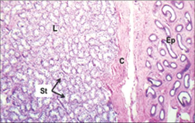
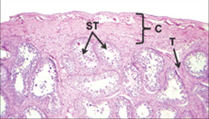
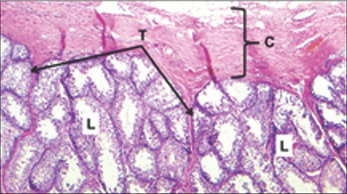
AbstractBackground: The testicles are located in a muscular, skin-covered, highly pigmented sac called the scrotum. The testicles produce both sperm and androgens, such as testosterone. Aim: The current study aimed to evaluate the immunohistochemical characteristics of the testis in three age groups (kitten, adult, and senior) of Scottish cat breeds (Felis catus) during the postnatal period, focusing on changes in Sertoli cells and Leydig cells. These two cell types play essential roles in male reproductive function, as Sertoli cells provide structural and metabolic support for spermatogenesis, while Leydig cells are responsible for testosterone production. Methods: Testicular samples were collected from clinically healthy cats and categorized into three age groups. The tissues were processed using routine histological techniques, followed by immunohistochemical staining to detect specific cellular markers for Sertoli and Leydig cells. The immunoreactivity and cellular distribution were evaluated and compared among the different age groups. Results: The findings demonstrated clear age-dependent variations in both cell populations. In kittens, weak immunoreactivity and incomplete development of seminiferous tubules were observed, indicating functional immaturity of the testis. Adult cats showed the highest immunoreactivity and well-organized seminiferous tubules, reflecting active spermatogenesis and optimal endocrine function. In contrast, senior cats exhibited a marked reduction in immunostaining intensity and structural degeneration of the seminiferous tubules, accompanied by a decrease in Leydig cell density. Conclusion: The study concludes that testicular structure and function in Scottish cats undergo significant age-related changes. Peak reproductive activity occurs in adulthood, while aging is associated with a decline in both cellular activity and structural integrity of the testis, affecting overall reproductive efficiency. Keywords: Cat, Immunohistochemistry, Leydig cell, Sertoli cell, Testis. IntroductionThe testicles are located in a muscular, skin-covered, highly pigmented sac called the scrotum. The testicles produce both sperm and androgens, such as testosterone. They are surrounded by two layers of protective connective tissue (tunica albuginea and tunica vaginalis). The tunica albuginea covers the outside of the testicle and also folds to form septa that divide the testicle into lobules. Within the lobules, sperm develop in structures called seminiferous tubules. The male gonads (testicles) are sex glands that have both an exocrine secretory function in the production of sperm and an endocrinological function as part of the hypothalamic–pituitary–gonadal axis in men through the production of androgens (Tsili et al., 2018; Yang et al., 2018). The immunoreactivity of FOXO1 and HSD3B2 was detected in Sertoli cells, Leydig cells, and spermatids. The intensity was increased every week. At first, the intensity of immunoreactive cells detected in spermatids and Sertoli was weak at 1 week after the first injection in the P1 group. The spermatogenic cells in 1 week after the first and the second injections were still not complete compared to 1 week after the third injection (Khasanah et al., 2021). Due to the lack of adequate studies describing the postnatal development of the testicles in Scottish Fold cats (Felis catus), the aim of this study was to provide a comprehensive assessment of these characteristics during the various postnatal stages, evaluating the immune-activity and cellular distribution of FOXO1 and HSD3B2 positive cells, and compare them among different age groups. Materials and MethodsFor the light microscopic investigation, 45 Scottish cats (Felis catus) of three different ages (15 kittens, 15 adults, and 15 seniors) were used in this study. The samples were fixed in 10% formalin for 48 hours. Then, they were washed under tap water. The sample was histologically examined by applying the main histological techniques (Al-Khafaji and Al-Kafagy, 2024), and stained with Hematoxylin and Eosin stain, Periodic Acid Schiff technique, and Masson’s trichrome stain. The immunohistochemical study of Sertoli cells and Leydig cell secretion was done by using FOXO1 Polyclonal Antibody (C29H4) and HSD3B1 Polyclonal Antibody (Ag7782), respectively. The blocking steps were as follows: Immerse the slices in Xylene for 10 minutes × 2 times; Remove excess liquid and immerse the slices in anhydrous ethanol for 3 minutes × 2 times; Remove excess liquid and immerse the slices in 95% ethanol for 3 minutes; Remove excess liquid and immerse the slices in 85% ethanol for 3 minutes. The deionized water rinses the slides for 1 minute, and phosphate buffered saline buffer rinses the slices for 3 minutes × 3 times. Light microscopy was used to analyze tissue slices. Future Win Joe's microscopic camera (INVENIO 12 EIV) was used for photomicrography, which was provided with analysis software to achieve the micro morphometric measurements. Ethical approvalThe current research has followed the accepted principles of ethical conduct by the College of Veterinary Medicine, University of Al-Qasim Green, qgec/73/2026 ResultsHistological resultsThe histological result shows that the testis is composed of a capsule that surrounds the trabeculae to form the testis lobules, which are filled by the seminiferous tubules. This result was the same in all ages in this study: Kitten, Adult, and Senior. The capsule was composed of tunica albuginea and tunica vasculosa. The tunica albuginea and vasculosa were thicker in kittens and adults. Underlining the capsule, between the capsule and the seminiferous tubule, there was the germinal epithelial layer. The germinal epithelial layer was thicker in adults and seniors, and thinner in kittens (Figs. 1–3).
Fig. 1. Cross-histological section of testis in Scottish Fold cats (2-month-old) showing epididymis (Ep), testicular capsule (C), testicular lobules (L), and seminiferous tubules (St). H&E stain.
Fig. 2. Cross-histological section of testis in Scottish Fold cats (9-month-old) showing seminiferous tubules (ST), testicular capsule (C).
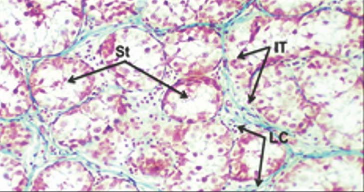
Fig. 3. Cross-histological section of testis in a Scottish Fold cat (4-year-old) showing capsula (C), trabecula (T), testicular lobule (L). H&E stain. 100×. The testis lobule was filled with a large number of seminiferous tubules composed of basement membrane and Sertoli cells with spermatogenic cells—spermatogonia, primary spermatocytes, secondary spermatocytes, and spermatid, and interstitial tissue—Leydig cells. The seminiferous tubules in kittens measured 106.42 ± 1.20 µm in diameter. The Sertoli cells and spermatogenic cells (spermatogonia) were present at the first age with absence of the primary spermatocytes, secondary spermatocytes, and spermatids. In adults, the seminiferous tubules have a typical histological structure that is composed of a basement membrane, Sertoli cells, and spermatogenic cells—spermatogonia, primary spermatocytes, secondary spermatocytes, and spermatids. The seminiferous tubules had the largest diameter of those in the kittens, and the seniors, which measured about 399.74 ± 6.33 µm. The seminiferous tubules of the seniors had the same histological structure as that in the adults, with a smaller diameter, which was measured about 274.78 ± 5.62 µm. This study shows that fertility is higher in adults than in seniors, which depends on the number of spermatogenic cells. Spermatogenic cell values increased when the diameter of the seminiferous tubules increased. The number of seminiferous tubules bearing fields (10×) in the adults was 8.0 ± 0.37 (10×) and in the seniors (15.0 ± 0.86 (10×), while in kittens, 49.0 ± 2.6 10×). The number was increased when the diameter was decreased. That gives an indicator of the larger fertility of the adult. That has a large diameter with a smaller number of seminiferous tubules (Figs. 4–6).
Fig. 4. Cross-histological section of testis in Scottish Fold cats at (2-month-old) showing seminiferous tubules (St), interstitial tissue (IT), and Leydig cells (LC). Masson’s trichrome. 200×.
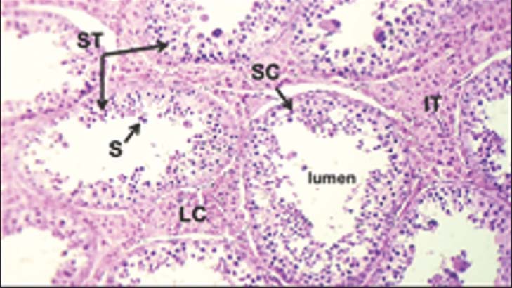
Fig. 5. Cross-histological section of testis in Scottish cat (9-month-old) showing seminiferous tubules (ST), Sertoli cells (SC), spermatids (S), interstitial cells (IT), Leydig cells (LC). H&E stain. 100×.
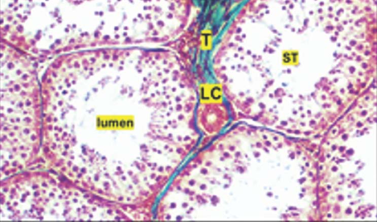
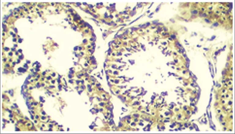
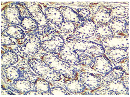
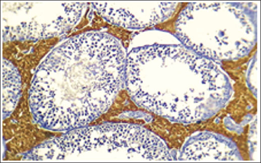
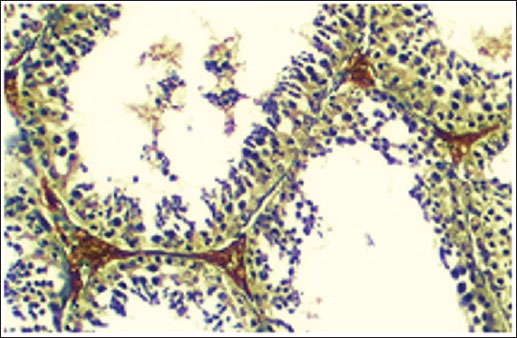
Fig. 6. Cross-histological section of testis in a Scottish cat (4-year-old) showing trabecula (T), seminiferous tubules (ST), Leydig cells (LC). Masson’s trichome. 200×. Immunohistochemical resultsDue to the lack of adequate studies describing the postnatal development of the testicles in Scottish Fold cats (Felis catus), the aim of this study was to provide a comprehensive assessment of these characteristics during the various postnatal stages, evaluating the immune-activity and cellular distribution of FOXO1 and HSD3B2 positive cells, and compare them among different age groups. Immunohistochemical results show that the number of Sertoli cells in the seminiferous tubules in the kitten was less in number, but increased in adults and in seniors was moderate. The results demonstrated clear age-dependent variations in both cell populations. In kittens, weak immune-activity (expression) and incomplete development of seminiferous tubules were observed, indicating functional immaturity of the testis. Adult cats showed the highest immune activity and well-organized seminiferous tubules, reflecting active spermatogenesis and optimal endocrine function. In contrast, senior cats exhibited a marked reduction in immune-staining intensity and structural degeneration of the seminiferous tubules, accompanied by a decrease in Leydig cell density (Figs. 7–12).
Fig. 7. Immunohistochemical staining of kitten Scottish cat testis (FOXO1) for Sertoli cells shows a small number of Sertoli cells, indicating reduced spermatogenesis inside the seminiferous tubules. 200×.
Fig. 8. Immunohistochemical staining of adult Scottish cat testis (FOXO1) for Sertoli cells shows a large number of Sertoli cells, indicating a large number of spermatogenic cells inside the seminiferous tubules. 200×.
Fig. 9. Immunohistochemical staining of senior scotch cat testis (FOXO1) for Sertoli cells shows a decreased number of Sertoli cells and a decreased number of spermatogenic cells inside the seminiferous tubules. 200×.
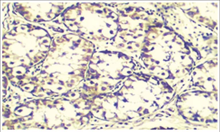
Fig. 10. Immunohistochemical staining of kitten scotch cat testis (HSD3B1) for Leydig cells shows a very weak antibody intensity, indicating a very low secretion level of testosterone hormone at this age. 200×.
Fig. 11. Immunohistochemical staining of adult scotch cat testis (HSD3B1) for Leydig cells shows a large amount of secretion of the testosterone hormone at this age. 200×.
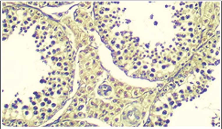
Fig. 12. Immunohistochemical staining of senior scotch cat testis (HSD3B1) for Leydig cells shows a decreased amount of secretion level of testosterone hormone at this age from that in adults. 200×. DiscussionThe result of a capsule that surrounds the trabeculae to form the testis lobules, filled by the seminiferous tubules Table 1. This result was the same in all ages in this study (Kitten, Adult, and Senior). The capsule was composed of (tunica albuginea and tunica Vasculosa), the finding agreement with (Homady Al-Jebori et al., 2021; Ganga Naik et al., 2023) how mention tubules and interstitial tissue. The tunica albuginea was the outermost covering of the testis. It contains a thick connective tissue capsule consisting of more collagen and fewer elastic fibers and myofibroblast internal to which was vascular layer of loose connective tissue called the tunica vasculosa. The finding is in agreement with Gofur et al. (2008); Eurell and Frappier (2006) in domestic animals, in boar (Ohanian et al., 1979); in bull (Ahmed and Sinowatz 2005; Gofur et al., 2008), in deer (Moonjit and Adcharatt, 2007), in horse (Shukla et al., 2013), in domestic pig by Reddy Dhyana et al. (2016)The result of the tunica albuginea agrees with França and Godinho (2003) and Silva et al. (2009) who studied the structure and function of testicles. They studied the seminiferous epithelium cycle in adult animals and found that cats aged over 8 years exhibited thicker tunica albuginea compared with other mammals and that it was formed by collagen fibers (França and Godinho, 2003; Silva et al., 2009; Al-Kafagy et al., 2022). Table 1. Thickness of capsule, tunica albuginea, tunica vasculosa, germinal layers, and the diameter and number of seminiferous tubules in the kitten, adult, and senior Scottish cat testis (Field, 2013).
Mehanna et al. (2018)in the horny fox (Lycalopex vetulus) mention that the seminiferous tubules, the most abundant component of the testicular parenchyma, consist of spermatogenic cells (which give rise to sperm) and Sertoli cells, surrounded by a basement membrane and connective tissue formed by fibroblasts. This was similar to the findings of Scottish cats in the result. Previous results of the diameter of seminiferous tubule disagree with Siemieniuch and Woclawek-Potoca (2007), who examined 118 pairs of testicles in 4-month-old to 10-year-old cats and found the following mean values for the diameter of seminiferous tubules: 191.83 μm in 8- to 12-month-old cats; 202.61 μm in 1- to 3-year-old cats; 193.38 μm in 3- to 6-year-old cats. (Siemieniuch and Woclawek-Potocka, 2007; Al-Khakani et al., 2020). ConclusionPostnatal aging has a significant influence on the immunohistochemical expression and functional status of Sertoli and Leydig cells in the Scottish cat testis. These findings provide valuable insight into the reproductive biology and age-related testicular changes in domestic cats. AcknowledgmentsThe authors express their sincere thankfulness for the support and facilities provided by Al-asim Green University/College of Veterinary Medicine. Conflict of interestThe authors declare that there is no conflict of interest. FundingNone. Authors' contributionsDura Faris Hashem: Conceptualization, Methodology, Validation, Writing – Original Draft, Writing – Review and Editing. Siraj Moner Al-Kafagy: Conceptualization, Methodology, Review & Editing, Supervisor. Data availabilityAll data were provided in the manuscript. ReferencesAhmed, A. and Sinowatz, F. 2005. Histological and ultrastructural studies on the testis of bull. Anat. Histol. Embryol. Al-Kafagy, S.M., Al-Jebori, A.K. and Alseady, Y.Y. 2022. Histochemical study of proventriculus in pre-hatch and post-hatch days in northern bobwhite quail (Colinus virginianus). Iraqi J. Vet. Sci. 36(Suppl 1), 161–166; doi:10.33899/ijvs.2022.135832.2529 Al-Khafaji, S.D.A. and Al-Kafagy, S.M. 2024. Histological and histochemical comparative study of the tongue in white-eared bulbul (Pycnonotus leucotis) and bronze fallow cockatiel (Nymphicus hollandicus). J. Anim. Health Prod. 12(s1), 319–325; doi:10.17582/journal.jahp/2024/12.s1.319.325 Al-Khakani, S.S.A., Zabiba, I.M.J., Al-Jebori, A.K.H., Al-Khafaji, S.M. and Neamah, D.A. 2020. Some histological study of the testes in male white-eared bulbul (Pycnonotus leucotis) during reproductive season. Biochem. Cell. Arch. 20, 3613–3617. Eurell, J.A.C., Frappier, B.L. and Dellmann, H. 2006. Dellmann’s textbook of veterinary histology. Blackwell Publishing. Field, 2013. Discovering statistics using IBM SPSS statistics, sage. França, L.R. and Godinho, C.L. 2003. Testis morphometry, seminiferous epithelium cycle length, and daily sperm production in domestic cats (Felis catus). Biol. Reprod. 68(5), 1554–1561; doi:10.1095/biolreprod.102.010652 Ganga Naik, S., Baddi, S.Y. and Rajashailesha, N. 2023. Gross morphology and histological details of testis and epididymis of the Hassan ram. Pharma. Innov. J. 12(3), 1514–1518. Available via https://www.thepharmajournal.com/archives/2023/vol12issue3/PartN/12-2-581-252.pdf Gofur, M.R., Khan, M.Z.I., Karim, M.R. and Islam, M.N. 2008. Histomorphology and histochemistry of testis of indigenous bull of Bangladesh. Bangladesh. J. Vet. Med. 6, 67–74. Homady Al-Jebori, A.K., Al-Khafaji, S.M. and Al-Zubaidi, K.A. 2021. Development of metanephrous in Awasi sheep (Ovis aries) fetuses during prenatal and postnatal periods. Biochem. Cell. Arch. 21(1), 285–290. Khasanah, L.M., Budipitojo, T. and Fibrianto, Y.H. 2021. Immunolocalization of steroidogenic enzymes (3β-hydroxysteroid dehydrogenase, 17β-hydroxysteroid dehydrogenase, and P450scc) in rats with testicular dysfunction treated with mesenchymal stem cells-conditioned medium. World. Vet. J. 11(3), 368–376; doi:10.54203/scil.2021.wvj48 Mehanna, M., Ferreira, A.L.S., Ferreira, A., Da Paz, R.C.R. and Morgado, T.O. 2018. Histology of the testis and the epididymal ducts from hoary fox (Lycalopex vetulus). Biosci. J. 34(6), 1697–1705; doi:10.14393/bj-v34n6a2018-39395 Moonjit, P. and Adcharatt, S. 2007. Histological structure of testes and ductus epididymis of Rusa deer (Cervus timorensis). Kasetsart J. Nat. Sci. 41, 86–90. Ohanian, C., Rodriguez, H., Piriz, H., Martino, I., Rieppi, G., Garófalo, E. G. and Roca, R.A. 1979. Studies on the contractile activity and ultrastructure of the boar testicular capsule. J. Reprod. Fertil. 57(1), 79–85. Reddy Dhyana, V., Rajendranath, N., Pramod Kumar, D. and Raghavender, K.B.P. 2016. Microanatomical studies on the testis of domestic pig. Int. J. Sci. Environ. Technol. 5(4), 2226–2231. Shukla, P., Bhardwaj, R.L. and Rajput, R. 2013. Histomorphology and micrometry of testis of Chamurthi horse. Indian J. Vet. Anat. Siemieniuch, M.J. and Woclawek-Potocka, I. 2007. Morphological features of the seminiferous epithelium in cat (Felis catus). J. Reprod. Dev. 53(5), 1125–1130; doi:10.1262/jrd.19053 Silva, C.A., Perri, S.H., Koivisto, M.B., Silva, A.M., Carvalho, R.G. and Monteiro, C.M. 2009. Histological and morphometric aspects of testes in domestic cats (Felis catus). Pesq. Vet. Bras. 29(4), 312–316; doi:10.1590/s0100-736x2009000400006 Tsili, A.C., Sofikitis, N., Stiliara, E. and Argyropoulou, M.I. 2018. MRI of testicular malignancies. Abdom. Radiol. 44(3), 1070–1082; doi:10.1007/s00261-018-1816-5 Yang, Y., Workman, S. and Wilson, M.J. 2018. The molecular pathways underlying early gonadal development. J. Mol. Endocrinol. 62(1), R47–R64; doi:10.1530/jme-17-0314 | ||

| How to Cite this Article |
| Pubmed Style Hashem DF, Al-kafagy SM. Immunohistochemical evaluation of Sertoli and Leydig cells of scotch cat testis (Felis catus) at diverse ages. Open Vet. J.. 2026; 16(5): 3229-3236. doi:10.5455/OVJ.2026.v16.i5.62 Web Style Hashem DF, Al-kafagy SM. Immunohistochemical evaluation of Sertoli and Leydig cells of scotch cat testis (Felis catus) at diverse ages. https://www.openveterinaryjournal.com/?mno=306601 [Access: June 26, 2026]. doi:10.5455/OVJ.2026.v16.i5.62 AMA (American Medical Association) Style Hashem DF, Al-kafagy SM. Immunohistochemical evaluation of Sertoli and Leydig cells of scotch cat testis (Felis catus) at diverse ages. Open Vet. J.. 2026; 16(5): 3229-3236. doi:10.5455/OVJ.2026.v16.i5.62 Vancouver/ICMJE Style Hashem DF, Al-kafagy SM. Immunohistochemical evaluation of Sertoli and Leydig cells of scotch cat testis (Felis catus) at diverse ages. Open Vet. J.. (2026), [cited June 26, 2026]; 16(5): 3229-3236. doi:10.5455/OVJ.2026.v16.i5.62 Harvard Style Hashem, D. F. & Al-kafagy, . S. M. (2026) Immunohistochemical evaluation of Sertoli and Leydig cells of scotch cat testis (Felis catus) at diverse ages. Open Vet. J., 16 (5), 3229-3236. doi:10.5455/OVJ.2026.v16.i5.62 Turabian Style Hashem, Durah Fares, and Siraj Muneer Al-kafagy. 2026. Immunohistochemical evaluation of Sertoli and Leydig cells of scotch cat testis (Felis catus) at diverse ages. Open Veterinary Journal, 16 (5), 3229-3236. doi:10.5455/OVJ.2026.v16.i5.62 Chicago Style Hashem, Durah Fares, and Siraj Muneer Al-kafagy. "Immunohistochemical evaluation of Sertoli and Leydig cells of scotch cat testis (Felis catus) at diverse ages." Open Veterinary Journal 16 (2026), 3229-3236. doi:10.5455/OVJ.2026.v16.i5.62 MLA (The Modern Language Association) Style Hashem, Durah Fares, and Siraj Muneer Al-kafagy. "Immunohistochemical evaluation of Sertoli and Leydig cells of scotch cat testis (Felis catus) at diverse ages." Open Veterinary Journal 16.5 (2026), 3229-3236. Print. doi:10.5455/OVJ.2026.v16.i5.62 APA (American Psychological Association) Style Hashem, D. F. & Al-kafagy, . S. M. (2026) Immunohistochemical evaluation of Sertoli and Leydig cells of scotch cat testis (Felis catus) at diverse ages. Open Veterinary Journal, 16 (5), 3229-3236. doi:10.5455/OVJ.2026.v16.i5.62 |


