





| Case Report | ||
Open Vet. J.. 2026; 16(8): 5555-5562 !
Open Veterinary Journal, (2026), Vol. 16(8): 5555–5562 Case Report Copper-associated chronic hepatitis in a geriatric mixed-breed female dogLarissa Nunes de Almeida1, Jakeline Martins Sanches1, Liliane Aparecida de Lara1, Jéssica Lima Mendes1, Jandra Pacheco dos Santos1 and Iago Martins Oliveira2*1Departamento de Medicina Veterinária, Centro Universitário de Goiás (UniGoiás), Goiânia, Brazil 2Escola de Ciências Médicas e da Vida, Pontifícia Universidade Católica de Goiás, Goiânia, Brazil *Corresponding Author: Iago Martins Oliveira. Escola de Ciências Médicas e da Vida, Pontifícia Universidade Católica de Goiás, Goiânia, Brazil. Email: iago.vetufg [at] gmail.com Submitted: 08/01/2026 Revised: 22/06/2026 Accepted: 07/07/2026 Published: 08/08/2026 © 2025 Open Veterinary Journal
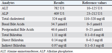
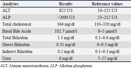
AbstractBackground: Excess hepatic copper can be either a cause or a consequence of chronic liver inflammation. The main mechanisms involved in copper accumulation include primary defects in hepatic copper metabolism, cholestasis leading to impaired biliary excretion, and excessive dietary copper intake. Copper overload may result in acute hepatic necrosis, often associated with hemolytic anemia. This study aimed to report a case of copper-associated hepatitis (CAH). Case Description: A 13-year-old mixed-breed spayed female dog was evaluated due to a progressive increase in serum liver enzyme activities and ultrasonographic liver abnormalities, with a clinical course of 6 months. Based on the initial diagnostic findings, a liver histopathological examination was performed, revealing hepatocellular hyperplasia associated with mild fibrosis and moderate diffuse vacuolar degeneration. Rhodanine staining demonstrated copper accumulation within hepatocytes, confirming hepatic copper accumulation consistent with CAH. Medical treatment and dietary management were instituted. Approximately 1 year and 6 months after diagnosis, the patient returned with recurrent emesis, food selectivity, residual head tilt, and ultrasonographic alterations involving the liver, gallbladder, and pancreas. Due to clinical deterioration, euthanasia was elected. Conclusion: This case report contributes to the limited literature on CAH in mixed-breed dogs, emphasizing the importance of histopathology and specific copper staining for definitive diagnosis, as well as the chronic and progressive nature of the disease despite therapeutic management. Keywords: Canine, Copper, Hepatopathy, Inflammation, Liver. IntroductionThe term chronic hepatitis has been used to describe inflammatory liver diseases that share similar historical, clinical, and possibly histopathological characteristics. Affected dogs exhibit abnormalities persisting for weeks or months, including clinical, laboratory, and imaging alterations. Clinical findings may include anorexia, weight loss, vomiting, lethargy, abdominal distension, and jaundice, while laboratory abnormalities commonly involve persistent elevation of liver enzymes and changes in bilirubin and bile acid concentrations. Serum liver enzyme activities, particularly alanine aminotransferase (ALT), remain elevated for a prolonged period, followed by evidence of hepatic insufficiency, which may include hypoalbuminemia, decreased urea synthesis, coagulopathies, ascites, and neurological signs associated with hepatic encephalopathy (Loan et al., 2025). Excess hepatic copper may be either a cause or a consequence of chronic hepatic inflammation, with the main mechanisms responsible for this accumulation including primary metabolic deficiencies in hepatic copper metabolism, cholestasis resulting in impaired biliary excretion of the metal, and increased dietary copper concentrations (Den Boer et al., 2025). Dietary copper in its cupric form (Cu2+) is absorbed in the small intestine after being reduced to cuprous copper (Cu+) by the enzyme metalloreductase. It enters enterocytes via copper transporter 1 (CTR1) or divalent metal transporter 1. To reach the portal circulation, the metal is exported by the alpha ATPase enzyme (ATP7A) and is transported to the liver bound to albumin or transcuprein, where it is subsequently internalized into hepatocytes through CTR1 (Den Boer et al., 2025). Copper is an essential trace element in canine nutrition, playing a key role in enzymatic activity, iron metabolism, connective tissue formation, and antioxidant defense. According to current nutritional guidelines, the recommended dietary copper concentration for dogs is approximately 7.3 mg/kg of dry matter, with safe upper limits reaching up to 25 mg/kg. However, it has been suggested that the copper content of some commercial dog foods may exceed biological requirements and potentially surpass the tolerance of certain animals (Center et al., 2021). When this micronutrient exceeds the hepatocellular transport capacity and the binding capacity to other molecules, free copper induces oxidative stress, leading to hepatocellular degeneration and cell death (Center et al., 2021). The definitive diagnosis of chronic hepatopathies associated with copper accumulation relies on histopathological evaluation using rhodanine or rubeanic acid staining, as well as the determination of copper concentration in hepatic tissue (Gupta and Al-Dissi, 2022). Differential diagnoses include infectious, toxic, and/or neoplastic causes, with potential involvement of pathogenic microorganisms, toxins, and hepatotoxic drugs (Webster et al., 2019). Treatment for copper-associated hepatitis (CAH) is based on reducing hepatic copper concentrations through dietary copper restriction and enhancement of urinary copper excretion using chelating agents such as D-penicillamine (DPA) (Rodrigues et al., 2020; Mutton et al., 2024). In addition, the use of hepatoprotective agents, including ursodeoxycholic acid and S-adenosyl-L-methionine, is essential (Mutton et al., 2024). This study aimed to report a case of CAH in a mixed-breed female dog, in order to contribute to the understanding of the clinical findings, complementary diagnostic examinations, and recommended therapeutic approach. Case DetailsA mixed-breed, spayed female dog weighing 10 kg and aged 13 years was presented for a routine check-up at a private veterinary clinic, with a history of progressive increases in serum ALT and alkaline phosphatase (ALP) activities, as well as ultrasonographically detectable hepatic morphological changes, including heterogeneous parenchymal echotexture and increased echogenicity, for approximately 6 months. During anamnesis, weight loss was reported, and it was also noted that the patient was being fed a commercial diet formulated as adjuvant therapy for obesity; however, the specific brand, copper content, and duration of administration before the onset of hepatic alterations were not available. Therefore, any potential contribution of the previously consumed diet to hepatic copper accumulation could not be determined. On physical examination, the patient presented normal vital parameters, including a body temperature of 38.4°C, heart rate within normal limits, and normal respiratory rate. The visible mucous membranes were pink and moist, with a normal capillary refill time. No ocular abnormalities suggestive of copper-associated changes, such as corneal opacity or scleral icterus, were observed at that time. The follow-up tests requested included serum cholesterol and the hepatic enzymes ALT and ALP, which were reported as increased; however, the exact numerical values from the initial evaluation were not available. At that time, the patient was not receiving any medications. Table 1. Serum biochemistry in a canine patient with copper-associated chronic hepatitis.
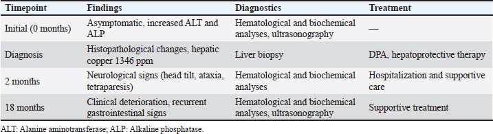
Table 2. Clinical timeline of a canine patient with copper-associated chronic hepatitis.
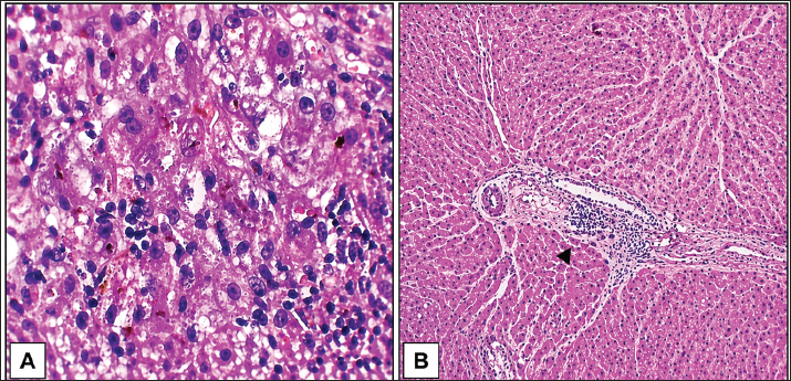
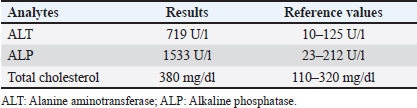
Fig. 1. Liver Doppler flowmetry. (A) Representation of blood flow in the caudal vena cava (CVC) with a laminar and phasic venous pattern. (B) Hepatobiliary morphology, liver parenchyma with mixed and heterogeneous echogenicity (blue arrow), and gallbladder with anechoic content and a moderate amount of hyperechoic points deposited (red arrow). (C) Morphometry of the abdominal aorta, caudal vena cava, and portal vein is represented by the blue, red, and yellow arrows, respectively. Based on these findings, the patient was referred for gastroenterological evaluation, and additional laboratory tests were subsequently requested, including basal and postprandial bile acids, total and fractionated bilirubin, and a coagulation profile, as well as imaging studies such as hepatic Doppler ultrasonography. The abnormalities observed in the performed tests are summarized in Table 1. The clinical course, diagnostic procedures, and therapeutic interventions are summarized in Table 2. On hepatic ultrasonographic examination, parenchymal changes were observed, suggesting a possible diffuse liver disease with impaired vascular compliance and moderate biliary sludge (Fig. 1). Histopathological examination was performed using hepatic samples obtained via videolaparoscopy to determine the etiology of the observed abnormalities. Two irregular tissue fragments were collected from the left lateral hepatic lobe, measuring together 0.4 × 0.3 × 0.2 cm. Gross evaluation of the material revealed a brownish, soft, and irregular surface. Histopathological analysis using hematoxylin and eosin (H&E) staining demonstrated foci of hepatocellular hyperplasia, resulting in compaction and compression of adjacent hepatic structures, associated with marked telangiectasia and mild foci of hemorrhagic necrosis peripheral to the portal region. Hepatocytes appeared typical, with mild foci of cholestasis. Representative histopathological findings are shown in Figure 2A and B. Based on the diagnostic findings, which characterized hepatocellular hyperplasia associated with fibrotic changes and moderate diffuse vacuolar degeneration, together with the morphological features of the tissue fragment, rhodanine staining was performed. This staining revealed reddish-brown cytoplasmic copper granules within hepatocytes. Quantification of hepatic copper concentration was performed at a reference laboratory (Cornell University, USA), using standardized methods for hepatic trace mineral analysis, yielding a result of 1,346 ppm (reference range: 120–500 ppm). Following the diagnosis of CAH, an initial therapeutic cycle for outpatient management was instituted, consisting of eicosapentaenoic acid and docosahexaenoic acid (Ograx-3®, 100 mg/kg, once daily [SID] for 45 days); ursodeoxycholic acid (Ursacol®, 15 mg/kg, SID for 45 days); DPA (Cuprimine®, 10 mg/kg, SID for 100 days); bezafibrate (Cedur®, 5 mg/kg, SID for 45 days); vitamin E (Tocoferol E®, 10 IU/kg, SID for 45 days); and S-adenosyl-L-methionine (SAMe®, 20 mg/kg, SID for 45 days). Due to the occurrence of emesis and inappetence during the first month of treatment, a second therapeutic cycle was established, replacing the chelating agent with compounded zinc sulfate (Nesh Zinco®, 8 mg/kg, twice daily [BID], continuous use), in addition to dietary management with a commercial hepatic diet (Royal Canin® Hepatic), formulated with restricted copper levels (approximately 7.2 mg/kg dry matter), aimed at reducing hepatic copper accumulation, following the manufacturer’s feeding guidelines. For the clinical signs presented, the following medications were prescribed: ondansetron hydrochloride (Vonau®, 0.5 mg/kg, three times daily [TID], for 5 days) and omeprazole (Gaviz V®; 1 mg/kg, BID for 5 days). Two months after diagnostic confirmation, the patient returned for veterinary evaluation due to the presence of neurological signs and frequent emesis. She was hospitalized and referred to the neurology service, where right-sided head tilt, tetraparesis, decreased proprioception in all four limbs, and ataxia were observed. During hospitalization, ondansetron hydrochloride was prescribed (Emedron® 1%, 0.5 mg/kg intravenous, TID for 5 days), and fluid therapy with lactated Ringer’s solution was instituted at 3 ml/kg/h. In addition, a complete blood count was evaluated and showed no abnormalities, whereas serum biochemistry revealed increased ALT (719 U/l), ALP (1,533 U/l), and total cholesterol (380 mg/dl) concentrations (Table 3).
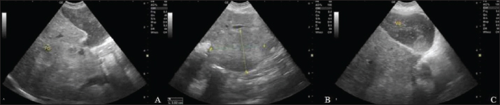
Fig. 2. Histopathological findings in the liver of a dog with copper-associated chronic hepatitis. (A) High-magnification photomicrograph of hepatic parenchyma showing hepatocytes with moderate to marked vacuolar degeneration, associated with cellular swelling and cytoplasmic rarefaction. Occasional intracytoplasmic brownish granular pigment is observed, consistent with bile pigment accumulation within hepatocytes (cholestasis). H&E stain, 400× objective. (B) Liver section demonstrating preserved lobular architecture with portal area expansion, mild fibrosis, and a moderate periportal inflammatory infiltrate composed predominantly of lymphocytes and plasma cells (arrowhead). Hepatocellular cords appear mildly disorganized, with evidence of hepatocellular degeneration. H&E stain, 100× objective. After 5 days of hospitalization, the patient showed clinical improvement and was discharged with the following home medications: meclizine (Meclin®, 25 mg/animal, SID for 5 days); bezafibrate (Cedur®, 5 mg/kg, SID for 45 days); and maropitant citrate (Cerenia®, 1 mg/kg, SID for 3 days), administered concomitantly with the previously prescribed copper chelator and hepatic diet. Approximately 18 months after diagnosis, the patient returned to the clinic presenting recurrent episodes of emesis, food selectivity, and residual head tilt. During this period, follow-up was inconsistent, with irregular clinical monitoring. She was hospitalized, and therapy was instituted with tramadol hydrochloride (Tramal®, 4 mg/kg, subcutaneous, BID for 3 days); maropitant citrate (Cerenia®, 1 mg/kg, SC, SID for 3 days); metoclopramide hydrochloride (Plasil®, 1 mg/kg, IV, TID for 3 days); cyproheptadine hydrochloride (Apevitin BC®, 0.1 ml/kg, orally, BID for 3 days); and fluid therapy with lactated Ringer’s solution at 3 ml/kg/h. At 18 months after diagnosis, a complete blood count revealed normocytic normochromic anemia and thrombocytopenia, and serum biochemistry demonstrated decreased total urea values and increased ALT, ALP, cholesterol, pre-prandial bile acids, and direct bilirubin concentrations (Table 4). Abdominal ultrasonography was performed, revealing a markedly enlarged liver with irregular contours, rounded margins, heterogeneous parenchymal echotexture, and increased echogenicity. In addition, multiple nodular structures with heterogeneous echotexture were distributed throughout the hepatic parenchyma, the largest measuring 4.54 × 3.52 cm, with irregular contours, heterogeneous echotexture, and mildly increased echogenicity. The gallbladder was dilated, with thin walls and filled with anechoic content suggestive of biliary sludge (Fig. 3). Other abnormalities observed included the presence of rounded structures in the left kidney, suggestive of renal cysts, as well as splenomegaly with increased dimensions, irregular contours, heterogeneous parenchymal echotexture, and nodular structures distributed throughout the parenchyma. Pancreatomegaly was also noted, along with abnormalities in the mesenteric tissue suggestive of an inflammatory process, including the presence of free fluid of low cellularity within the cystocolic window. Due to the patient’s clinical deterioration, unresponsive to the established therapeutic measures, and considering the history of inconsistent follow-up and irregular clinical monitoring during the course of the disease, euthanasia was elected. The owner signed the informed consent form. Table 3. Serum biochemistry in a canine patient with copper-associated chronic hepatitis, 2 months after diagnosis.
Table 4.Serum biochemistry in a canine patient with copper-associated chronic hepatitis. Changes observed 1 year and 6 months after the diagnostic confirmation.
Fig. 3. Ultrasound of the liver (LG) and gallbladder (GB). (A) Liver parenchyma with heterogeneous echotexture and increased echogenicity. (B) Heterogeneous nodular formation with slightly increased echogenicity in the liver parenchyma, measuring 4.54 × 3.52 cm. (C) Dilated gallbladder filled with anechoic content. DiscussionCertain canine breeds, including Bedlington terriers, Doberman pinschers, Labrador retrievers, and West Highland white terriers, exhibit a genetic predisposition to the development of CAH. Although genetic mechanisms have been described, including mutations in genes involved in copper metabolism such as COMMD1 in Bedlington terriers and ATP7B in other predisposed breeds, the disease may develop in any breed (Webster et al., 2019). The condition observed in this patient differs from most reports in the literature, as CAH is more commonly described in predisposed purebred dogs, whereas reports in mixed-breed dogs remain limited. CAH may represent either a primary disorder of copper metabolism or a secondary phenomenon associated with chronic hepatobiliary disease. Risk factors for hepatic copper accumulation are multifactorial and include excessive dietary copper intake and chronic cholestatic processes that impair biliary excretion (Gori et al., 2021). In the present case, although hepatic copper accumulation was confirmed histologically and by quantitative analysis, the available findings did not allow definitive differentiation between primary copper-associated hepatopathy and secondary copper retention resulting from impaired biliary excretion. Increased ALT, ALP, bilirubin, and bile acid concentrations may support hepatocellular injury and cholestasis; however, these findings are not specific and may also occur in chronic hepatitis. The patient was initially asymptomatic and subsequently developed hyporexia, emesis, weight loss, and neurological signs, including head tilt, ataxia, and proprioceptive deficits. Although these findings may be compatible with hepatic encephalopathy, this diagnosis could not be objectively confirmed because ammonia concentrations were not measured, and no additional diagnostic investigations were performed to exclude other neurological disorders. In dogs with chronic hepatopathies, neurological manifestations may be multifactorial and not exclusively related to hepatic encephalopathy, particularly in advanced disease stages. Considering the advanced age of the patient, alternative conditions such as vestibular disorders, inflammatory diseases, and intracranial neoplasia should also be considered. These clinical signs may be associated with progressive hepatic dysfunction; however, a definitive causal relationship could not be established in this case (Rodrigues et al., 2020). Although dietary copper has been proposed as a contributing factor to hepatic copper accumulation in dogs (Center et al., 2021), the contribution of dietary factors in this case could not be determined because information regarding the composition and copper content of the previously consumed diet was unavailable. Hematological abnormalities are not specific; however, they may include thrombocytopenia, anemia, leukocytosis, neutropenia, and lymphopenia (Loan et al., 2025). The release of copper into the bloodstream leads to hemolysis, and in more severe cases of CAH, gastrointestinal hemorrhage may occur (Mazaro et al., 2019). The patient’s complete blood count did not show clinically significant abnormalities at the initial evaluation or at 2 months after diagnosis; however, at 18 months after diagnosis, normocytic normochromic anemia and thrombocytopenia were observed; these findings are nonspecific and may not be directly related to copper accumulation. Considering the presence of nodular structures in the liver and spleen, further diagnostic investigation, such as cytology or biopsy, would be required for definitive characterization, but was not performed in this case. On the initial ultrasonographic examination, parenchymal alterations were identified as the diagnostic impression, supporting the diagnosis of liver disease. On the second examination, nodular structures distributed throughout the hepatic parenchyma were detected. The presence of nodular hepatic changes on abdominal ultrasonography has been described in dogs with copper-associated hepatopathy (Rodrigues et al., 2020). However, in the present case, no cytological or histopathological evaluation of the hepatic and splenic nodular structures was performed. Consequently, their biological nature could not be determined, and important differential diagnoses, including regenerative nodular hyperplasia, nodular cirrhosis, primary hepatic neoplasms, metastatic disease, and splenic proliferative disorders, could not be excluded. Therefore, it was not possible to correlate the ultrasonographic findings with histopathological features. Confirmation of CAH is established by the American College of Veterinary Internal Medicine and is essentially histological (Webster et al., 2019). Multiple tissue samples should be collected for histopathological evaluation and determination of hepatic copper concentration. In the present case, histopathological examination revealed hepatocellular hyperplasia associated with mild fibrosis, moderate diffuse vacuolar degeneration, focal cholestasis, and a predominantly periportal lymphoplasmacytic inflammatory infiltrate. Rhodanine staining demonstrated cytoplasmic copper granules within hepatocytes, confirming hepatic copper accumulation (Gori et al., 2021). After the identification of copper accumulation, its quantification is important, as the total copper burden may influence the duration of chelation therapy. Hepatic injury occurs when hepatic copper concentrations exceed 1,000 ppm (Dirksen and Fieten, 2017). Hepatic copper concentrations in dogs have been reported to increase over time (Johnston et al., 2013; Ullal et al., 2022). Population studies have suggested that this trend may be associated with changes in dietary copper recommendations and commercial food formulation practices over the years (Center et al., 2021; Ullal et al., 2022). In addition, the physiological reference limit for hepatic copper concentration (dry weight) in dogs has progressively increased from 7 µg/g in 1929 to 80 µg/g in 1956 and finally to 400 µg/g since the late 1970s (Center et al., 2021; Ullal et al., 2022). For initial treatment, a therapeutic protocol with different substances was instituted. S-adenosyl-L-methionine has anti-inflammatory and antioxidant effects and is involved in cellular replication and protein synthesis (Loan et al., 2025). Ursodeoxycholic acid has a choleretic effect that promotes the excretion of endogenous toxins through bile and is beneficial when concomitant cholestatic disease is present (Den Boer et al., 2025; Loan et al., 2025). Vitamin E acts as an antioxidant supplement, protecting hepatocytes against damage caused by copper and free radicals (Loan et al., 2025). Bezafibrate reduces cholesterol levels, and omega-3 fatty acids exert anti-inflammatory effects and help prevent cellular aging. Following the definitive diagnosis, DPA was instituted as pharmacological therapy. DPA remains the most commonly used copper chelator in dogs because of its effectiveness in promoting urinary copper excretion (Gupta and Al-Dissi, 2022). However, urinary copper excretion was not measured in this patient. It also has anti-inflammatory effects and increases glutathione levels, which play an important role in non-enzymatic defense against oxidative stress (Center et al., 2021). After the patient developed intolerance to DPA, zinc sulfate was prescribed to reduce copper absorption in the gastrointestinal tract, as it induces increased metallothionein synthesis by enterocytes. This protein has a higher binding affinity for copper and, for this reason, prevents the metal from entering the circulation, leading to its elimination in the feces (Den Boer et al., 2025). Approximately one-third of dogs with chronic hepatitis have copper-associated disease, and the prevalence of hepatic copper accumulation has increased over time, possibly related to changes in dietary copper content in commercial dog foods (Ullal et al., 2022, 2025). Treatment with DPA combined with dietary copper restriction is recommended and may be associated with clinical improvement and stabilization of hepatic copper concentrations (Webster et al., 2019; Mutton et al., 2024). Moreover, dogs fed copper-replete diets have a higher risk of hepatic copper accumulation compared with those receiving copper-restricted diets (Center et al., 2021). Although dietary restriction is an important component of treatment, it does not replace copper chelation in clinically affected patients (Center et al., 2021). A copper-restricted diet is recommended when hepatic copper concentrations exceed 600 µg/g dry weight (Webster et al., 2019). The dietary management established for the patient consisted of a hepatic diet (Royal Canin®), with the aim of supporting liver function and promoting dietary copper reduction. Den Boer et al. (2025) state that appropriate dietary management is as important as pharmacological therapy during treatment. Clinical improvement after treatment may take weeks to months, and marked individual variability is observed regarding chelator efficacy. Periodic liver biopsies should be performed to assess the extent of hepatic copper removal during chelation therapy (Gupta and Al-Dissi, 2022); however, in the patient described in this report, these examinations were not performed during treatment at the owner’s discretion. After 1 year and 6 months following the diagnosis, the patient returned to the clinic with worsening of her clinical condition, despite the previously instituted therapeutic management. Persistent increases in ALT, ALP, cholesterol, bile acids, and direct bilirubin were observed, indicating hepatic injury and cholestasis (Webster et al., 2019; Center et al., 2021). Although hepatic copper accumulation was confirmed, it was not possible to determine whether it represented a primary etiological factor or a secondary consequence of cholestatic disease, since copper retention may occur as a result of impaired biliary excretion (Webster et al., 2019; Center et al., 2021). Therefore, the diagnosis of primary copper-associated hepatopathy should be interpreted with caution. Furthermore, the absence of serial histopathological evaluations and repeated hepatic copper quantification limited the assessment of disease progression, therapeutic response, and the contribution of copper to the development of the hepatic lesions observed in this patient. The presence of hepatic and splenic nodular structures raises differential diagnoses such as nodular cirrhosis, regenerative hyperplasia, or neoplastic processes; however, their nature could not be confirmed, as no cytological or histopathological evaluation was performed. This report does not represent the first description of copper-associated chronic hepatitis in dogs; however, it describes an atypical presentation due to the advanced age and mixed-breed status of the patient, since most reports involve younger animals from predisposed breeds. Therefore, this case highlights a less commonly reported clinical scenario rather than a novel condition. Based on the findings of this case, early histopathological evaluation combined with hepatic copper quantification remains essential for establishing the diagnosis and guiding clinical management in dogs with persistent hepatic enzyme elevation. In addition, therapeutic management should include dietary copper restriction, use of copper chelating agents or zinc supplementation when indicated, and regular clinical and biochemical monitoring to assess disease progression and treatment response. This case report has some limitations that should be taken into account. Follow-up evaluations were inconsistent, which may have influenced clinical progression and the interpretation of findings. Furthermore, although nodular structures were identified in the liver, spleen, and pancreas on ultrasound examination, no cytological, histopathological, or postmortem evaluation was performed. Therefore, the biological nature of these lesions could not be determined, limiting the interpretation of disease progression and preventing exclusion of concomitant proliferative or neoplastic disorders. Furthermore, serial liver biopsies were not performed during follow-up, which limited the assessment of disease progression and response to therapy. ConclusionThis case illustrates the occurrence of chronic hepatitis associated with marked hepatic copper accumulation in a geriatric mixed-breed dog without known genetic predisposition. However, based on the available findings, it was not possible to determine whether copper accumulation represented a primary etiological factor or a secondary phenomenon associated with chronic cholestatic liver disease. Therefore, histopathological evaluation and hepatic copper quantification remain essential tools for diagnosis and clinical management in dogs with chronic hepatopathies. Conflicts of interestThe authors declare that there are no conflicts of interest in this study. FundingThis research did not receive any specific grants. Authors’ contributionConceptualization L.N.A., J.M.S., L.A.L., J.L.M., J.P.S., and I.M.O.; methodology I.M.O. and J.P.S.; formal analysis L.N.A., J.M.S., and L.A.L.; data curation I.M.O. and J.P.S.; conducted the clinical case I.M.O.; drafted the manuscript L.N.A., J.M.S., L.A.L., and J.L.M.; revised the manuscript and provided guidance on the clinical case J.P.S.; translated the manuscript I.M.O. All the authors have read and agreed with the published version of the manuscript. Data availabilityAll data supporting the findings of this study are available within the manuscript. ReferencesCenter, S.A., Richter, K.P., Twedt, D.C., Wakshlag, J.J., Watson, P.J. and Webster, C.R.L. 2021. Is it time to reconsider current guidelines for copper content in commercial dog foods?. J. Am. Vet. Med. Assoc. 258(4), 357–364; doi:10.2460/javma.258.4.357 Den Boer, E.R., Fieten, H. and Aicher, K.M. 2025. Copper-associated chronic hepatitis in dogs. Vet. Clin. North. Am. Small. Anim. Pract. 55(4), 25–54; doi:10.1016/j.cvsm.2025.08.010 Dirksen, K. and Fieten, H. 2017. Canine copper-associated hepatitis. Vet. Clin. North Am. Small Anim. Pract. 47(3), 631–644; doi:10.1016/j.cvsm.2016.11.011 Gori, E., Pierini, A., Meucci, V., Abramo, F., Muscatello, L.V. and Marchetti, V. 2021. Hepatic lead and copper concentrations in dogs with chronic hepatitis and their relationship with hematology, serum biochemistry, and histopathology. J. Vet. Intern. Med. 35(4), 1773–1779; doi:10.1111/jvim.16149 Gupta, A. and Al-Dissi, A. 2022. Correlation of hepatic copper levels, rhodanine scores and histological diagnosis in archived canine liver samples. Can. J. Vet. Res. 86(4), 300–305. Johnston, A.N., Center, S.A., McDonough, S.P., Wakshlag, J.J. and Warner, K.L. 2013. Hepatic copper concentrations in Labrador Retrievers with and without chronic hepatitis: 72 cases (1980-2010). J. Am. Vet. Med. Assoc. 242(3), 372–378; doi:10.2460/javma.242.3.372 Loan, N.V.T.H., Chao, N.V. and Nhung, T.T. 2025. Clinical features and hematological and biochemical blood parameters of dogs with hepatobiliary disorders. Vet. World 18(4), 986–993; doi:10.14202/vetworld.2025.986-993 Mazaro, R.D., Da Luz, F.S., Herbichi, A.P., Paz, M.C. and Fighera, R.A. 2019. Hemolytic crisis in a dog with copper-associated chronic hepatitis. Acta. Sci. Vet. 47(1), 1–6; doi:10.22456/1679-9216.97546 Mutton, J., Yeomans, S. and White, J. 2024. Copper hepatopathies in Australian dogs. Aust. Vet. J. 102(8), 385–391; doi:10.1111/avj.13338 Rodrigues, A., Leal, R.O., Girod, M., Dally, C., Guery, E., Gomes, E. and Hernandez, J. 2020. Canine copper-associated hepatitis: a retrospective study of 17 clinical cases. Open. Vet. J. 10(2), 128–134; doi:10.4314/ovj.v10i2.2 Ullal, T., Choi, E.A., Gui, D. and Medici, V. 2025. Comparative review of copper-associated chronic hepatitis in dogs and Wilson disease in humans. Front. Vet. Sci. 12, 1692603; doi:10.3389/fvets.2025.1692603 Ullal, T.V., Lakin, S., Gallagher, B., Sbardellati, N., Abdo, Z. and Twedt, D.C. 2022. Demographic and histopathologic features of dogs with abnormally high concentrations of hepatic copper. J. Vet. Intern. Med. 36(2), 678–687; doi:10.1111/jvim.16580 Webster, C.R.L., Center, S.A., Cullen, J.M., Penninck, D.G., Richter, K.P., Twedt, D.C. and Watson, P.J. 2019. ACVIM consensus statement on the diagnosis and treatment of chronic hepatitis in dogs. J. Vet. Intern. Med. 33(3), 1173–1200; doi:10.1111/jvim.15467 | ||

| How to Cite this Article |
| Pubmed Style Almeida LND, Sanches JM, Lara LAD, Mendes JL, Santos JPD, Oliveira IM. Copper-associated chronic hepatitis in a geriatric mixed-breed female dog. Open Vet. J.. 2026; 16(8): 5555-5562. doi:10.5455/OVJ.2026.v16.i8.49 Web Style Almeida LND, Sanches JM, Lara LAD, Mendes JL, Santos JPD, Oliveira IM. Copper-associated chronic hepatitis in a geriatric mixed-breed female dog. https://www.openveterinaryjournal.com/?mno=306162 [Access: August 08, 2026]. doi:10.5455/OVJ.2026.v16.i8.49 AMA (American Medical Association) Style Almeida LND, Sanches JM, Lara LAD, Mendes JL, Santos JPD, Oliveira IM. Copper-associated chronic hepatitis in a geriatric mixed-breed female dog. Open Vet. J.. 2026; 16(8): 5555-5562. doi:10.5455/OVJ.2026.v16.i8.49 Vancouver/ICMJE Style Almeida LND, Sanches JM, Lara LAD, Mendes JL, Santos JPD, Oliveira IM. Copper-associated chronic hepatitis in a geriatric mixed-breed female dog. Open Vet. J.. (2026), [cited August 08, 2026]; 16(8): 5555-5562. doi:10.5455/OVJ.2026.v16.i8.49 Harvard Style Almeida, L. N. D., Sanches, . J. M., Lara, . L. A. D., Mendes, . J. L., Santos, . J. P. D. & Oliveira, . I. M. (2026) Copper-associated chronic hepatitis in a geriatric mixed-breed female dog. Open Vet. J., 16 (8), 5555-5562. doi:10.5455/OVJ.2026.v16.i8.49 Turabian Style Almeida, Larissa Nunes De, Jakeline Martins Sanches, Liliane Aparecida De Lara, Jéssica Lima Mendes, Jandra Pacheco Dos Santos, and Iago Martins Oliveira. 2026. Copper-associated chronic hepatitis in a geriatric mixed-breed female dog. Open Veterinary Journal, 16 (8), 5555-5562. doi:10.5455/OVJ.2026.v16.i8.49 Chicago Style Almeida, Larissa Nunes De, Jakeline Martins Sanches, Liliane Aparecida De Lara, Jéssica Lima Mendes, Jandra Pacheco Dos Santos, and Iago Martins Oliveira. "Copper-associated chronic hepatitis in a geriatric mixed-breed female dog." Open Veterinary Journal 16 (2026), 5555-5562. doi:10.5455/OVJ.2026.v16.i8.49 MLA (The Modern Language Association) Style Almeida, Larissa Nunes De, Jakeline Martins Sanches, Liliane Aparecida De Lara, Jéssica Lima Mendes, Jandra Pacheco Dos Santos, and Iago Martins Oliveira. "Copper-associated chronic hepatitis in a geriatric mixed-breed female dog." Open Veterinary Journal 16.8 (2026), 5555-5562. Print. doi:10.5455/OVJ.2026.v16.i8.49 APA (American Psychological Association) Style Almeida, L. N. D., Sanches, . J. M., Lara, . L. A. D., Mendes, . J. L., Santos, . J. P. D. & Oliveira, . I. M. (2026) Copper-associated chronic hepatitis in a geriatric mixed-breed female dog. Open Veterinary Journal, 16 (8), 5555-5562. doi:10.5455/OVJ.2026.v16.i8.49 |


