





| Case Report | ||
Open Vet. J.. 2026; 16(8): 5531-5537 !
Open Veterinary Journal, (2026), Vol. 16(8): 5531–5537 Case Report First report of canine leishmaniasis in an urban area of Medellín, ColombiaFelipe Amador-Luján, Isabel Montoya-Álvarez, Leidy Alejandra Giraldo-Martínez, Diego Edison Sánchez-Ávila, Mariana Martínez-Aristizábal and César Orlando Muñoz-Cadavid*On Site Research Group, On Site Diagnostic SAS, Medellín, Colombia *Corresponding Author: César Orlando Muñoz-Cadavid. On Site Research Group, On Site Diagnostic SAS, Medellín, Colombia. Email: direccioncientifica [at] osdiagnostic.com Submitted: 20/12/2025 Revised: 27/06/2026 Accepted: 09/07/2026 Published: 08/08/2026 © 2025 Open Veterinary Journal
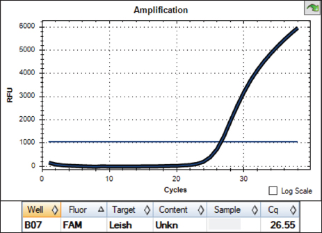
AbstractBackground: Leishmaniasis is an infectious zoonotic disease caused by protozoa of the genus Leishmania, which are transmitted by hematophagous sand flies. This disease is endemic in Colombia; it is more frequently reported in rural areas, and dogs are the main domestic reservoirs. This case report describes the first confirmed case of canine leishmaniasis in an urban area of Medellín, Colombia. Case Description: A 6-year-old intact male Belgian Malinois dog, residing in the urban area of Medellín, was presented for consultation due to lameness, lethargy, and inappetence. The initial clinical presentation was acute, but 1 month later its condition worsened with the appearance of subcutaneous nodules, weight loss, a hirsute coat, sublingual ulcers, and joint inflammation. Laboratory analysis revealed anemia, thrombocytopenia, and hyperglobulinemia. Abdominal ultrasound showed splenomegaly, lymphadenomegaly, and signs of chronic kidney disease. A complete real-time PCR (qPCR) panel for hemoparasites was positive for Leishmania spp. with a high parasitic load, and sequencing identified Leishmania infantum. The diagnosis was confirmed by cytology of subcutaneous nodules, which revealed amastigotes, and serology detected anti-Leishmania antibodies. Treatment with allopurinol (11 mg/kg PO q24h) was administered for 8 months, supplemented with domperidone (0.5 mg/kg PO q24h) for 4 weeks and marbofloxacin (3 mg/kg SC once, then PO for 15 days). Clinical signs resolved progressively, thrombocytopenia and anemia normalized, and follow-up qPCR tests at 7 and 9 months were negative. Conclusion: This is the first reported case of canine leishmaniasis caused by L. infantum in the urban area of Medellín, diagnosed by qPCR, cytology, and serology. The treatment protocol based on prolonged administration of allopurinol, domperidone, and marbofloxacin achieved clinical remission, with negative follow-up qPCR results at months 7 and 9. This case highlights the need for surveillance in urban areas due to the potential displacement of vectors and the close contact between people and domestic reservoirs. Keywords: Allopurinol, Canine leishmaniasis, Dog, Domperidone, Leishmania infantum. IntroductionLeishmaniasis is an infectious disease caused by protozoa of the genus Leishmania, transmitted by the hematophagous activity of dipteran insects of the genera Phlebotomus and Lutzomyia (Torres-Guerrero et al., 2017; Ribeiro et al., 2018). Around 53 species of Leishmania have been identified, of which 31 species parasitize mammals, and 20 are pathogenic to humans, domestic animals, and wildlife (Akhoundi et al., 2016). These species are grouped into complexes based on tropism for specific tissues and can infect macrophages in the bone marrow, lymph nodes, spleen, liver, kidneys, gastrointestinal tract, skin, and mucous membranes (Morales-Yuste et al., 2022). The diagnostic approach is initially based on history and physical examination. Signs found in a typical case may include anorexia, lethargy, cachexia, peripheral lymphadenomegaly, skin lesions, splenomegaly, epistaxis, ocular lesions, and onychogryphosis (Güran, 2019). A complete blood count may reveal non-regenerative normocytic normochromic anemia, thrombocytopenia, or changes in the leukogram. Blood biochemistry and urinalysis may indicate renal dysfunction and an inflammatory response with increased acute-phase proteins and gammaglobulins (Paltrinieri et al., 2016). These signs are variable and constitute multiple differential diagnoses, requiring parasitological, serological, and molecular tests to confirm leishmaniasis. Leishmaniasis is an endemic disease in Colombia, and the cutaneous form is the most prevalent clinical presentation. It is distributed in rural areas throughout the national territory, with higher reports in the departments of Antioquia, Santander, Meta, Tolima, and Guaviare (Instituto Nacional de Salud de Colombia (INS), 2024). Nine species have been reported: Leishmania panamensis, Leishmania braziliensis, Leishmania guyanensis, Leishmania infantum, Leishmania colombiensis, Leishmania amazonensis, Leishmania equatoriensis, Leishmania lansoni, and Leishmania mexicana (Ramírez et al., 2016; Salgado-Almario et al., 2019). The specific involvement of pets, particularly dogs, in the epidemiological cycle of leishmaniasis is unknown in Colombia. Active epidemiological surveillance based on the detection and treatment of infected animals may be an initial step to establish appropriate control measures for this zoonosis. Therefore, in this case report, we describe the diagnostic and therapeutic process of a dog with leishmaniasis from an urban area of Medellín, Colombia. Case DetailsA 6-year-old intact male Belgian Shepherd dog weighing 27 kg, from the urban area of Medellín city, was presented for consultation by his owner. The reason for consultation was lameness, lethargy, and inappetence after returning from a boarding kennel. The boarding kennel is located in the eastern region, an urban-peripheral zone that borders semi-rural areas. The dog had no history of travel to rural or traditionally endemic areas of Colombia. Clinical findings included edema and pain during flexion-extension movements of the elbows; rectal temperature was normal (38.2°C). Initial treatment consisted of dipyrone (20 mg/kg SC), dexamethasone (0.6 mg/kg SC), firocoxib (½ tablet of 227 mg PO every 24 hours for 7 days), omeprazole (20 mg PO for 7 days), and a joint supplement (glucosamine, chondroitin, methylsulfonylmethane). Hygroma was suspected, and it was recommended to wait for evolution to perform ultrasound and radiography. One month later, there was an acute worsening of the clinical condition, with firm subcutaneous nodules, weight loss, hirsute coat, sublingual ulcers, and joint inflammation at the tarsi and carpi. Figure 1 shows some physical examination findings observed in this second consultation. A complete blood count revealed anemia, moderate thrombocytopenia, absolute neutrophilia, hyperglobulinemia, and elevated CPK Creatine Phosphokinase. An abdominal ultrasound was performed, showing moderate splenomegaly, severe jejunal lymphadenomegaly, a mild amount of anechoic free fluid in the spleno-renal and hepatodiaphragmatic regions, and changes suggestive of mild-to-moderate bilateral chronic nephropathy (Fig. 2). A hemoparasitic agent was suspected, so blood and joint fluid were taken to evaluate the "Complete Hemotropic" real-time PCR (qPCR) panel with two additional tests (On Site Diagnostic, Colombia), which includes the following pathogens: Anaplasma spp., Rickettsia spp., Ehrlichia spp., Mycoplasma spp., Bartonella spp., Hepatozoon spp., Babesia spp., Dirofilaria immitis, Dirofilaria striata, Dirofilaria repens, Brugia pahangi, Trypanosoma spp., Leptospira spp., Toxoplasma spp., Brucella spp., and Leishmania spp. The qPCR was performed on a pooled sample of blood and joint fluid. The result of this test was positive for Leishmania spp., with a Ct value of 26.55 (Fig. 3). A Ct value of 26.55 is below the threshold of 30 and is indicative of high parasite load in the tested pooled sample, consistent with the severe multisystemic clinical presentation observed in this patient. Subsequently, Sanger sequencing of the qPCR product was performed, identifying L. infantum. Treatment was indicated with human-use allopurinol (11 mg/kg PO every 24 hours for 30 days), a multivitamin supplement, and vitamin E.
Fig. 1. Physical examination findings in the patient. (A) Fracture of the nail in the lateral phalanx of the left pelvic limb, with mild underlying hemorrhage. (B) Alopecic exfoliative nodule approximately 2 cm in diameter, located in the region of the left ischiatic tuberosity. (C) Periorbital alopecia and hirsute coat.
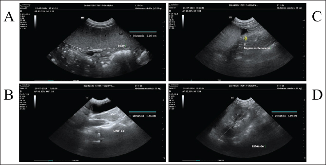
Fig. 2. Abdominal ultrasound findings in the patient. (A) Moderate splenomegaly, with mild presence of adjacent free fluid. (B) Severe jejunal lymphadenomegaly. (C) Anechoic free fluid in the spleno-renal region (yellow arrow). (D) Mild-to-moderate renal dystrophy in the right kidney.
Fig. 3. Amplification curve of qPCR with FAM hydrolysis probe. Amplification was observed in the patient sample (blood and joint fluid pool); the test target was a 171-bp fragment of the 18S gene of Leishmania sp. In order to confirm the diagnosis for the veterinary staff of the boarding kennel the patient frequented, 2 weeks later, a qPCR was performed by sending a counter-sample of blood to a different laboratory than the one used for the first test, which resulted in a negative. Given this contradictory finding, it was decided to perform cytology of subcutaneous nodules and lymph nodes, where amastigotes compatible with Leishmania spp. were found (image not available). Furthermore, a serological test was performed, detecting anti-L. infantum IgG antibodies at 1/160. The follow-up complete blood count found mild anemia, elevated total proteins, hypoalbuminemia, and hyperglobulinemia. The patient continued with inflammation at the carpi, presented periorbital alopecia, and a decrease in the size of the subcutaneous nodules. Two weeks later, it had to be taken to an emergency consultation as it was lethargic, lame, and vomiting. Physical examination revealed fever, periorbital alopecia, and exfoliative dermatitis, reactive popliteal and inguinal lymph nodes, left pelvic limb inflamed from the phalanges to the knee, with 4/4 lameness, and a fracture with hemorrhage in the nail of the lateral phalanx of this same limb. Dipyrone IV (25 mg/kg), maropitant (1 mg/kg), and ranitidine (1 mg/kg) were administered. After 10 minutes of monitoring, the body temperature normalized, and the patient regained his spirits. At that time, it was indicated to extend the administration of allopurinol to 8 months, and domperidone was added for 4 weeks (0.5 mg/kg PO every 24 hours). A dose of marbofloxacin (3 mg/kg SC) was also administered, which was extended orally for 15 days, and the affected nail was treated. Allopurinol acts as a leishmanistatic agent by interfering with purine metabolism of the parasite (Morales-Yuste et al., 2022), domperidone exerts an immunomodulatory effect that stimulates the Th1 immune response (Cavalera et al., 2021), and marbofloxacin has an indirect leishmanicidal effect by activating macrophages and inducing nitric oxide (NO) production (Oliveira, 2023). An ultrasound control was performed, showing moderate hepatomegaly and splenomegaly, mild-to-moderate bilateral renal dystrophy, and mild jejunal lymphadenomegaly, with a decrease in lymph node thickness compared to the previous ultrasound. A urinalysis revealed proteinuria, for which a renal protector was indicated. During the last months of treatment and once completed, the patient was stable with improvement of clinical signs and normalization of the complete blood count; thrombocytopenia and anemia resolved, and platelet counts returned to the reference range. However, medium- and long-term follow-up of renal function was recommended due to sequelae of leishmaniasis. At months 7 and 9 after the start of treatment, control qPCR for Leishmania spp. was performed, resulting in negative results on both occasions. Figure 4 presents a chronological summary of the clinical evolution, diagnostic steps, treatment, and follow-up.
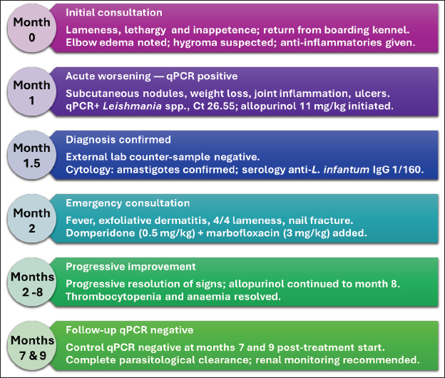
Fig. 4. Chronological summary of the clinical evolution, diagnostic steps, treatment, and follow-up of the patient. (Month 0) Initial consultation for lameness and lethargy after returning from a boarding kennel. (Month 1) Acute clinical worsening; qPCR was positive for Leishmania spp.; allopurinol was initiated. (Month 1.5) A negative counter-sample at external laboratory; cytology and serology confirmed the diagnosis. (Month 2) Emergency consultation; treatment intensified with domperidone and marbofloxacin. (Months 2–8) Progressive clinical improvement and complete blood count normalization. (Months 7 and 9) Follow-up qPCR negative on both occasions. DiscussionLeishmaniasis in Colombia is mainly distributed in rural areas with a high prevalence in the departments of Antioquia, Santander, Meta, Tolima, and Guaviare. The incidence of cutaneous leishmaniasis in the department of Antioquia for 2024 was 68.90 per 100,000 inhabitants (Instituto Nacional de Salud de Colombia (INS), 2024), representing a significant risk of exposure for both humans and animals. Case presentation occurs mainly in the rural population at 81.6% (Instituto Nacional de Salud de Colombia (INS), 2024); however, in this case, it was found in a dog in an urban setting. Dogs are the main domestic reservoirs of the pathogen and constitute an important epidemiological element in the control of leishmaniasis. The prevalence of this parasite in dogs has not been reported in the city of Medellín; however, there are reports in Bucaramanga and Ibagué with a prevalence of 4.3% (9/207) and 91.33% (158/173), respectively (Giraldo-Martínez et al., 2022; Jaimes-Dueñez et al., 2023). This wide range of prevalence could be due to the fact that in Bucaramanga, only the species L. infantum was detected, while in Ibagué, the presence of the genus Leishmania spp. was studied. In addition, isolated cases in dogs have been reported in Valle del Cauca, Risaralda, Tolima, Cundinamarca, and Meta (Arbeláez et al., 2019; González-Colonia et al., 2021; Pérez-Ramírez et al., 2023; Lopez-Medina and Rondón-Barragán, 2024). The differential diagnosis initially considered inflammatory joint diseases such as systemic or cutaneous lupus erythematosus, as the first reason for consultation was joint pain and edema, and it was later consistent with renal and dermatological lesions (Amerman et al., 2023). Similarly, cutaneous polyautoimmunity could be related to signs of alopecia and exfoliative dermatitis (Levy et al., 2020). The lack of response to non-steroidal anti-inflammatory drugs and immunosuppressants guided the diagnosis toward a hemoparasitic infection caused by an infectious agent such as Ehrlichia spp., compatible with anemia, lymphadenopathy, and thrombocytopenia in the clinical picture (Ramakant et al., 2020). This was ruled out by a broad qPCR panel for hemoparasites, which was positive only for Leishmania spp. The clinical presentation of leishmaniasis in dogs can involve various alterations, as evidenced in this case with ulcerative skin lesions, alopecia, splenomegaly, lymphadenomegaly, and renal dystrophy, while other signs, such as nail fracture, have been very poorly documented (Morales-Yuste et al., 2022), although dogs can also be asymptomatic reservoirs (García-Castro et al., 2022). Infection by Leishmania spp. can cause joint problems, especially at the carpi and tarsi, due to the accumulation of immune complexes that generate inflammation (Astigarraga and Selich, 2022; Silva et al., 2022). For this reason, if a dog presents with arthritis and has been in endemic areas of leishmaniasis, it is important to consider this disease as a possible cause (Sbrana et al., 2014). Regarding the diagnosis of leishmaniasis, microscopy via blood smear remains a widely used tool. However, in the present case, the direct implementation of qPCR was chosen to obtain a faster diagnosis and estimate the parasitic load. The qPCR was performed on a pool of blood and joint fluid, targeting a 171 bp fragment of the 18S rRNA gene of Leishmania sp. The Ct value of 26.55 reflects high parasite load in the tested sample, consistent with the severe and multisystemic clinical presentation, as lower Ct values are generally associated with active and high-level infection in qPCR assays for Leishmania (Martínez et al., 2011). In parallel, a counter-sample of blood was sent to an external laboratory for confirmation of the result, which was negative. This discrepancy is likely explained by one or more of the following factors: first, a possible reduction in parasite load following initiation of allopurinol treatment in the interval between collections, given that the second qPCR was performed 2 weeks after the first; second, the use of a different commercial kit with a distinct molecular target in the other laboratory (specific commercial kit, molecular target, and protocol are not available to the authors); or third, the fact that the second qPCR was performed on a blood sample alone rather than on a pooled sample of blood and joint fluid as in the first test. This last factor may have reduced the probability of parasite detection, since synovial fluid has been shown to harbor Leishmania parasites in dogs with articular involvement (Martínez et al., 2011; Silva et al., 2022). Given this discrepancy, cytology of the affected masses and lymph nodes was performed, revealing intracellular amastigotes in macrophages, thus confirming the presence of the parasite. The combination of qPCR with cytological studies increases diagnostic certainty, as both techniques can yield complementary results, with their combined use being fundamental for definitive diagnostic confirmation (Paltrinieri et al., 2016; Morales-Yuste et al., 2022). A limitation of our molecular approach is that blood and joint fluid were pooled for qPCR analysis; separate analysis of each sample type would have provided more informative data on comparative diagnostic sensitivity. Clinical remission of canine leishmaniasis is potentially achievable through the administration of miltefosine, a leishmanicidal agent administered orally. Although the study by Gonçalves et al. (2024) did not conclusively demonstrate the clinical efficacy of the drug, other reports, such as that of Dos Santos Dos Santos Nogueira et al. (2019) showed a significant reduction in parasite infectivity following its administration over a period of 4 weeks. In Colombia, a favorable response to the combination of meglumine antimoniate (100 mg/kg) and allopurinol (10 mg/kg) for 8 weeks has also been described, where a negative result for Leishmania spp. by qPCR was found at the end of treatment (Arbeláez et al., 2019). This therapeutic strategy could not be replicated in the present clinical case, as meglumine antimoniate and miltefosine could not be acquired at the time due to their scarce availability; moreover, an attempt was made to import them from other countries, but shipment was unviable due to logistical restrictions. For this reason, the extension of allopurinol to 8 months was chosen, along with domperidone for 1 month and marbofloxacin for 15 days, where a decrease in parasitic load and improvement of clinical signs were observed, supporting the effectiveness of this combination as part of the therapeutic protocol for the control of L. infantum in dogs. Allopurinol acts as a leishmanistatic agent by interfering with purine metabolism of the parasite and is the most widely recommended drug for long-term management of canine leishmaniasis (Morales-Yuste et al., 2022). This is similar to the literature reports, where domperidone has shown an immunomodulatory effect, stimulating the Th1 immune response and improving serological and clinical status (Cavalera et al., 2021), while marbofloxacin has an indirect leishmanicidal effect by activating macrophages, inducing the production of NO through the L-arginine-NO pathway, which enhances its activity against the parasite (Oliveira, 2023). ConclusionThis is the first report of canine leishmaniasis caused by L. infantum in the urban area of Medellín, Colombia, with a diagnostic approach that combined qPCR, cytology, and serology, where complete remission of the patient was observed after applying the indicated treatment. Detection in this context reinforces the need for surveillance in urban and peri-urban areas, given the potential displacement of vectors, coupled with the close contact between humans and domestic animals as reservoirs of the parasite. AcknowledgmentsWe thank the entire On-Site Diagnostic team for their support. We also extend our gratitude to the patients’ owners for providing informed consent for this report. Conflict of interestThe authors declare that there is no conflict of interest. FundingThis report received financial support for its publication from On Site Diagnostic SAS. Authors' contributionsFA: conceptualization and writing (original draft preparation). IM: case management and data interpretation. LAG: writing (original draft preparation, review, and editing), sequence analysis, and visualization. DES: writing (review and editing) and data interpretation. MM: data collection and writing (review and editing). COM: writing (review and editing), supervision, and funding acquisition. All authors have read and agreed to the published version of the manuscript. Data availabilityThe partial sequence obtained is available in the European Nucleotide Archive with the accession code OZ387670 (http://www.ebi.ac.uk/ena/data/view/OZ387670-OZ387670). ReferencesAkhoundi, M., Kuhls, K., Cannet, A., Votýpka, J., Marty, P., Delaunay, P. and Sereno, D. 2016. A historical overview of the classification, evolution, and dispersion of Leishmania parasites and sandflies. PLos Negl. Trop. Dis. 10(3), 4349. Amerman, H.K., Cianciolo, R.E., Casal, M.L. and Mauldin, E. 2023. German shorthaired pointer dogs with exfoliative cutaneous lupus erythematosus develop immune-complex membranous glomerulonephropathy. Vet. Pathol. 60(6), 843–848. Arbeláez, N., Moreno, J., Murillo, J., Montoya, A., Robledo, S.M., Vélez, A. and Vélez, I.D. 2019. First report of an urban case of canine visceral leishmaniasis in the municipality of Cali, Colombia. Am. J. Trop. Med. Hyg. 102(2), 289–292. Astigarraga, M.J.T. and Selich, V.P.A. 2022. Artrosis erosiva canina asociada con Leishmania spp: canine erosive arthrosis associated with Leishmania spp. Stud. Health. Sci. 3(1), 477–498. Cavalera, M.A., Gernone, F., Uva, A., D'Ippolito, P., Roura, X., Paltrinieri, S. and Zatelli, A. 2021. Effect of domperidone (leisguard®) on antibody titers, inflammatory markers and creatinine in dogs with leishmaniosis and chronic kidney disease. Parasites. Vectors. 14(1), 1–9. Dos Santos Nogueira, F., Avino, V.C., Galvis-Ovallos, F., Pereira-Chioccola, V.L., Moreira, M.A.B., Romariz, A.P.P.L., Molla, L.M. and Menz, I. 2019. Use of miltefosine to treat canine visceral leishmaniasis caused by Leishmania infantum in Brazil. Parasites. Vectors. 12(1), 79. García-Castro, A., Egui, A., Thomas, M.C. and López, M.C. 2022. Humoral and cellular immune response in asymptomatic dogs with visceral leishmaniasis: a review. Vaccines 10(6), 947. Giraldo-Martínez, L.A., Petano-Duque, J.M., Uribe-García, H.F., Chacón-Novoa, R.A., Guzmán-Barragán, B.L. and Rondón-Barragán, I. 2022. High prevalence of Leishmania spp. in dogs from Central West Colombia. Vet. Ital. 58(4), 379–389. Gonçalves, G., Campos, M.P.D., Gonçalves, A.S. and Figueiredo, F.B. 2024. Therapeutic success and failure in using miltefosine to treat dogs naturally infected with Leishmania infantum. Rev. Bras. Parasitol. Vet. 33(1), 15023. González-Colonia, L.V., Bonilla-Aldana, D.K., Álvarez-Serrano, M.P., Granados-Hincapie, L.Y., Pinilla-Ticora, L.C. and Rodríguez-Morales, A.J. 2021. An urban case of canine visceral leishmaniasis in the municipality of Pereira, Colombia. World's. Vet. J. 11(1), 115–118. Güran, M. 2019. An overview of leishmaniasis: historic to future perspectives. In Vectors and Vector-Borne Zoonotic Diseases. Ed., Savić, S. London, UK: IntechOpen, pp: 82–98. Instituto Nacional de Salud de Colombia (INS). 2024. Informe de evento: Leishmaniasis, Periodo Epidemiológico 12. Available via https://www.ins.gov.co/buscador-eventos/Informesdeevento/LEISHMANIASIS%20PE%20XII%202024.pdf Jaimes-Dueñez, J., Castillo-Castañeda, A., Jiménez-Leaño, A., Duque, J.E., Cantillo-Barraza, O., Cáceres-Rivera, D.I., Granada, Y., Triana-Chávez, O. and Ramírez, J.D. 2023. Epidemiological features of Leishmania infantum in dogs (Canis lupus familiaris) suggest a latent risk of visceral leishmaniasis in the metropolitan area of Bucaramanga, Santander, Eastern Colombia. Prev. Vet. Med. 219, e106021. Levy, B.J., Linder, K.E., Mamo, L.B., Herrmann, I. and Bizikova, P. 2020. Cutaneous polyautoimmunity in two unrelated dogs: pemphigus foliaceus and generalized discoid lupus erythematosus. Vet. Dermatol. 31(4), 325. Lopez-Medina, N.K. and Rondón-Barragán, I.S. 2024. Identificación de Leishmania spp en caninos de tres departamentos del centro de Colombia y su correlación clínica. Rev. Investig. Vet. Perú. 35(4), e26389. Martínez, V., Quilez, J., Sanchez, A., Roura, X., Francino, O. and Altet, L. 2011. Canine leishmaniasis: the key points for qPCR result interpretation. Parasit. Vectors 4(1), 57. Morales-Yuste, M., Martín-Sánchez, J. and Corpas-Lopez, V. 2022. Canine leishmaniasis: update on epidemiology, diagnosis, treatment, and prevention. Vet. Sci. 9(8), 387. Oliveira, J.D. 2023. Monitoramento terapêutico de cães com infecção natural por Leishmania infantum tratados com marbofloxacina combinada ao alopurinol. Ph.D. thesis, Universidade Federal Rural de Pernambuco, Recife, Brazil. Paltrinieri, S., Gradoni, L., Roura, X., Zatelli, A. and Zini, E. 2016. Laboratory tests for diagnosing and monitoring canine leishmaniasis. Vet. Clin. Pathol. 45(4), 552–578. Pérez-Ramírez, R.D., Lugo-Vargas, R., Petano-Duque, J.M., Cruz-Méndez, J.S. and Rondón-Barragán, I.S. 2023. First study on microscopic and molecular detection of Acanthocheilonema reconditum and Leishmania infantum coinfection in dogs in Southwest Colombia. Vet. World 16(1), 94. Ramakant, R.K., Verma, H.C. and Diwakar, R.P. 2020. Canine ehrlichiosis: a review. J. Entomol. Zool. Stud. 8(2), 1849–1852. Ramírez, J.D., Hernández, C., León, C.M., Ayala, M.S., Flórez, C. and González, C. 2016. Taxonomy, diversity, temporal and geographical distribution of cutaneous leishmaniasis in Colombia: a retrospective study. Sci. Rep. 6, 28266. Ribeiro, R.R., Michalick, M.S.M., Da Silva, M.E., Dos Santos, C.C.P., Frézard, F.J.G. and Da Silva, S.M. 2018. Canine leishmaniasis: an overview of the current status and strategies for control. Biomed Res. Int. 2018, 3296893. Salgado-Almario, J., Hernández, C.A. and Ovalle, C.E. 2019. Geographical distribution of Leishmania species in Colombia, 1985-2017. Biomedica 39, 278–290. Sbrana, S., Marchetti, V., Mancianti, F., Guidi, G. and Bennett, D.L.H. 2014. Retrospective study of 14 cases of canine arthritis secondary to Leishmania infection. Small. Anim. Pract. 55(6), 309–313. Silva, A.R.S., Gomes, A.A.D., Aquino, M.C.C., Almeida, B.F.M., Lima, V.M.F., Ciarlini, P.C., Pinoti, L.D.R., Marcondes, M. and Vieira, R.F.C. 2022. Synovial fluid and radiographic evaluation of joints from dogs with visceral leishmaniasis. Parasites. Vectors. 15(1), 320. Torres-Guerrero, E., Quintanilla-Cedillo, M.R., Ruiz-Esmenjaud, J. and Arenas, R. 2017. Leishmaniasis: a review. F1000Research 6(750), F1–F15. | ||

| How to Cite this Article |
| Pubmed Style Amador-luján F, Montoya-Álvarez I, Giraldo-martínez LA, SánchezÁvila DE, Martínez-aristizábal M, Muñoz-cadavid CO. First report of canine leishmaniasis in an urban area of Medellín, Colombia. Open Vet. J.. 2026; 16(8): 5531-5537. doi:10.5455/OVJ.2026.v16.i8.46 Web Style Amador-luján F, Montoya-Álvarez I, Giraldo-martínez LA, SánchezÁvila DE, Martínez-aristizábal M, Muñoz-cadavid CO. First report of canine leishmaniasis in an urban area of Medellín, Colombia. https://www.openveterinaryjournal.com/?mno=303917 [Access: August 08, 2026]. doi:10.5455/OVJ.2026.v16.i8.46 AMA (American Medical Association) Style Amador-luján F, Montoya-Álvarez I, Giraldo-martínez LA, SánchezÁvila DE, Martínez-aristizábal M, Muñoz-cadavid CO. First report of canine leishmaniasis in an urban area of Medellín, Colombia. Open Vet. J.. 2026; 16(8): 5531-5537. doi:10.5455/OVJ.2026.v16.i8.46 Vancouver/ICMJE Style Amador-luján F, Montoya-Álvarez I, Giraldo-martínez LA, SánchezÁvila DE, Martínez-aristizábal M, Muñoz-cadavid CO. First report of canine leishmaniasis in an urban area of Medellín, Colombia. Open Vet. J.. (2026), [cited August 08, 2026]; 16(8): 5531-5537. doi:10.5455/OVJ.2026.v16.i8.46 Harvard Style Amador-luján, F., Montoya-Álvarez, . I., Giraldo-martínez, . L. A., SánchezÁvila, . D. E., Martínez-aristizábal, . M. & Muñoz-cadavid, . C. O. (2026) First report of canine leishmaniasis in an urban area of Medellín, Colombia. Open Vet. J., 16 (8), 5531-5537. doi:10.5455/OVJ.2026.v16.i8.46 Turabian Style Amador-luján, Felipe, Isabel Montoya-Álvarez, Leidy Alejandra Giraldo-martínez, Diego Edison SánchezÁvila, Mariana Martínez-aristizábal, and César Orlando Muñoz-cadavid. 2026. First report of canine leishmaniasis in an urban area of Medellín, Colombia. Open Veterinary Journal, 16 (8), 5531-5537. doi:10.5455/OVJ.2026.v16.i8.46 Chicago Style Amador-luján, Felipe, Isabel Montoya-Álvarez, Leidy Alejandra Giraldo-martínez, Diego Edison SánchezÁvila, Mariana Martínez-aristizábal, and César Orlando Muñoz-cadavid. "First report of canine leishmaniasis in an urban area of Medellín, Colombia." Open Veterinary Journal 16 (2026), 5531-5537. doi:10.5455/OVJ.2026.v16.i8.46 MLA (The Modern Language Association) Style Amador-luján, Felipe, Isabel Montoya-Álvarez, Leidy Alejandra Giraldo-martínez, Diego Edison SánchezÁvila, Mariana Martínez-aristizábal, and César Orlando Muñoz-cadavid. "First report of canine leishmaniasis in an urban area of Medellín, Colombia." Open Veterinary Journal 16.8 (2026), 5531-5537. Print. doi:10.5455/OVJ.2026.v16.i8.46 APA (American Psychological Association) Style Amador-luján, F., Montoya-Álvarez, . I., Giraldo-martínez, . L. A., SánchezÁvila, . D. E., Martínez-aristizábal, . M. & Muñoz-cadavid, . C. O. (2026) First report of canine leishmaniasis in an urban area of Medellín, Colombia. Open Veterinary Journal, 16 (8), 5531-5537. doi:10.5455/OVJ.2026.v16.i8.46 |


