





| Research Article | ||
Open Vet. J.. 2026; 16(8): 5383-5396 !
Open Veterinary Journal, (2026), Vol. 16(8): 5383–5396 Research Article Acute and subchronic oral toxicity of fermented Crescentia cujete (L) preparation in ratsYos Adi Prakoso1*, Achmadi Susilo2, Sitarina Widyarini3, Oscar Maulana Pribadi1 and Paskalis Guntur Widya Mahendra11Department of Pharmacology, Faculty of Veterinary Medicine, University of Wijaya Kusuma Surabaya, Surabaya, Indonesia 2Department of Agrotechnology, Faculty of Agriculture, University of Wijaya Kusuma Surabaya, Surabaya, Indonesia 3Department of Pathology, Faculty of Veterinary Medicine, Universitas Gadjah Mada, Yogyakarta,Indonesia *Corresponding Author: Yos Adi Prakoso. Department of Pharmacology, Faculty of Veterinary Medicine, University of Wijaya Kusuma Surabaya, Indonesia, Email: yos.vet.docter [at] gmail.com Submitted: 19/12/2025 Revised: 26/06/2026 Accepted: 13/07/2026 Published: 08/08/2026 © 2025 Open Veterinary Journal
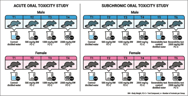
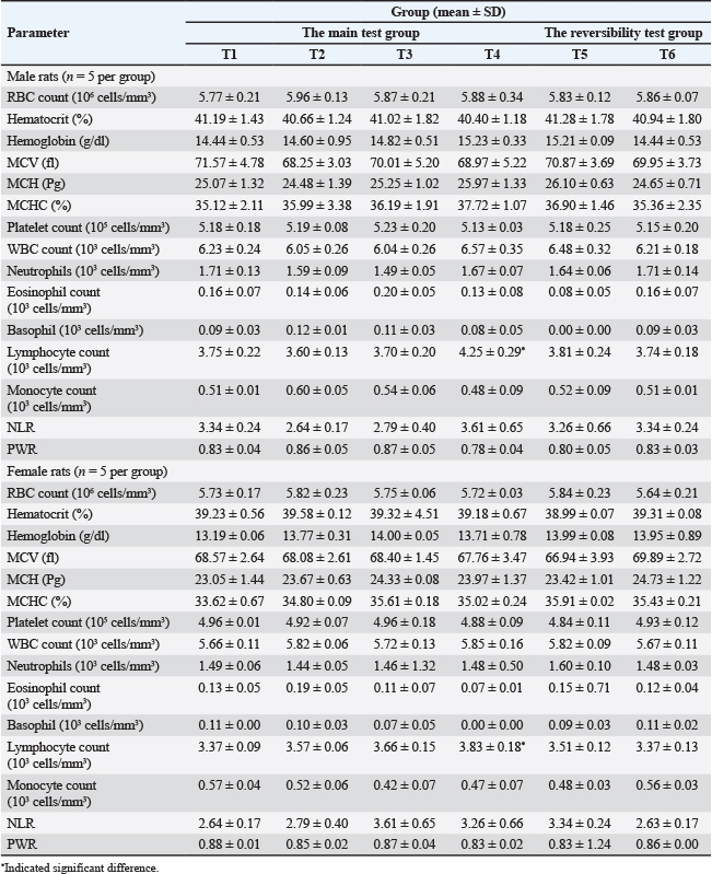
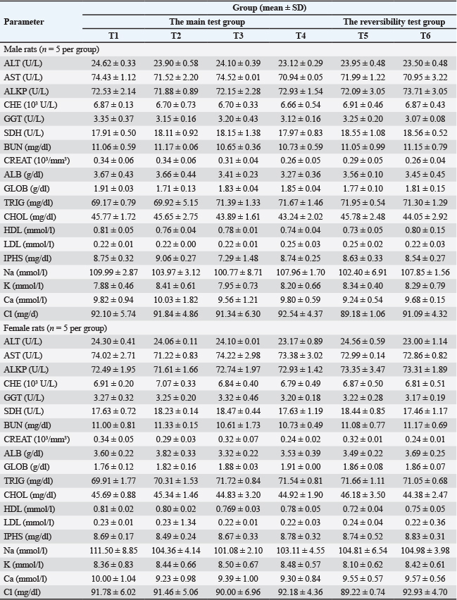
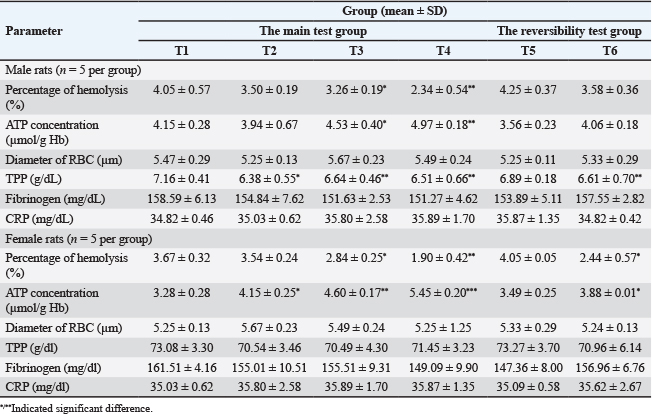
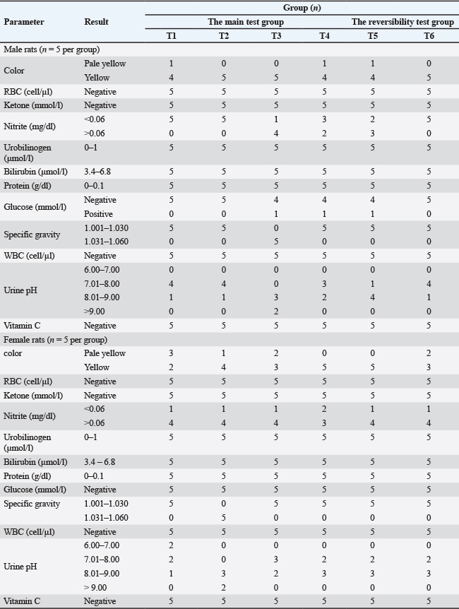
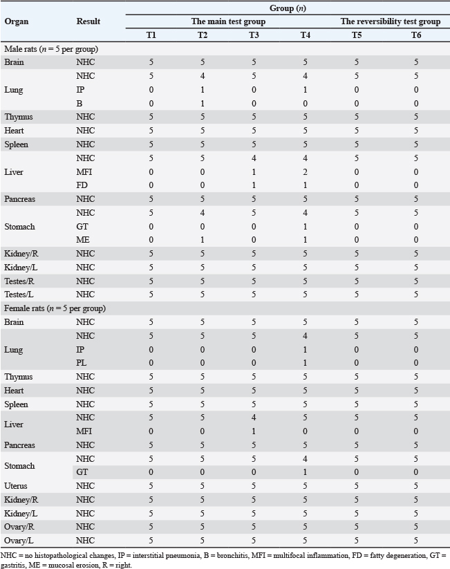
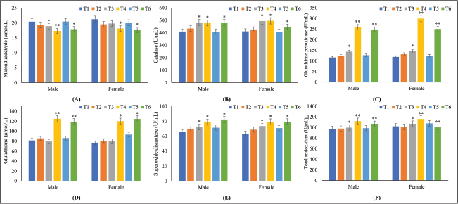
AbstractBackground: Fermented Crescentia cujete L (FC-C) has significant potential as a multimodal therapeutic agent. However, its toxicological profile remains undefined and requires comprehensive elucidation. Aim: This study assessed the acute and 90-day subchronic oral toxicity of the FC-C preparation in rat models. Methods: The C. cujete fruit was fermented for 30 days before toxicity testing, and the resulting product was subsequently evaluated for acute and subchronic toxicity. A total of 100 Sprague–Dawley rats were employed in this study, comprising 40 rats (20 of each sex) for the acute toxicity test and 60 rats (30 of each sex) for the subchronic evaluation. The study utilized three doses of FC-C: high (2,000 mg/kg BW, equivalent to 2 ml/kg BW), medium (1,000 mg/kg BW, 1 ml/kg BW), and low (500 mg/kg BW, 0.5 ml/kg BW). The control group received 2 ml of distilled water. The acute toxicity observation period lasted for 14 days, whereas the subchronic assessment spanned 90 days, followed by a 30-day recovery period to monitor reversibility. The parameters assessed included body weight, hematology, urinalysis, histopathology, malondialdehyde levels, and antioxidant activity. All data were statistically analyzed using SPSS version 26 at a p-value of 0.05. Results: Findings from both acute and subchronic assays indicate that FC-C possesses a high safety margin, with an estimated LD50 exceeding 2,000 mg/kg body weight. Long-term administration at doses up to 2,000 mg/kg BW did not induce significant adverse alterations in BW, organ integrity, serum biochemistry, or tissue histopathology relative to the control group. Conversely, FC-C administration conferred beneficial effects by significantly augmenting systemic antioxidant capacity and enhancing erythrocyte stability. Conclusion: This study substantiates that the 90-day FC-C administration does not induce any adverse toxicological effects. Furthermore, the preparation positively modulates antioxidant defense mechanisms and erythrocyte quality in rat models. Keywords: Antioxidant capacity, Erythrocyte stability, Fermented Crescentia cujete, Oral toxicity, Safety assessment. IntroductionThe growing global prevalence of chronic diseases has spurred significant interest in complementary and alternative medicine (CAM), particularly in plant-derived herbal preparations. These natural products are valued for their perceived safety, lower cost, and complex array of bioactive compounds (Lajolo et al., 2025). Despite their traditional use, modern research must confirm their clinical efficacy and safety. Focus has been placed on the fermented Crescentia cujete (FC-C) preparation (Wilujeng et al., 2023), where fermentation increases the bioavailability and potency of compounds such as choline (Prakoso et al., 2024a). FC-C demonstrates considerable pharmacological potential through multiple mechanisms of action, specifically its neuroprotective, antioxidant, and anti-inflammatory effects, which are driven by neuroprotective choline and other components (Hidayah et al., 2023). FC-C has immunological and anti-inflammatory effects. It acts as an immunostimulant by increasing the number of circulatory differentiation-4+ (CD4+) T-cells and the CD4+/CD8+ ratio (Wijayanti et al., 2024), while lowering inflammatory markers, such as cyclooxygenase-2 (COX-2) and C-reactive protein (CRP), and maintaining hematological balance (Prakoso et al., 2025). In a rat model of acute pneumonia, FC-C combined with antibiotics enhanced lung repair and increased local immunity via increased CD4+ expression and BALT activity (Prakoso et al., 2024b). Studies using rat models of ischemic stroke showed that FC-C significantly reduced infarct size and accelerated neurological recovery by modulating Granulocyte-Macrophage Colony-Stimulating Factor and Vascular Endothelial Growth Factor (Prakoso et al., 2024a). Ischemic stroke is a disease that requires long-term management (Boehme et al., 2021). Despite these promising therapeutic benefits, insufficient safety data restrict the clinical adoption of FC-C. A toxicological profile is necessary to establish safe dosages and identify adverse effects from short- and long-term exposure before human consumption can be confidently recommended (OECD, 2001). Given the lack of rigorous safety data, this study evaluated the acute and 90-day subchronic oral toxicity of FC-C in rats to establish a safety profile for its potential use as a complementary medicine. Materials and MethodsFermented Crescentia cujeteCrescentia cujete fruit was collected from UWKS, Indonesia. The fruit was processed into a liquid fermented product, designated as FC-C, following a previously established procedure (Wilujeng et al., 2023). Experimental animalsThe study used Sprague–Dawley rat models from the Laboratory of Pharmacology, UWKS. a. A total of 40 rats (20 male and 20 female), aged 3 months and weighing 247.25 ± 7.11 g, were used for the acute oral toxicity study. b. Furthermore, 60 rats (30 male and 30 female), aged 3 months and weighing 253.14 ± 8.92 g, were used for the subchronic oral toxicity study. All rats were acclimatized for seven days before the study. They were individually housed in metabolic acrylic cages with oven husk bedding. The following environmental conditions were strictly maintained: room temperature at 25°C and humidity at 65%. Rats were provided pellet feed (RatBio, Indonesia) and water ad libitum. Oral toxicity test resultsThe acute and subchronic oral toxicity study was conducted following the National Agency of Drug and Food Control of the Republic of Indonesia guidelines for oral toxicity testing (BPOM, 2021). Acute oral toxicity testThe study utilized three doses of FC-C: high (2,000 mg/kg BW, equivalent to 2 mL/kg BW), medium (1,000 mg/kg BW, 1 ml/kg BW), and low (500 mg/kg BW, 0.5 ml/kg BW). The control group received 2 ml of distilled water (Fig. 1). The FC-C dose volumes for the treatment groups were supplemented with distilled water to reach a final volume of 2 ml to standardize administration volume across all groups. Rats were fasted for 18 hours before administration of FC-C via a gastric tube. Initial observations for behavioral changes, clinical signs, and mortality were conducted 14 days after administration. On day 14, the final body weights were recorded, and a macroscopic organ analysis was performed.
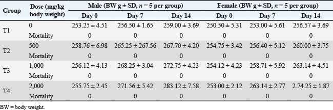
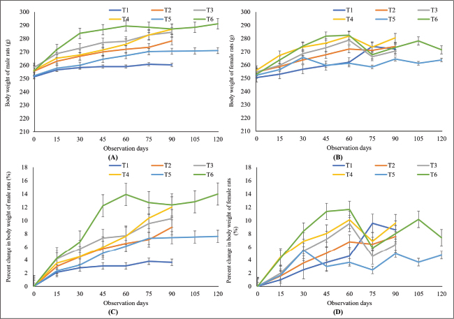
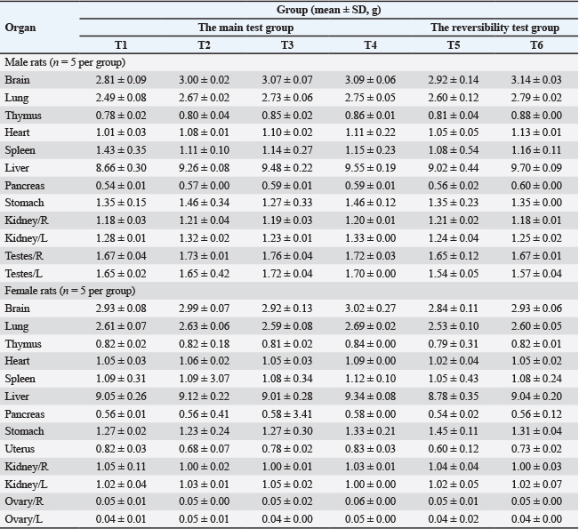
Fig. 1. Grouping of animal models in the oral toxicity study. Subchronic oral toxicity test resultsThe subchronic study employed the same dosage levels (500, 1,000, and 2,000 mg/kg BW). Sixty rats (30 males and 30 females) were divided into six groups (Fig. 1), which included two additional groups (control and high-dose groups, T5 and T6) designated for the reversibility test. Treatment was administered once daily for 13 weeks (90 days) to groups T1–T6. End point analysisOn Day 91, groups T1–T4 were anesthetized (ketamine, 50 mg/kg body weight), and blood and serum were collected. Subsequently, the rats were euthanized (lethal ketamine dose, 150 mg/kg BW), necropsied, and organs were collected. Reversibility testGroups T5 and T6 underwent a 4-week (28-day) recovery period without treatment, receiving only normal feed and water. On day 119, the rats were anesthetized, blood/serum was collected, euthanized, and necropsied. Observation and measurementAll rats were monitored daily for toxicological effects, including changes in skin, fur, eyes, mucous membranes, excretion, secretion, autonomic reflexes, respiration, behavior, feed/water intake, and body weight. HematologyBlood and serum samples were collected from the retro-orbital plexus under general anesthesia (50 mg/kg BW ketamine) on day 91 (T1–T4) or day 119 (T5–T6). Blood was collected into ethylenediaminetetraacetic acid tubes (1 ml for hematology), plain tubes (1 ml for serum collection), and 100 µl for blood smears. The following hematological parameters were measured using an automatic hematology analyzer (VetScan® HM5, Zoetis, US): red blood cells (RBC), hematocrit, hemoglobin, platelet count, mean corpuscular volume (MCV), mean corpuscular hemoglobin (MCH), mean corpuscular hemoglobin concentration (MCHC), white blood cells (WBC), differential counts (neutrophil, eosinophil, basophil, lymphocyte, monocyte), neutrophil-to-lymphocyte ratio (NLR), and platelet-to-white blood cell ratio (PWR). Clinical chemistry and electrolytesClinical chemistry tests were performed using a photometer (MicroLab 300, Netherlands). The parameters included alanine transaminase (ALT), aspartate aminotransferase (AST), alkaline phosphatase (ALKP), cholinesterase (CHE), gamma-glutamyl transferase (GGT), sorbitol dehydrogenase (SDH), blood urea nitrogen (BUN), creatinine (CREAT), albumin (ALB), globulin (GLOB), triglycerides (TRIG), cholesterol (CHOL), high-density lipoprotein (HDL), low-density lipoprotein (LDL), inorganic phosphorus (IPHS), sodium (Na), potassium (K), calcium (Ca), and chloride (Cl). The procedure was conducted following the manufacturer’s instructions. Quality of erythrocytesThe quality of erythrocytes was measured following published methods for RBC diameter (Wijayanti et al., 2024), ATP concentration (Almizraq et al., 2013), and the percentage of hemolysis (Sowemimo-Coker, 2002). Circulating proteinThe following specific circulating proteins were quantified: total plasma protein (TPP) via refractometry (Hunsaker et al., 2016), fibrinogen using the coagulation method (Chen et al., 2014), and CRP via ELISA (E-MSEL-M0059, Elabscience, US). UrinalysisUrine samples were collected on days 91 (T1–T4) and 119 (T5–T6) and analyzed using a Urine Analyzer (Urite-50, China). The semiquantitative parameters measured included color, RBC, ketones, nitrite, urobilinogen, bilirubin, protein, glucose, specific gravity, WBC, pH, and vitamin C. Macroscopy and organ histopathologyDuring necropsy, various organs were collected from both sexes, including the brain, lung, thymus, heart, spleen, liver, pancreas, stomach, kidney, testes, uterus, and ovary. All harvested organs were weighed. Organs were fixed in 10% NBF, dehydrated, cleared, embedded in paraffin, sectioned using a microtome, and stained with H&E for histopathology. All slides were analyzed by a pathologist, and histopathological changes were reported as the frequency of affected animals. Malondialdehyde and antioxidantsMDA levels were measured using a colorimetric assay (Thermo Fisher, US). Antioxidant capacity was assessed by measuring the activities of catalase (CAT) (Farman and Hadwan, 2021), glutathione peroxidase (GPx), superoxide dismutase (SOD), and glutathione (GSH) (Bahrami et al., 2016) and total antioxidant capacity (TAC) (Horváth-Szalai et al., 2023). Analysis dataData normality was verified using the Kolmogorov–Smirnov test, and the Levene test was used to test homogeneity. Because all data were normally distributed and homogeneous, ANOVA was applied. Subsequently, significant differences were analyzed using a Duncan post hoc test. The statistical significance level (α) was 0.05. Ethical approvalThis research was conducted between May and November 2025 at the Laboratory of Pharmacology, UWKS, Indonesia. The ethical clearance committee of the Faculty of Veterinary Medicine, UWKS, approved the study protocols for animal experimentation under approval number 234-KKE-2025. ResultsAcute oral toxicity of FC-CNone of the tested doses (500, 1,000, and 2,000 mg/kg BW of FCC) produced clinical signs, changes in BW, or mortality in the rats during the acute toxicity study (Table 1). These results indicate that the LD50 of FCC in both sexes exceeds 2,000 mg/kg BW. Therefore, the same dose levels were used for the 90-day subchronic oral toxicity study. Subchronic oral toxicity of FC-CBody weightNo animal exhibited clinical symptoms or experienced mortality after FC-C exposure. Body weights increased across all groups, with T4 and T6 showing significantly greater gains than the remaining groups (p < 0.05, Fig. 2a and b). The proportional increase in body weight was significantly higher in males than in females (Fig. 2c and d). Absolute organ weight (%)The absolute organ weights remained statistically consistent across all groups for both sexes. Although minor variations were observed in certain male organs, the differences were not significant (p > 0.05, Table 2). HematologyThe hematological parameters generally showed minimal variation across all groups (Table 3). Most metrics, including RBC, hematocrit, hemoglobin, and platelet count, in the treatment groups remained stable compared with the control (p > 0.05). The only statistically significant difference was an increase in lymphocyte count in the T4 groups for both sexes compared with the control (p < 0.05). Clinical chemistry and electrolytesClinical chemistry and electrolyte analysis revealed no significant toxicological effects on hepatic, renal, or metabolic function. Key markers, including liver enzymes, BUN, CREAT, electrolytes, and lipid profiles, remained stable across all treated groups compared with the control (p > 0.05, Table 4). Erythrocyte quality and circulating proteinFC-C significantly lowered TPP in most treated groups compared with the control (p < 0.05). The T3 and T4 groups in both sexes demonstrated improved erythrocyte stability, as evidenced by a significant decrease in hemolysis and a simultaneous increase in ATP concentration (p < 0.05). Fibrinogen and CRP remained stable (Table 5). UrinalysisUrinalysis predominantly indicated normal renal function, with consistent negative results for major markers such as RBC, ketone, urobilinogen, bilirubin, protein, WBC, and vitamin C. Although the specific gravity in the male T3 group was transiently elevated, it normalized in T4. The isolated variations in glucose and pH were not dose-dependent, confirming the absence of compound-related nephrotoxicity. The urinalysis data can be found in Table 6. HistopathologyHistopathology revealed minimal, noncritical changes in both sexes (Table 7). Most organs, including the brain, thymus, heart, spleen, pancreas, and kidneys, remained unaffected. Liver and stomach alterations observed in T3 and T4 males were non-dose-dependent and absent in the reversibility groups, indicating that they were incidental. FC-C administration did not induce irreversible organ toxicity. Malondialdehyde and antioxidant levelsGroups treated with FC-C showed a gradual decrease in MDA concentrations compared with the control group (T1). Specifically, T4 exhibited a statistically significant reduction in MDA. This effect was maintained at 1,000 and 2,000 mg/kg BW and was not reversed following treatment cessation in the T6 reversibility groups (p < 0.05). FC-C significantly reduced MDA levels (T4), indicating that oxidative stress was mitigated. Moreover, FC-C boosted antioxidant defenses, with the T3 and T4 groups showing statistically significant increases (p < 0.05) in CAT, GPx, SOD, GSH, and TAC (Fig. 3). DiscussionFermentation enhances the bioavailability of plant-derived products. One of the essential roles of fermentation is the degradation of toxic metabolites, such as cyanogenic glycosides, found in the fresh pulp of C. cujete (Adebayo and Ogunlade, 2014). While fresh fruit compounds can induce histotoxic hypoxia by inhibiting cytochrome-C oxidase within the mitochondria (Baud, 2007), fermentation of C. cujete preserves essential nutrients such as choline, α-tocopherol, phytonadione, and retinol in the FC-C product (Prakoso et al., 2024b). During fermentation, microbial pectinase releases bioactive antioxidants (Knez et al., 2023; Zhao et al., 2021). However, integrating FC-C into clinical use requires rigorous scientific validation, particularly a toxicological profile, as natural ingredients are not universally safe for long-term application. Table 1. Body weight and mortality data in rats with acute oral toxicity of FC-C.
Fig. 2. Rat body weight and percent change in body weight during the subchronic and reversibility tests of oral FCC toxicity. This study indicated that the acute toxicity evaluation established a pseudo-LD50 exceeding 2,000 mg/kg BW, classifying FC-C as safe according to Organisation for Economic Co-operation and Development, Food and Drug Administration (FDA), and National Agency of Drug and Food Control of the Republic of Indonesia guidelines. Moreover, the subchronic study noted significant lymphocytosis at 2,000 mg/kg BW (T4). Lymphocytosis occurs as adaptive immune stimulation rather than toxicity due to the absence of elevated serum CRP (Pepys and Hirschfield, 2003), neutrophilia, or oxidative stress markers (Cetin et al., 2018). This mirrors the effects of herbal immunomodulators, such as Panax ginseng, which increase the proliferation of naive immune cells, such as lymphocytes (Kim et al., 2018). The mechanism is likely driven by the high choline content of FC-C, which facilitates biosynthesis via the Kennedy pathway to enhance proliferation (Yao et al., 2023). Moreover, choline can activate the non-neuronal cholinergic system in lymphocytes (Lu and Wu, 2021) and triggers proliferation pathways via acetylcholine (ACh) receptors (Resende and Adhikari, 2009). Thus, high choline intake provided a dual stimulus as structural support for new cells and a chemical signal to trigger proliferation. Additionally, fermentation-derived postbiotics may interact with GALT receptors (Hamdi et al., 2025). Metabolites from FC-C fermentation interacting with macrophages and dendritic cells in GALT may promote lymphocyte maturation and proliferation through the release of cytokines, such as interleukin-2 (IL-2) or interleukin-7 (IL-7), while avoiding excessive induction of pro-inflammatory cytokines such as tumor necrosis factor-alpha (TNF-α) or interleukin-6 (IL-6) (Subramanian et al., 2024). This pattern is similar to that of fermented Houttuynia cordata, which enhances infection resistance in rats by increasing CD4+ T lymphocyte and natural killer (NK) cell populations without provoking autoimmunity (Sumneang et al., 2025). Table 2. Absolute weight of the rats’ organs in subchronic oral toxicity of FC-C.
FC-C treatment improved erythrocyte quality, as evidenced by reduced hemolysis and increased ATP levels. Because erythrocytes rely on anaerobic glycolysis for membrane pump maintenance, oxidative damage typically depletes ATP (Orrico et al., 2023). The observed ATP accumulation demonstrated an ATP-sparing effect (Yamaguchi and Fukuzaki, 2019), where antioxidant-stabilized membranes require less energy for ion homeostasis (Gou et al., 2021). Furthermore, erythrocytes are rich in PUFAs, which are vulnerable to lipid peroxidation by free radicals. The results of this study indicated a sharp decrease in MDA levels in the treatment groups. This is direct evidence that antioxidants in FC-C (such as α-tocopherol) effectively neutralize reactive oxygen species (ROS) in the erythrocyte membrane, preventing lipid peroxidation (Traber and Stevens, 2011). The normal liver markers (ALT, AST, ALKP, and GGT) and histopathology confirmed the hepatoprotective effects of FC-C. The most convincing data regarding the hepatoprotective effect of FC-C is the consistent and dose-dependent increase in antioxidant activities such as SOD, CAT, GPx, miGSH, and TAC. Such an enzymatic pattern is a typical sign of the activation of the nuclear factor erythroid 2-related factor 2 (Nrf2) pathway (Saha et al., 2020). Nrf2 is a major transcription factor that regulates the expression of cytoprotective genes. Histopathologically, the liver showed normal histological architecture without any signs of abnormality. At the molecular level, Nrf2 is kept inactive in the cytoplasm by the Kelch-like ECH-associated protein 1 (Keap1) inhibitor protein, which continuously targets it for degradation (Adinolfi et al., 2023). Table 3. Hematology of rats in subchronic oral toxicity of FC-C.
Table 4. Clinical chemistry and electrolytes in rats with subchronic oral toxicity of FC-C.
Table 5. Rats’ percentage of hemolysis, ATP concentration, RBC diameter and circulating protein in FC-C subchronic oral toxicity.
FC-C phytochemicals likely modify Keap1 cysteine residues (Tossetta et al., 2025), releasing Nrf2 to bind the Antioxidant Response Element (ARE) and trigger cytoprotective gene transcription-encoded antioxidant enzymes (SOD, CAT, GPx) and phase II detoxification enzymes (such as heme oxygenase-1/HO-1) (Ngo and Duennwald, 2022). This hormetic effect aligns with previous findings on C. cujete hepatoprotection. Gonzales et al. (2023) revealed that C. cujete contains flavonoid compounds, such as naringenin, pinocembrin, and eriodictyol. These 3 compounds can inhibit Nrf2-related inflammatory pathways and suppress oxidative proteins during in silico studies. Reversibility groups (T5–T6) showed no delayed toxicity. Notably, CAT activity remained elevated after cessation, demonstrating persistent enzyme induction or epigenetic modifications (Busch et al., 2015). This result also implies that the protective benefits of FC-C can be long-lasting, providing residual protection against oxidative stress. Other parameters that changed, such as lymphocytosis, tended to return toward control values or remained stable within the normal range, confirming that the immune modulation is responsive to the presence of the compound and can be re-regulated by the body. A limitation of this study is the lack of evaluation regarding the impact of FC-C on the gut microbiota in the animal model. Investigating this effect is crucial because of the choline content in the FC-C preparation (Prakoso et al, 2024b). Long-term, high-choline diets can significantly alter the gut microbiota profile (Zhan et al., 2023). These alterations occur because intestinal bacteria use choline as a substrate to produce TMA (Cho et al., 2020). TMA is subsequently converted into trimethylamine-N-oxide (TMAO) in the liver, a compound known to increase cardiovascular risk (Amaritei et al., 2025). Furthermore, it is suspected that high choline levels reduce the colonization of beneficial bacteria, such as Bacteroidetes (Eslami et al., 2024). Therefore, comprehensive studies on how FC-C affects the gut microbiota, in addition to standard toxicity assessments, are warranted prior to clinical translation in humans. ConclusionThis study establishes a no-observed-adverse-effect level (NOAEL) of >2,000 mg/kg BW in Sprague–Dawley rats. The observed lymphocytosis, stabilization of erythrocytes, and upregulation of antioxidants reflect safe immunomodulatory adaptation and cytoprotective priming. Hence, FC-C is a promising and safe candidate for clinical development. Table 6. Urinalysis of rats in subchronic oral toxicity of FC-C.
Table 7. Histopathology of rats in subchronic oral toxicity of FC-C.
Fig. 3. Malondialdehyde, catalase, glutathione peroxidase, glutathione, superoxide dismutase, and total antioxidant levels in rats during the subchronic toxicity study. AcknowledgmentThe authors would like to thank the Kemdiktisaintek of the Republic of Indonesia for their support in funding this study through the Fundamental Research Grant 2025. FundingThis study was funded by Kemdiktisaintek of the Republic of Indonesia through the Fundamental Research Grant 2025 (Decree number: 0070/C3/AL.04/2025, National grand number: 128/C3/DT.05.00/PL/2025, derivative grand number: 007/LL7/DT.05.00/PL/2025, 193/PL/LPPM/UWKS/V/2025). Authors’ contributionYAP supervised the study. AS collected and determined the plant specimens. YAP, AS, SW, OMP, and PGWM conducted the study. AS and PGWM analyzed the compounds of the FC-C preparation. YAP and SW performed the histopathology and immunohistochemistry analyses. YAP and OMP performed hematological, serological, and antioxidant analyses. Mouth and AS conducted the statistical analysis. All authors equally contributed to the manuscript preparation. Conflict of interestThe authors declare no conflict of interest. Data availabilityAll data in this study are included in the manuscript. ReferencesAdebayo, M.A. and Ogunlade, I. 2014. The chemical constituents of calabash (Crescentia cujete). Afr. J. Biotechnol. 13(28), 2824–2830. Adinolfi, S., Patinen, T., Jawahar, D.A., Pitkänen, S., Härkönen, J., Kansanen, E., Küblbeck, J. and Levonen, A.L. 2023. The KEAP1-NRF2 pathway: therapeutic targets and role in cancerRedox Biol. (Basel) 63, 102726. Almizraq, R, Tchir, JD, Holovati, J.L. and Acker, J.P. 2013. Storage of red blood cells affects membrane composition, microvesiculation, and in vitro quality. Transfus 53, 2258–2267. Amaritei, O., Mierlan, O.L., Dinu, C.A., Chiscop, I., Matei, M.N., Gutu, C. and Gurau, G. 2025. TMAO and cardiovascular disease: exploring its potential as a biomarker. Med (Kaunas) 61(10), 1767; doi:10.1016/j.medicina.2025.07.067. Badan Pengawas Obat dan Makanan 2021. Peraturan Badan Pengawas Obat dan Makanan Nomor 20 Tahun 2021 tentang Pedoman Uji Toksisitas Produk Obat Tradisional [Regulation of the National Agency of Drug and Food Control Number 20 Year 2021 concerning Guidelines for Toxicity Testing of Traditional Medicine Products]. Republik Indonesia. Bahrami, S., Shahriari, A., Tavalla, M., Azadmanesh, S. and Hamidinejat, H. 2016. Blood levels of oxidant/antioxidant parameters in rats infected with Toxoplasma gondii. Oxid. Med. Cell. Long. 2016, 8534604. Baud, F.J. 2007. Cyanide: critical issues in diagnosis and treatment. Hum. Exp. Toxicol. 26(3), 191–201. Boehme, C., Toell, T., Lang, W., Knoflach, M. and Kiechl, S. 2021. Longer term patient management following stroke: a systematic review. Int. J. Stroke 16(8), 917–926. Busch, C, Burkard, M, Leischner, C, Lauer, UM, Frank, J. and Venturelli, S. 2015. Epigenetic activities of flavonoids in the prevention and treatment of cancer. Clin. Epigenetics 7, 64. Cetin, M, Koc, I. and Savas, O. 2018. Neutrophil to lymphocyte ratio as a predictive marker for oxidative stress and inflammation in patients with stable coronary artery disease. J. Clin. Exp. Invest. 9(3), 85–91; doi:10.1016/j.jci.2018.09.010. Chen, W, Wang, D, Ni, N, Li, M, Liu, Y. and Wang, B. 2014. A fast and simple approach to the quantitative evaluation of fibrinogen coagulation. Biotechnol Lett. 36(2), 337–340. Cho, C.E., Aardema, N.D.J., Bunnell, M.L., Larson, D.P., Aguilar, S.S., Bergeson, J.R., Malysheva, O.V., Caudill, M.A. and Lefevre, M. 2020. Effect of choline forms and gut microbiota composition on trimethylamine-N-Oxide response in healthy men. Nutrients 12(8), 2220. Eslami, M., Alibabaei, F., Babaeizad, A., Banihashemian, S.Z., Mazandarani, M., Hoseini, A., Ramezankhah, M., Oksenych, V. and Yousefi, B. 2024. The importance of gut microbiota on choline metabolism in neurodegenerative diseases. Biomolecules 14(11), 1345; doi:10.1016/j.biomoes.2024.01345. Farman, A.A. and Hadwan, M.H. 2021. A simple kinetic method for assessing catalase activity in biological samples was developed. MethodsX 8, 101434. Gonzales, A.L., Huang, S.K., Sevilla, U.T.A., Hsieh, C.Y. and Tsai, P.W. 2023. In silico analysis of anti-inflammatory and antioxidant properties of bioactive compounds from Crescentia cujete L. Molecules 28(8), 3547. Gou, Z, Zhang, H, Abbasi, M. and Misbah, C. 2021. Red blood cells under flow show maximal ATP release for specific hematocrit. Biophys J. 120, 4819–4831. Hamdi, A., Lloyd, C., Eri, R. and Van, T.T.H. 2025. Postbiotics: a promising approach to combat age-related diseases. Life (Basel) 15(8), 1190. Hidayah, J.H., Prakoso, Y.A. and Widyarini, S. 2023. Brain histopathological changes after treatment using calabash fruit (Crescentia cujete l.) In rat model with artificially induced ischemic stroke. Adv. Anim. Vet. Sci. 11(12), 2003–2009; doi: 10.1016/j.advvs.2023.09.010. Horváth-Szalai, Z., Jakabfi-Csepregi, R., Szirmay, B., Ragán, D., Simon, G., Kovács-Ábrahám, Z., Szabó, P., Sipos, D., Péterfalvi, Á., Miseta, A., Csontos, C., Kőszegi, T. and Tóth, I. 2023. Serum total antioxidant capacity (TAC) and TAC/lymphocyte ratio as promising predictive markers in COVID-19. Int. J. Mol. Sci. 24(16), 12935. Hunsaker, J.J.H., Wyness, S.P., Snow, T.M. and Genzen, J.R. 2016. Clinical performance evaluation of total protein measurement by digital refractometry and characterization of nonprotein solute interferences. Prac. Lab. Med. 6, 14–24. Kim, M., Yi, Y.S., Kim, J., Han, S.Y., Kim, S.H., Seo, D.B., Cho, J.Y. and Shin, S.S. 2018. Effect of polysaccharides from a Korean ginseng berry on the immunosenescence of aged mice. J. Ginseng. Res. 42(4), 447–454. Knez, E, Kadac-Czapska, K. and Grembecka, M. 2023. Effect of fermentation on the nutritional quality of the selected vegetables and legumes and their health effects. Life (Basel) 13, 655. Lajolo, P.P., Martins, E.P., de Souza, B.H.P., Pereira, G.A. and de Oliveira, R.S. 2025. Medicinal plants: bioactive compounds, biological activities, combating multidrug-resistant microorganisms, and human health benefits—a comprehensive review Front. Immunol. 16, 1491777. Lu, J. and Wu, W. 2021. Cholinergic modulation of the immune system - A novel therapeutic target for myocardial inflammation. Int. Immunopharmacol. 93, 107391; doi: 10.1016/j.intimmunopharmacol.2021.107391. Ngo, V. and Duennwald, M.L. 2022. Nrf2 and oxidative stress: a general overview of mechanisms and implications in human disease. Antioxidants (Basel) 11(12), 2345–2346; doi:10.1016/j.antioxide.2022.02.345. Organization for Economic Co-operation and Development. 2001. OECD Guideline for the Testing of Chemicals, Section 4: Health Effects, Test Guideline 407: Repeated Dose 28-day Oral Toxicity Study in Rodents. OECD Publishing. Orrico, F., Laurance, S., Lopez, A.C., Lefevre, S.D., Thomson, L., Möller, M.N. and Ostuni, M.A. 2023. Oxidative stress in healthy and pathological red blood cells. Biomolecules 13(8), 1262; doi:10.1016/j.biom.2023.12.62 Pepys, M.B. and Hirschfield, G.M. 2003. C-reactive protein: a critical update. J. Clin. Invest. 111(12), 1805–1812. Prakoso, YA, Hidayah, J.H. and Widyarini, S. 2024a. Fermented calabash fruit-derived choline (Crescentia cujete L.) against artificial-induced ischemic stroke in rat models: analysis of N/LR, PWR, histopathology, GM-CSF, and VEGF. J. Appl. Pharm. Sci. 14(11), 082–092. Prakoso, YA, Susilo, A. and Widyarini, S. 2024b. The standardization and efficacy of fermented Crescentia cujete (L.) in combination with enrofloxacin against artificially induced pneumonic pasteurellosis in rat models. Open Vet. J. 14(12), 3404–3416; doi:10.1016/j.ovj.2024.04.004. Prakoso, Y.A., Susilo, A., Widyarini, S., Wahyuningtyas, P.A. and Hidayah, H.H. 2025. Effects of fermented calabash fruit (Crescentia cujete L.) on the Nissl’s body, C-RP and COX-2 in rat models with artificial-induced ischemic stroke. J. Respir Pharmacology 29(5), 2017–2022. Resende, R.R. and Adhikari, A. 2009. Cholinergic receptor pathways involved in apoptosis, cell proliferation, and neuronal differentiation. Cell Commun Signal. 7, 20. Saha, S, Buttari, B, Panieri, E, Profumo, E. and Saso, L. 2020. An overview of Nrf2 signaling pathway and its role in inflammation. Molecules 25(22), 5474. Sowemimo-Coker, S.O. 2002. Red blood cell hemolysis during processing. Transfus. Med. Rev. 16(1), 46–60; doi:10.1016/j.transfusmedrev.2002.01.013. Subramanian, A., T.T., Kumarasamy, V., Begum, M.Y., Sekar, M., Subramaniyan, V., Wong, L. S. and Al Fatease, A. 2024. Exploring the connections: autophagy, gut microbiota, and pathogenesis of inflammatory bowel disease. J. Inflamm. Res. 17, 10453–10470. Sumneang, N, Kobroob, A, Phungphong, S, Boonhoh, W, Punsawad, C. and Kangwan, N. 2025. Fermented Houttuynia cordata juice exerts cardioprotective effects by reducing cardiac inflammation and apoptosis in rats with lipopolysaccharide-induced sepsis. Nutrients 17(3), 501. Tossetta, G., Fantone, S., Olivieri, F., Mazzucchelli, R., Togni, L., Santarelli, A., Marzioni, D. and Rippo, M.R. 2025. Effect of natural compounds on NRF2/KEAP1 signaling in periodontitis: a potential use to prevent age-related disorders. Mol. Biol. Rep. 52(1), 771; doi:10.1016/S0150-0003 Traber, M.G. and Stevens, J.F. 2011. Vitamins C and E: beneficial effects from a mechanistic perspective. Free Radic. Biol. Med. 51(5), 1000–1013. Wijayanti, A.D., Prakoso, Y.A. and Isla, K.J.V. 2024. Effects of fermented Crescentia cujete L. on the profile of hematology, clinical chemistry, and circulatory CD4+/CD8+ in Sprague Dawley rats. Open Vet. J. 14(9), 2475–2483; doi:10.1016/j.openvj.2024.01.0 Wilujeng, S., Prakoso, Y.A. and Wirjaatmadja, R. 2023. Effects of extraction, fermentation, and storage processes on the level of choline derived from calabash fruit (Crescentia cujete L.). J. Res. Pharmaceuticals 27(2), 620–626. Yamaguchi, T. and Fukuzaki, S. 2019. ATP effects on response of human erythrocyte membrane to high pressure. Biophys. Physicobiol. 16, 158–166. Yao, N, Li, W, Xu, G, Duan, N, Yu, G. and Qu, J. 2023. Choline metabolism and its implications in cancer. Front. Oncol. 13, 1234887. Zhan, X., Fletcher, L., Huyben D., Cai, H., Dingle, S., Qi, N., Huber, L.A., Wang, B. and Li, J. 2023. Choline supplementation regulates gut microbiome diversity, gut epithelial activity, and the cytokine gene expression in gilts. Front. Nutr. 10, 1101519. Zhao, Y.S., Eweys, A.S., Zhang, J.Y., Zhu, Y., Bai, J., Darwesh, O.M., Zhang, H.B. and Xiao, X. 2021. Fermentation affects the antioxidant activity of plant-based food material through the release and production of bioactive components. Antioxidants (Basel) 10(12), 2004; doi:10.1016/j.antioxidant.2001.0. | ||
| How to Cite this Article |
| Pubmed Style Prakoso YA, Susilo A, Widyarini S, Pribadi OM, Mahendra PGW. Acute and subchronic oral toxicity of fermented Crescentia cujete (L) preparation in rats. Open Vet. J.. 2026; 16(8): 5383-5396. doi:10.5455/OVJ.2026.v16.i8.32 Web Style Prakoso YA, Susilo A, Widyarini S, Pribadi OM, Mahendra PGW. Acute and subchronic oral toxicity of fermented Crescentia cujete (L) preparation in rats. https://www.openveterinaryjournal.com/?mno=303736 [Access: August 08, 2026]. doi:10.5455/OVJ.2026.v16.i8.32 AMA (American Medical Association) Style Prakoso YA, Susilo A, Widyarini S, Pribadi OM, Mahendra PGW. Acute and subchronic oral toxicity of fermented Crescentia cujete (L) preparation in rats. Open Vet. J.. 2026; 16(8): 5383-5396. doi:10.5455/OVJ.2026.v16.i8.32 Vancouver/ICMJE Style Prakoso YA, Susilo A, Widyarini S, Pribadi OM, Mahendra PGW. Acute and subchronic oral toxicity of fermented Crescentia cujete (L) preparation in rats. Open Vet. J.. (2026), [cited August 08, 2026]; 16(8): 5383-5396. doi:10.5455/OVJ.2026.v16.i8.32 Harvard Style Prakoso, Y. A., Susilo, . A., Widyarini, . S., Pribadi, . O. M. & Mahendra, . P. G. W. (2026) Acute and subchronic oral toxicity of fermented Crescentia cujete (L) preparation in rats. Open Vet. J., 16 (8), 5383-5396. doi:10.5455/OVJ.2026.v16.i8.32 Turabian Style Prakoso, Yos Adi, Achmadi Susilo, Sitarina Widyarini, Oscar Maulana Pribadi, and Paskalis Guntur Widya Mahendra. 2026. Acute and subchronic oral toxicity of fermented Crescentia cujete (L) preparation in rats. Open Veterinary Journal, 16 (8), 5383-5396. doi:10.5455/OVJ.2026.v16.i8.32 Chicago Style Prakoso, Yos Adi, Achmadi Susilo, Sitarina Widyarini, Oscar Maulana Pribadi, and Paskalis Guntur Widya Mahendra. "Acute and subchronic oral toxicity of fermented Crescentia cujete (L) preparation in rats." Open Veterinary Journal 16 (2026), 5383-5396. doi:10.5455/OVJ.2026.v16.i8.32 MLA (The Modern Language Association) Style Prakoso, Yos Adi, Achmadi Susilo, Sitarina Widyarini, Oscar Maulana Pribadi, and Paskalis Guntur Widya Mahendra. "Acute and subchronic oral toxicity of fermented Crescentia cujete (L) preparation in rats." Open Veterinary Journal 16.8 (2026), 5383-5396. Print. doi:10.5455/OVJ.2026.v16.i8.32 APA (American Psychological Association) Style Prakoso, Y. A., Susilo, . A., Widyarini, . S., Pribadi, . O. M. & Mahendra, . P. G. W. (2026) Acute and subchronic oral toxicity of fermented Crescentia cujete (L) preparation in rats. Open Veterinary Journal, 16 (8), 5383-5396. doi:10.5455/OVJ.2026.v16.i8.32 |


