





| Research Article | ||
Open Vet. J.. 2026; 16(5): 2714-2721 Open Veterinary Journal, (2026), Vol. 16(5): 2714-2721 Research Article Ovarian gonadal histomorphometric as bioindicators of lead contamination in stray cats from Jakarta and Surabaya, IndonesiaRagil Angga Prastiya1*, Raket Wonggo Murgiyana2, Tutus Multianingsih2, Trilas Sardjito1, Amung Logam Saputro1, Nagia Musa Alghoul3 and Samira Musa Sasi41Department of Health and Life Science, Faculty of Health, Medicine, and Life Sciences (FIKKIA), Universitas Airlangga, Banyuwangi, , Indonesia 2Bachelor of Veterinary Medicine, Faculty of Health, Medicine, and Life Sciences (FIKKIA), Universitas Airlangga, Banyuwangi, Indonesia 3Cell and Tissue Culture Department, Libyan Biotechnology Center, Tripoli, Libya 4Department of Zoology, Faculty of science, University of Tripoli, Tripoli, Libya *Corresponding Author: Ragil Angga Prastiya. Department of Health and Life Science, Faculty of Health, Medicine, and Life Sciences (FIKKIA), Universitas Airlangga, Banyuwangi, Indonesia. Email: ragilap [at] fkh.unair.ac.id Submitted: 15/12/2025 Revised: 11/04/2026 Accepted: 22/04/2026 Published: 31/05/2026 © 2025 Open Veterinary Journal
AbstractBackground: Rapid urbanization in Jakarta and Surabaya has intensified lead (Pb) contamination in the urban environment, and stray female cats, as effective bioindicators, provide a valuable model for assessing the accumulation of this persistent heavy metal and its potential adverse effects on ovarian health and reproductive function. Aim: This research was conducted to assess the effect of environmental exposure to lead on reproductive parameters in stray female cats in Jakarta and Surabaya as a sign of urban environmental pollution. Methods: We measured blood lead levels, and ovarian weights and follicular sizes were assessed to determine the effects on reproduction. Results: There were no significant differences in lead levels or reproductive parameters between the two cities, indicating a similar burden across these urban areas. Furthermore, the lack of discernible changes in ovarian morphological structure implies that current levels of exposure to lead may not be immediately affecting reproductive structures. However, the possibilities of sub-clinical, long-term endocrine and reproductive influences cannot be ignored, making continued surveillance and rigorous cell-based investigations a priority. Overall, the prevailing pollution conditions did not cause stray cats to reach toxic levels of lead, but rather revealed a pattern of chronic exposure. Conclusions: This work highlights the need for targeted environmental health policies to reduce pollution risks, protect urban animal populations, and consequently preserve human health. The results make very important contributions to the knowledge of urban. Keywords: Environmental bioindicators, Lead exposure, Reproductive health, Stray cats, Urban pollution. IntroductionUrbanization in Jakarta and Surabaya has caused an increase in environmental pollution, mainly from heavy metals like lead (Pb), which is harmful to both humans and animals (Lestiani et al., 2024; Husniyah et al., 2024; Prastiya et al., 2024). Lead is one of the pollutants which is environmentally persistent and bioaccumulates, thereby it may cause long time effect (Sholehhudin et al., 2021; Collin et al., 2022). Stray cats living in human-inhabited cities can serve as good bioindicators to determine the effects of lead pollution on the environment. The presence of heavy metals in the animal body can be very harmful to their reproductive organs, especially the ovaries, which are a key determinant of the animal's general health. Different studies have demonstrated that lead exposure can disrupt hormonal balance, resulting in impaired reproductive function in various species, including mammals (Balali‐Mood et al., 2021). There is evidence that lead is capable of causing oxidative stress and DNA damage, both of which are detrimental to reproductive health. Heavy metals have been connected to changes in hormone levels, such as follicle-stimulating hormone, which is essential for reproduction (Lee et al., 2019). Furthermore, lead accumulation in the body can disrupt the normal operations of reproductive organs, leading to infertility and other reproductive complications (Kabakçi et al., 2023). Research on the effects of lead on the reproductive health of urban female stray cats remains limited, highlighting a critical gap in understanding the long-term consequences of environmental pollutant exposure in this population. Besides that, the urban settings can get complicated, and that is why lead-bound health risks are honed even more since it can be considered synergistic pollution of different kinds that work together (Ali et al., 2024). For example, previous studies have demonstrated that heavy metals can adversely affect the reproductive systems of various animal models, suggesting that cats, as biologically comparable mammals, may be similarly affected (Hardneck et al., 2018). Thus, the offspring of the polluted urban stray cats would be fewer in number, which could result in bad consequences not only for the cat populations but also for the nature that exists in the cities. Therefore, it is very important to carry out more work that will help understand how lead and other heavy metals influence the reproductive systems of urban stray cats so that the information obtained may be used in public health and environmental pollution reduction programs. We do not have many comprehensive studies, even though the risks are well known; that is why we do not know much about how lead affects the reproductive systems of non-human species. There are a lot of studies relating to lead being harmful to humans and lab animals, but not so much on how it affects pets, particularly stray cats living in highly polluted cities such as Jakarta and Surabaya. These cats, which may be exposed to lead via contaminated air, water, and soil, function as practical bioindicators for urban environmental health risks; however, data regarding their blood lead levels and the associated reproductive health implications are insufficient. This study aims to address this deficiency by examining the blood lead concentrations in urban stray female cats and their possible reproductive health consequences. This study evaluates blood lead concentrations in stray female cats from Jakarta and Surabaya and examines their potential association with ovarian health indicators. This research intends to seek the possible relationship between lead exposure and reproductive organ health, thereby revealing the use of these cats as bioindicators of environmental toxicity. The expected results could provide insight into the health risks of heavy metals for urban animal populations and help to develop methods for reducing environmental threats in urban areas. This study proposes two significant contributions to the existing body of knowledge. First, it looks at the use of stray cats as bioindicators of lead exposure in Indonesia's highly urbanized and polluted areas, which is not something that happens very often. Second, it focuses on the negative effects of lead on these cats' ovarian health in a way that no other study has. This method not only fills a big hole in the current research, but it also shows how important it is to have focused environmental health policies that protect urban animal populations, which in turn protect human health. The research that is to be carried out aims to examine the levels of lead in the blood of stray female cats in the cities of Jakarta and Surabaya in the country of Indonesia. Such an examination would provide an insight into the severity of heavy metal contamination and also allow assessment of the extent to which gonadal health, especially the ovaries, may be affected by it. Since they are part of the city’s ecosystem, stray cats consequently come into contact with various kinds of pollutants released into the environment. Thus, they can be considered as living indicators of the state of the environment and, more specifically, of the chronic effects this environment has on the health of reproductive organs due to the exposure to pollutants like lead (Pb). Like lead, some other heavy metals will get accumulated in organisms. Furthermore, lead exposure has been linked to a variety of reproductive disorders, including the disruption of the hormonal system and the dysfunction of the ovaries (Balali‐Mood et al., 2021; Obasi et al., 2022). The main focus of this research is on whether and how the ovulatory system is influenced by lead, thus underscoring the function of these animals in detecting hazardous environmental conditions. This study enhances the current body of knowledge in two substantial ways. First, it looks at the use of stray cats as bioindicators of lead exposure in Indonesia's highly urbanized and polluted areas, which is not something that happens very often. Due to the scarcity of literature regarding the health effects of heavy metals on urban stray cats, this study addresses a significant deficiency by concentrating on the impact of lead on reproductive organs (Lee et al., 2019). Second, it specifically focuses on the detrimental effects of lead on the ovarian health of these cats, a subject that has not been thoroughly investigated in relation to urban pollution (Kabakçi et al., 2023). The expected results may elucidate the health hazards that heavy metals present to urban animal populations and guide the development of strategies to reduce environmental risks in urban areas. The research elucidates the consequences of lead exposure on feral cats, highlighting the necessity for targeted environmental health policies that safeguard urban animal populations, consequently protecting human health as well (Hardneck et al., 2018). In spite of the fact that lead poisoning in humans and lab animals is well documented, there is a lack of information about free-roaming urban cats as bioindicators, especially in the case of linking blood lead levels with ovarian histomorphometry in Southeast Asia. This is why our work can be considered as one of the first to present evidence in environmental toxicology and reproductive morphometrics of urban stray cats from two major Indonesian cities, in a way, presenting a unique One Health approach. It is important to point out that the main objective of this research was to determine blood lead levels in female stray cats from Jakarta and Surabaya and to correlate them with their ovarian morphometric parameters. Materials and MethodsResearch designThe research takes a lead from a prevalence-based design in its assessment of lead exposure in female cats that are living freely in industrial areas of the big cities of Indonesia, especially Jakarta and Surabaya. The samples were collected using the accidental sampling method. The blood and ovarian tissues of each subject were examined to find out the presence of lead toxicity, its level, and any changes in the blood cells and the ovarian histomorphometric due to the lead toxicity. Population and sampleA total of 9 adult female stray cats were included in this study. Inclusion criteria were adult female cats living freely in industrial zones and considered clinically suitable for blood sampling and ovariohysterectomy. Body weight and estimated age were recorded at the time of sampling. Due to ethical and logistical constraints associated with capturing and surgically sampling free-roaming animals, a convenience (accidental) sampling approach was applied, which is commonly used in field-based veterinary studies involving stray populations. Although no a priori sample size calculation was performed, the sample size was considered sufficient for exploratory comparisons between study locations. Sample collection procedureThe study involved the collection of samples from female cats obtained through accidental sampling. All animals underwent preliminary physical examinations, including inspection, palpation, percussion, auscultation, and vital sign measurement (temperature and pulse rate). Blood samples were obtained via the cephalic vein (3 ml per cat), collected using a venoject containing EDTA anticoagulant. A single blood smear was prepared using a drop of blood spread on a slide, stained with eosin and methylene blue, to evaluate the presence of lead-induced morphological changes in erythrocytes, including basophilic stippling, elliptocytosis, and microcytic erythrocytes. The remaining blood was used for lead concentration testing, conducted at the Jakarta Central Health Laboratory. Following blood collection, each cat was premedicated with atropine sulfate (subcutaneous) and acepromazine (intramuscular), followed by ketamine HCl anesthesia (intramuscular). Aseptic procedures were applied, including 70% alcohol and povidone iodine. Measurement of blood lead levelsLead concentration in blood samples was used as the standard for assessing exposure in free-roaming female cats in industrial areas. Blood samples were collected in EDTA-containing tubes and maintained under controlled conditions until analysis. The lead concentrations were found through atomic absorption spectrophotometry (AAS) at the Jakarta Central Health Laboratory. The AAS method is very specific and sensitive to trace amounts of lead in the whole blood; therefore, it is a direct and unequivocal indicator of the cats' environmental exposure to lead and its accumulation in the blood circulation. Gonad extractionOvarian tissues were sourced from the gonads of stray female cats after an ovariohysterectomy operation that was carried out following strict professional standards. In order to reduce the risk of aspiration during an anesthetic procedure, the cats were initially held off from food for 8 hours. As pre-medication, xylazine was given intramuscularly for sedation and muscle relaxation, and then an injection of ketamine HCL was given to induce general anesthesia. Thus, the animals were still unconscious and pain-free during the surgery. In order to reach the ovaries and uterus, a careful midline abdominal incision was made, exposing the reproductive organs. After that, the reproductive organs were removed, the least destruction of the tissues was ensured, and they were immediately fixed in 10% buffered formalin to keep their structural integrity. After the operation, the cats were allowed to recuperate and were given appropriate analgesia to make sure they were comfortable (Kostenko et al., 2023). The fixed ovarian tissues were later prepared for histological studies, which included dehydration, paraffin embedding, sectioning, and hematoxylin and eosin staining. Such detailed preparations enable detailed histomorphometric studies, which can be used to measure the effect of environmental pollutants on reproductive health. Morphometric measurement of gonadsGonadal morphology was thoroughly analyzed through macroscopic as well as microscopic measurements. In addition, the testes and ovaries were weighed and measured for their gross dimensions (length and width). The first step was to rinse each gonad in physiological saline and blot it dry to ensure accuracy. After that, the weight was taken with an Ohaus analytical balance of 0.0001 g precision. For length and width, a digital caliper of 0.1 mm accuracy was used to measure the extent of lead exposure in the gonads (Sellman, 2024). Additionally, the ovarian structures, especially the follicles, were studied and measured under the microscope for a more detailed evaluation. The preparation for microscopic analysis of ovarian tissues included the steps of sectioning and staining, after which the examination was carried out under a trinocular microscope with a micrometer scale (Westhauser et al., 2018). In order to achieve exact scaling, the image analysis software used for measuring follicle diameter and other parameters was calibrated with a micrometer. This method allowed for very precise measurements of follicular morphology and, hence, a more detailed understanding of the histological changes that might have been caused by the lead exposure. Statistical analysisStatistical analysis was performed using SPSS version 22 (IBM Corp., Armonk, NY, USA). Data distribution was assessed for normality using the Shapiro–Wilk test. For variables that followed a normal distribution, comparisons between groups were conducted using an independent samples t-test. Variables that did not meet the assumption of normality were analyzed using the Mann–Whitney U test. Data are presented as mean ± SD. A p-value of < 0.05 was considered statistically significant. Ethical approvalThe research adhered to the highest ethical standards for animal experiments and received the green light from the Ethics Committee of the Faculty of Veterinary Medicine, Universitas Airlangga. Before the commencement of the study, the protocols involving animals were thoroughly scrutinized and given the go-ahead to conform to the local and national regulations. The go-ahead for the study was given bearing the certificate number 1.KEH.114.07.2023. The well-being of animals was a priority, and thus, the number of animals used was kept to a minimum. ResultsBlood lead levelsThe comparative analysis of blood lead levels between Jakarta and Surabaya did not reveal any statistically significant differences. The mean blood lead concentration in Jakarta was 1.24 ± 0.15 mg/dl, slightly lower than in Surabaya, where it was 1.36 ± 0.24 mg/dl (Table 1). Table 1. Blood lead levels and ovarian weight in stray female cats.
Ovarian organ weightMeasurements of ovarian organ weight also showed no significant differences between the two locations. The right ovarian weight averaged 0.47 ± 0.61 g in Jakarta and 0.38 ± 0.11 g in Surabaya. Similarly, the left ovarian weight was 0.49 ± 0.70 g in Jakarta and 0.41 ± 0.70 g in Surabaya, indicating no notable variation between the sites (Table 1). Follicular dimensionsTable 2 presents the measurements for ovarian follicle width and area at different stages of development. Let us break down what the numbers show. Primordial follicles, which are the earliest stage, do not show any real difference in width or area between Jakarta and Surabaya. In Jakarta, the first width measurement comes in at 44.62 ± 2.28 µm, while Surabaya has 39.81 ± 11.42 µm. Second measurements: 52.47 ± 1.91 µm for Jakarta, 74.04 ± 63.76 µm for Surabaya. The areas: Jakarta 1869.93 ± 141.76 µm², Surabaya 1608.39 ± 724.08 µm². Table 2. Comparative follicular dimensions in stray female cats from Jakarta and Surabaya.
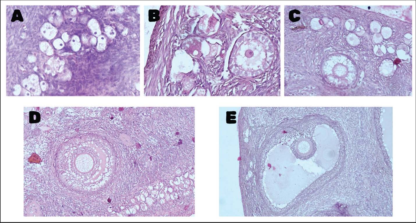
Primary follicles paint the same picture. Width 1 is 94.99 ± 12.34 µm in Jakarta and 74.33 ± 20.88 µm in Surabaya. Width 2 is 107.38 ± 12.96 µm and 65.47 ± 51.08 µm. For the area, Jakarta posts 8389.49 ± 2067.65 µm², Surabaya 5236.57 ± 3000.68 µm². No significant difference here either. Secondary follicles also do not stand out between the two cities. In Jakarta, the widths are 178.85 ± 105.33 µm and 195.98 ± 100.49 µm; Surabaya has 116.06 ± 15.76 µm and 134.84 ± 15.76 µm. The areas: 34329.71 ± 40457.64 µm² in Jakarta, 12625.78 ± 3119.42 µm² in Surabaya. Moving on to de Graaf follicles, still no significant differences. Jakarta’s widths were 245.22 ± 58.41 µm and 332.50 ± 148.52 µm; Surabaya’s were 450.64 ± 233.23 µm and 501.47 ± 191.53 µm. Their areas were as follows: 418686.64 ± 521791.57 µm² for Jakarta, 207926.10 ± 148981.14 µm² for Surabaya. As for tertiary follicles, there just weren’t enough samples, only five in total across both cities. That’s not enough for a meaningful statistical test, so no analysis there. All in all, across primordial, primary, secondary, and de Graaf follicles, the dimensions are pretty consistent between Jakarta and Surabaya. Tertiary follicles, though, remain a question mark due to the limited data. Histological examination of ovarian tissues revealed well-preserved structural organization across all follicular stages, including primordial, primary, secondary, and antral to de Graaf follicles. The follicles exhibited normal morphological characteristics, with clearly defined oocytes, granulosa cell layers, and theca structures. No apparent histopathological alterations, such as follicular degeneration, stromal fibrosis, or inflammatory cell infiltration, were observed. Overall, the ovarian architecture appeared intact, and no notable differences were identified between samples from Jakarta and Surabaya. Representative histological sections illustrating the different follicular stages are presented in Figure 1.
Fig. 1. Representative histological sections of ovarian follicles in stray female cats (H&E staining): (A) primordial, (B) primary, (C) secondary, (D) antral, and (E) de Graaf follicles. DiscussionThe outcomes of this study reveal no significant differences in blood lead levels and ovarian organ weights between stray female cats in Jakarta and Surabaya. This uniformity suggests a similar distribution of environmental lead exposure across these industrialized cities. Moreover, the consistent follicular dimensions across various stages further imply that the observed levels of lead exposure may not be associated with detectable alterations in ovarian morphology of these animals. Blood lead levels found in stray cats from cities like Jakarta and Surabaya reveal a concerning situation. These cats experience ongoing exposure to lead, even if their blood levels don’t reach the threshold for acute poisoning. Chronic lead exposure accumulates quietly in tissues, causing harm over time without immediate signs (Rajpoot et al., 2024). The scientific literature supports this; researchers repeatedly emphasize the importance of regular monitoring and further research, since the full impact of long-term lead exposure on health, both in urban wildlife and ultimately in humans, is not yet fully understood. The dangers are significant. Hwang et al. (2020) demonstrate that even low levels of lead can disrupt hormonal balance and reproductive health in mammals. Prolonged exposure results in infertility and developmental issues. Balali-Mood et al. (2021) explore these cumulative effects, highlighting that neurological and reproductive harm can develop gradually, often without clear symptoms, but the long-term consequences for populations are profound. Additionally, Mielcarek and Kaczmarek (2022) underscore the importance of ongoing environmental monitoring, especially in urban areas where pollution levels are constantly changing. Regular assessments of heavy metals in urban wildlife help not only the animals themselves but also provide insight into the broader ecological situation and inform public health strategies (Nkwunonwo et al., 2020). However, these broader toxicological findings should be interpreted cautiously in the context of the present study, as no direct physiological or endocrine parameters were measured. Continuous surveillance is essential. Without it, we risk overlooking the chronic impact of lead on stray cats, and we lose valuable information about the overall health of our urban environments. When examining ovarian organ weights and follicular size in stray female cats that have been exposed to environmental lead, the findings reveal little to no significant difference compared to cats that have not encountered such exposure. Both groups display remarkably similar reproductive organ structures, suggesting that, at least at the exposure levels observed, lead does not impart any overt or easily detectable changes to ovarian anatomy. This aligns with current scientific understanding, as mild or low-level exposure to environmental toxins like lead frequently fails to produce dramatic anatomical alterations that would be evident through gross morphological assessment (Dane and Şi̇şman, 2020). In other words, simply looking at the organs or measuring their size may not tell the whole story when it comes to the impact of environmental contaminants. However, the absence of visible anatomical differences should not be mistaken for a lack of underlying effects on reproductive health. Lead, in particular, has a well-established reputation for interfering with endocrine function. Nevertheless, in the context of the present findings, such effects cannot be confirmed and should be considered as potential mechanisms reported in other species rather than direct evidence in cats. This can have profound consequences for critical ovarian processes such as folliculogenesis, the development and maturation of ovarian follicles, and steroidogenesis, the production of hormones like estrogen and progesterone that regulate reproductive cycles (Javorac et al., 2023). In this study, however, the absence of hormonal or biochemical measurements limits the ability to draw conclusions regarding these pathways. Disruption of these processes may not immediately manifest as changes in organ size or weight but could have significant long-term implications for fertility, hormonal balance, and overall reproductive success. It is important to note that the studies referenced do not specifically investigate the effects of lead exposure on cats. Instead, they provide a broader context, demonstrating that environmental toxins can exert subtle influences on ovarian function and endocrine regulation across various species. Therefore, extrapolation to feline populations should be approached with caution. For instance, Martínez-Peña and González (2022) highlight how environmental factors such as THC can alter ovarian processes, even though their research does not directly address lead. This underscores a recurring theme in toxicology: many detrimental effects of environmental contaminants are not immediately visible and may only be detectable through more nuanced biochemical or physiological assessments. Beyond the lack of gross anatomical changes, lead exposure is also known to induce oxidative stress within ovarian tissues. This form of cellular stress can damage DNA, proteins, and lipids, undermining the health and function of ovarian cells even in the absence of outwardly visible alterations. Such biochemical disruptions can impair reproductive function by affecting oocyte quality, hormonal synthesis, and cellular signaling pathways essential for normal ovarian activity. Ma et al. (2019) reinforce this perspective, demonstrating that low-level exposure to environmental pollutants can compromise reproductive health without necessarily altering the size or external appearance of ovaries. This suggests that the true extent of lead’s impact may be masked at the macroscopic level, with significant effects only becoming apparent upon closer cellular or molecular examination. Despite these insights, there remains a notable gap in the literature regarding the direct impact of lead on ovarian health in felines. Most existing research focuses on other species or different environmental toxins, leaving questions about the specific mechanisms and outcomes of lead exposure in cats largely unanswered. As such, while gross anatomical assessments provide valuable initial information, they should be complemented by more sensitive techniques—such as histological analysis, hormone profiling, and molecular assays—to fully elucidate the potential for subtle or cumulative damage. In summary, although the reproductive anatomy of lead-exposed cats may appear unremarkable upon superficial examination, the potential for hidden, subclinical disturbances in ovarian function remains a significant concern. The absence of visible changes does not preclude the possibility of biochemical or physiological disruption, and future research should prioritize uncovering these less obvious but potentially consequential effects of environmental lead exposure on feline reproductive health. ConclusionThis research confirms that the stray cats are regularly exposed to lead in the environment. Even though their lead levels in blood were not within the range of acute toxicity, the exposure was evident and suggested old contamination in the environment. Ovarian weights and follicular sizes were similar in both groups, meaning there were no differences in the structure of these organs during this study. On the other hand, these results are limited only to the structure of the organs, and there was no direct measurement of hormonal or biochemical functions. Hence, it is not possible to identify the potential impact on endocrine or reproductive functions only by existing data. As with other species, chronic exposure may be linked with subtle or subclinical effects; however, these points were not considered in this work. Regular monitoring combined with detailed research employing hormonal, histological, and molecular techniques is needed for the understanding of the effect of chronic lead exposure on the reproductive systems of urban animals. Besides animal welfare, this study is a significant step towards the formulation of environmental health policies to safeguard both animals residing in the city and humans from contamination with heavy metals. AcknowledgmentsWe are deeply grateful to Universitas Airlangga for providing us with the research facilities we required; this study would not have been possible without their support. We also extend our sincere thanks to Yayasan Peduli Lingkungan Indonesia (YPLI), particularly Diah Ayu, DVM, for offering their expertise. They assisted us in selecting the cats and conducted the veterinary procedures during gonadal extraction. Their practical support and commitment to animal welfare and environmental health made this research achievable, and we sincerely appreciate it. FundingThis research received no specific grant. Authors' contributionsAll authors contributed to the conception and overall design of the study. Ragil Angga Prastiya supervised the research project, coordinated the study, and led the manuscript preparation. Raket Wonggo Murgiyana and Tutus Multianingsih were responsible for conducting the experiments and collecting the primary data. Trilas Sardjito and Amung Logam Saputro provided methodological guidance and technical support throughout the research process. Nagia Musa Alghoul contributed to data analysis and interpretation. Samira Musa Sasi assisted in drafting, critical revision, and refinement of the manuscript. All authors have read and approved the final version of the manuscript. Conflict of interestNo potential conflict of interest relevant to this article was reported. Data availabilityAll data that underpin the findings of this study are included within the manuscript. REFERENCESAli, S., Naseer, S., Rehman, M. and Wei, Z. 2024. Recent trends and sources of lead toxicity: a review of state-of-the-art nano-remediation strategies. J. Nanoparticle Res. 26(7), 168. Balali-Mood, M., Naseri, K., Tahergorabi, Z., Khazdair, M.R. and Sadeghi, M. 2021. Toxic mechanisms of five heavy metals: mercury, lead, chromium, cadmium, and arsenic. Front. Pharmacol. 12, 643972; doi:10.3389/fphar.2021.643972 Collin, M.S., Venkatraman, S.K., Vijayakumar, N., Kanimozhi, V., Arbaaz, S.M., Stacey, R.G.S., Anusha, J., Choudhary, R., Lvov, V., Tovar, G.I., Senatov, F., Koppala, S. and Swamiappan, S. 2022. Bioaccumulation of lead (Pb) and its effects on human: a review. J. Hazardous Mater. Adv. 7, 100094. Dane, H. and Şi̇şman, T. 2020. A morpho-histopathological study in the digestive tract of three fish species influenced with heavy metal pollution. Chemosphere 242, 125212. Hardneck, F., Israel, G., Pool, E., and Maree, L. (2018). Quantitative assessment of heavy metal effects on sperm function using computer‐aided sperm analysis and cytotoxicity assays. Andrologia, 50, e13141. https://doi.org/10.1111/and.1314 Husniyah, A., Nurika, G. and Ellyke, E. 2024. Lead Contamination in Pangasius Djambal in the Dam of Kalitidu District, Bojonegoro District: an Environmental Health Risk Study. J. Environ. Health 16(3). Hwang, H.J., Kim, J.H. and Lee, S.H. 2020. Chronic lead exposure and its effects on reproductive health: a review. Environ. Toxicol. Pharmacol. 78, 103367; doi:10.1016/j.etap.2020.103367 Javorac, D., Baralić, K., Marić, Đ., Mandić-Rajčević, S., Đukić-Ćosić, D., Bulat, Z. and Djordjevic, A.B. 2023. Exploring the endocrine disrupting potential of lead through benchmark modelling–study in humans. Environ. Pollut. 316, 120428. Kabakçi, M., Yilmaz, A. and Çelik, M. 2023. Determination of blood heavy metal concentrations and oxidant-antioxidant capacities in Angora cats at different age and gender. Ankara Üniversitesi. Veteriner. Fakültesi. Dergisi. 70(1), 1–10; doi:10.33988/auvfd.931057 Kostenko, E., Ius, A. and Maknickas, A. 2023. Histomorphometry and μCT scan analysis of osteoporosis in spayed female dogs. Open. Vet. J. 13(1), 1–10; doi:10.5455/ovj.2023.v13.i1.1 Lee, T.W., Kim, D.H. and Ryu, J.Y. 2019. The effects of exposure to lead, cadmium and mercury on follicle-stimulating hormone levels in men and postmenopausal women: data from the Second Korean National Environmental Health Survey (2012–2014). Ann. Occupational. Environ. Med. 31(1), 21; doi:10.35371/aoem.2019.31.e21 Lestiani, D.D., Santoso, M., Kijin, S., Ikuji, T., Kurniawati, S., Syahfitri, W.Y.N. and Damastuti, E. 2024. Toxic elements, sources and health risk assessment of PM2. 5 in an industrial area of Surabaya, Indonesia. Int. J. Environ. Anal. Chem. 1–19. Ma, Y., He, X., Qi, K., Wang, T., Qi, Y., Cui, L., Wang, F. and Song, M. 2019. Effects of environmental contaminants on fertility and reproductive health. J. Environ. Sci. 77, 210–217. Martínez-Peña, F.C. and González, M.A. 2022. Environmental factors influencing ovarian processes: implications for reproductive health. Reproductive. Toxicol. 107, 1–10; doi:10.1016/j.reprotox.2022.01.001 Mielcarek, K. and Kaczmarek, K. 2022. The importance of environmental monitoring for urban wildlife health. Ecotoxicol. Environ. Saf. 241, 113898; doi:10.1016/j.ecoenv.2022.113898 Nkwunonwo, U.C., Odika, P.O., and Onyia, N.I., 2020. A review of the health implications of heavy metals in food chain in Nigeria. Sci. World J. 2020(1), 6594109. Obasi, C.N., Frazzoli, C. and Orisakwe, O.E. 2022. Heavy metals and metalloids exposure and in vitro fertilization: critical concerns in human reproductive medicine. Front. Reprod. Health 4, 1037379; doi:10.3389/frph.2022.1037379 Prastiya, R.A., Sardjito, T., Abdillah, A.M., Murgiyana, R.W., Multianingsih, T., Andris, A.J. and Sasi, S.M. 2024. Impact of urban lead pollution on the hematology and reproductive histopathology of stray cats in Indonesian megacities. J. Anim. Behav. Biometeorol. 12(4), 2024028; doi:10.31893/jabb.2024028 Rajpoot, A., Aggarwal, T. and Sharma, V. 2024. Unraveling the enigma of cardiac damage caused by lead: understanding the intricate relationship between oxidative stress and other multifactorial mechanisms. Toxicology, 153984. Sellman, L., Tong, X., Burton, I.S. and Kröger, H. 2024. Retrospective characterization of bone histomorphometric findings in clinical patient specimens. J. Bone. Metab. 31(2), 132–140; doi:10.11005/jbm.2024.31.2.132 Sholehhudin, M., Azizah, R., Sumantri, A., Sham, S.M., Zakaria, Z.A. and Latif, M.T. 2021. Analysis of Heavy Metals (Cadmium, Chromium, Lead, Manganese, and Zinc) in Well Water in East Java Province. Malaysian J. Med. Health Sci. 17(2), 146–153. Westhauser, F., Reible, B., Höllig, M., Heller, R., Schmidmaier, G. and Moghaddam, A. 2018. Combining advantages: direct correlation of two-dimensional microcomputed tomography datasets onto histomorphometric slides to quantify three-dimensional bone volume in scaffolds. J. Biomed. Mater. Res. Part. A. 106(5), 1241–1250; doi:10.1002/jbm.a.36377 | ||

| How to Cite this Article |
| Pubmed Style Prastiya RA, Murgiyana RW, Multianingsih T, Sardjito T, Saputro AL, Alghoul NM, Sasi SM. Ovarian gonadal histomorphometric as bioindicators of lead contamination in stray cats from Jakarta and Surabaya, Indonesia. Open Vet. J.. 2026; 16(5): 2714-2721. doi:10.5455/OVJ.2026.v16.i5.14 Web Style Prastiya RA, Murgiyana RW, Multianingsih T, Sardjito T, Saputro AL, Alghoul NM, Sasi SM. Ovarian gonadal histomorphometric as bioindicators of lead contamination in stray cats from Jakarta and Surabaya, Indonesia. https://www.openveterinaryjournal.com/?mno=303258 [Access: June 26, 2026]. doi:10.5455/OVJ.2026.v16.i5.14 AMA (American Medical Association) Style Prastiya RA, Murgiyana RW, Multianingsih T, Sardjito T, Saputro AL, Alghoul NM, Sasi SM. Ovarian gonadal histomorphometric as bioindicators of lead contamination in stray cats from Jakarta and Surabaya, Indonesia. Open Vet. J.. 2026; 16(5): 2714-2721. doi:10.5455/OVJ.2026.v16.i5.14 Vancouver/ICMJE Style Prastiya RA, Murgiyana RW, Multianingsih T, Sardjito T, Saputro AL, Alghoul NM, Sasi SM. Ovarian gonadal histomorphometric as bioindicators of lead contamination in stray cats from Jakarta and Surabaya, Indonesia. Open Vet. J.. (2026), [cited June 26, 2026]; 16(5): 2714-2721. doi:10.5455/OVJ.2026.v16.i5.14 Harvard Style Prastiya, R. A., Murgiyana, . R. W., Multianingsih, . T., Sardjito, . T., Saputro, . A. L., Alghoul, . N. M. & Sasi, . S. M. (2026) Ovarian gonadal histomorphometric as bioindicators of lead contamination in stray cats from Jakarta and Surabaya, Indonesia. Open Vet. J., 16 (5), 2714-2721. doi:10.5455/OVJ.2026.v16.i5.14 Turabian Style Prastiya, Ragil Angga, Raket Wonggo Murgiyana, Tutus Multianingsih, Trilas Sardjito, Amung Logam Saputro, Nagia Musa Alghoul, and Samira Musa Sasi. 2026. Ovarian gonadal histomorphometric as bioindicators of lead contamination in stray cats from Jakarta and Surabaya, Indonesia. Open Veterinary Journal, 16 (5), 2714-2721. doi:10.5455/OVJ.2026.v16.i5.14 Chicago Style Prastiya, Ragil Angga, Raket Wonggo Murgiyana, Tutus Multianingsih, Trilas Sardjito, Amung Logam Saputro, Nagia Musa Alghoul, and Samira Musa Sasi. "Ovarian gonadal histomorphometric as bioindicators of lead contamination in stray cats from Jakarta and Surabaya, Indonesia." Open Veterinary Journal 16 (2026), 2714-2721. doi:10.5455/OVJ.2026.v16.i5.14 MLA (The Modern Language Association) Style Prastiya, Ragil Angga, Raket Wonggo Murgiyana, Tutus Multianingsih, Trilas Sardjito, Amung Logam Saputro, Nagia Musa Alghoul, and Samira Musa Sasi. "Ovarian gonadal histomorphometric as bioindicators of lead contamination in stray cats from Jakarta and Surabaya, Indonesia." Open Veterinary Journal 16.5 (2026), 2714-2721. Print. doi:10.5455/OVJ.2026.v16.i5.14 APA (American Psychological Association) Style Prastiya, R. A., Murgiyana, . R. W., Multianingsih, . T., Sardjito, . T., Saputro, . A. L., Alghoul, . N. M. & Sasi, . S. M. (2026) Ovarian gonadal histomorphometric as bioindicators of lead contamination in stray cats from Jakarta and Surabaya, Indonesia. Open Veterinary Journal, 16 (5), 2714-2721. doi:10.5455/OVJ.2026.v16.i5.14 |


