Open Veterinary Journal, (2026), Vol. 16(5): 2704-2713
Short Communication
10.5455/OVJ.2026.v16.i5.13
Effects of Oral Nanocurcumin on Uterine Caspase-3 and IL-10 Expression in Noise-Stressed Pregnant Mice
Viski Fitri Hendrawan1*, Ropi Faiha2, Dwi Kristianto3, Handayu Untari4, Dwi Rahmawati5 ,
and Ertika Fitri Lisnanti6,
1Department of Animal Reproduction, Faculty of Veterinary Medicine, Universitas Brawijaya, Malang, Indonesia
2Undergraduate Program in Veterinary Medicine, Faculty of Veterinary Medicine, Universitas Brawijaya, Malang, Indonesia
3Department of Internal Medicine, Faculty of Veterinary Medicine, Universitas Brawijaya, Malang, Indonesia
4Department of Veterinary Pathology, Faculty of Veterinary Medicine, Universitas Brawijaya, Malang, Indonesia
5Department of Orthodontics, Faculty of Dentistry, Universitas Brawijaya, Malang, Indonesia
6Program of Animal Husbandry, Faculty of Agriculture, Universitas Islam Kediri, Kediri, Indonesia
*Corresponding Author: Viski Fitri Hendrawan. Department of Animal Reproduction, Faculty of Veterinary Medicine, Malang, Indonesia. Email: viski [at] ub.ac.id
Submitted: 10/12/2025 Revised: 26/03/2026 Accepted: 06/04/2026 Published: 31/05/2026
© 2025 Open Veterinary Journal
This is an Open Access article distributed under the terms of the Creative Commons Attribution-Non Commercial-No Derivatives License (http://creativecommons.org/licenses/by-nc-nd/4.0/), which permits non-commercial re-use, distribution, and reproduction in any medium, provided the original work is properly cited, and is not altered, transformed, or built upon in any way.
ABSTRACT
Background: Exposure to environmental noise during pregnancy has been linked to the disruption of immune and apoptotic balance in uterine tissues. Caspase-3 (Casp-3) and interleukin-10 (IL-10) are key apoptosis and anti-inflammatory signaling biomarkers, respectively. Nanocurcumin has shown promise as an antioxidant and immunomodulator.
Aim: This study aimed to evaluate the effects of oral nanocurcumin on Casp-3 and IL-10 expression in gestational noise-exposed pregnant mice.
Methods: Twenty-five pregnant BALB/c mice were divided into five groups: negative control (K–), positive control with noise (K+), and three treatment groups exposed to noise and given nanocurcumin at 14 (P1), 21 (P2), and 24.5 mg/kg body weight (BW) (P3). Oral treatments were given from gestational day (GD) 6–18. Uterine tissues were harvested for immunohistochemical analysis. Data were analyzed using analysis of variance, Tukey’s HSD, and linear regression (p < 0.05).
Results: Noise exposure increased Casp-3 and reduced IL-10 expression (p < 0.05). Nanocurcumin reversed these effects in a dose-dependent manner; Casp-3 decreased by 20.7%, whereas IL-10 increased by 41.7% in the P3 group compared with the K+ group. Linear regression analysis showed strong correlations between the dose and expression levels of both markers.
Conclusion: Oral nanocurcumin effectively reduced uterine apoptotic signaling and improved anti-inflammatory responses in pregnancies with noise stress, supporting its potential role as a non-hormonal therapeutic in reproductive stress conditions. This study also aligns with the Sustainable Development Goal (SDG) 3: Good Health and Well-being, particularly in advancing maternal health through nutraceutical interventions during pregnancy under environmental stress.
Keywords: Caspase-3, Good health and well-being, IL-10, Nanocurcumin, Noise stress.
Introduction
Environmental noise is increasingly recognized as a pervasive stressor with adverse effects on physiology and reproduction in both humans and experimental animals (Ristovska et al., 2014; Bronzaft, 2017). Epidemiologic syntheses link chronic exposure above ~85 dB to higher risks of miscarriage, preterm birth, and fetal growth restriction in women (Ristovska et al., 2014). In laboratory models, repeated exposure to high-intensity sound (90–120 dB) perturbs neuroendocrine and immune homeostasis, impairs uterine receptivity, and compromises pregnancy maintenance (Jafari et al., 2017; Mehrizi et al., 2022; Clancy et al., 2023). These observations show that acoustic stress adversely affects gestational success through coordinated endocrine, vascular, and inflammatory pathways.
Mechanistically, stress activates the hypothalamic–pituitary–adrenal (HPA) axis and sympathetic outputs, elevating glucocorticoids and catecholamines and shifting metabolism toward a catabolic state (Sherwood, 2016; Wirtz and von Känel, 2017). Sustained glucocorticoid exposure alters cytokine profiles and mitochondrial integrity, thereby tipping the balance toward apoptosis in susceptible tissues (Yates and Cidlowski, 2013). At the cellular level, mitochondrial injury promotes cytochrome-c release and assembly of the apoptosome, leading to caspase-9 and Casp3 activation, the canonical executioner of apoptosis (Jia et al., 2015). Consistent with this framework, elevated cortisol levels under occupational noise exposure have been repeatedly documented (Naderyan Fe’Li et al., 2022), and noise-exposed gravid rodents display histologic uterine injury and cell death markers (Jafari et al., 2017; Clancy et al., 2023).
However, a successful pregnancy requires not only controlled tissue remodeling but also an immune milieu that favors tolerance at the maternal–fetal interface. Interleukin-10 (IL10) is a central anti-inflammatory cytokine that restrains excessive Th1-type responses, supports regulatory T-cell function, and contributes to placental development. Reduced IL10 levels are associated with gestational complications (Azizieh and Raghupathy, 2017). Experimental stressors and inflammatory challenges can suppress IL-10 while upregulating proinflammatory mediators, thereby creating conditions that promote apoptosis and impaired decidual function (Yates and Cidlowski, 2013; Mehrizi et al., 2022; Farahbakht et al., 2023). In this context, simultaneous assessment of uterine Casp3 and IL10 levels provides an informative window on the convergence of apoptotic and immunoregulatory processes under noise stress.
Curcumin, a polyphenolic constituent of Curcuma longa, exhibits antioxidant and anti-inflammatory properties relevant to stress-related tissue injury (Hewlings and Kalman, 2017; Rahiman et al., 2022). However, native curcumin demonstrates poor solubility and limited bioavailability. Nanoformulations enhance their stability, cellular uptake, and tissue distribution, thereby improving pharmacodynamic effects across diverse experimental models (Gera et al., 2017; Nikdad et al., 2020).
Experimental studies demonstrate that nanocurcumin mitigates oxidative stress and regulates apoptotic signaling in ovarian and other reproductive tissues (Syarifah et al., 2021). Clinical investigations further indicate that oral nanocurcumin exerts therapeutic effects in inflammatory conditions (Kia et al., 2020). Collectively, these properties position nanocurcumin as a promising countermeasure against gestational noise stress (GNS).
Despite accumulating evidence that loud noise provokes uterine dysfunction and that nanocurcumin can modulate oxidative-inflammatory cascades, the direct impact of nanocurcumin on uterine apoptotic execution (as indexed by Casp3) and local anti-inflammatory tone (as indexed by IL10) during pregnancy under acoustic stress has not been defined. This gap needs to be addressed because the dual regulation of apoptosis and immune tolerance is likely required to preserve uterine integrity and implantation sites under chronic stress.
In this study, we tested the hypothesis that oral nanocurcumin administered during midgestation attenuates noise-induced uterine injury by downregulating Casp3 and upregulating IL-10 in pregnant BALB/c mice. Mice were exposed to high-intensity white noise during gestational days (GD) 6–18, an interval spanning postimplantation remodeling through late gestation, and uterine Casp3 and IL10 levels were quantified by immunohistochemistry with image-based morphometry. We further examined dose–response relationships to determine whether the observed effects scale with nanocurcumin dosing (Ristovska et al., 2014; Sherwood, 2016; Jafari et al., 2017). This research holds translational relevance by highlighting nanocurcumin as a candidate intervention to promote maternal uterine resilience, aligning with the Sustainable Development Goal (SDG) 3: Good Health and Well-being, particularly in the domain of maternal reproductive health under environmental challenges.
Although 135 dB exceeds typical community exposure, this intensity has been employed to model occupational or military noise scenarios (Bronzaft, 2017; Naderyan Fe’Li et al., 2022), where extreme noise levels may be experienced by pregnant individuals. The use of such high-intensity stimuli in animal models helps simulate the upper range of human exposure and provokes measurable uterine responses.
Materials and Methods
Animals used and experimental design
A total of 25 hormonally induced pregnant BALB/c mice (Mus musculus), aged ≥3 months with an average body weight of 36.5 ± 1.2 g, were used in this study. The animals were housed in polycarbonate cages under standard laboratory conditions (12 hours light/dark cycle, temperature 23°C ± 2°C), with food and water ad libitum. After acclimatization, the mice were randomly assigned (computer-assisted randomization) into five groups (n=5 per group) as follows:
- K(–): Negative control (no stress or treatment),
- K(+): Positive control (exposed to noise stress only),
- P1: Noise stress + 14 mg/kg BW nanocurcumin,
- P2: Noise stress + 21 mg/kg BW nanocurcumin,
- P3: Noise stress + nanocurcumin 24.5 mg/kg body weight. The investigators performing the analysis were blinded to the group allocation.
The noise exposure protocol
From gestational day (GD) 6 to GD 18, the mice in groups K(+), P1, P2, and P3 were exposed to 135 dB white noise for 40 min per day (09:00–10:00) inside a soundproof acrylic chamber (30 × 20 × 15 cm) equipped with a calibrated amplifier and speaker system. The background noise was maintained at <40 dB in the absence of experimental noise.
Nanocurcumin administration
Nanocurcumin was obtained in a nano-emulsified formulation and administered orally via gavage from GD 6 to GD 18, concurrently with the noise exposure. Each treatment group received a daily dose according to their respective allocation: 14 mg/kg BW (P1), 21 mg/kg BW (P2), or 24.5 mg/kg BW (P3). The dosage was selected based on previous toxicological and pharmacodynamic evaluations. Dosages between 14–25 mg/kg BW were selected based on previous pharmacokinetic and toxicological data in rodent models indicating safety and optimal tissue penetration (Nainggolan et al., 2012; Syarifah et al., 2021).
Euthanasia and tissue collection
On GD 18, the animals were anesthetized with ketamine (100 mg/kg) and xylazine (10 mg/kg) via intraperitoneal injection. Uterine tissues were collected postmortem, and the presence of vaginal plugs and uterine enlargement confirmed pregnancy status. Uteri were fixed in 10% neutral-buffered formalin for histological and immunohistochemical analysis.
Immunohistochemistry (IHC)
Formalin-fixed, paraffin-embedded (FFPE) uterine samples were sectioned at 5 µm thickness and mounted onto poly-L-lysine-coated glass slides. Antigen retrieval was performed after deparaffinization and rehydration using citrate buffer (pH 6.0). Endogenous peroxidase activity was quenched using 3% hydrogen peroxide. The primary antibodies used were rabbit polyclonal anti-caspase-3 (Abcam, ab13847, 1:200 dilution) and rat monoclonal anti-IL-10 (Invitrogen, JES5-2A5, 1:200 dilution). Biotinylated secondary antibodies, streptavidin–HRP conjugate, and DAB chromogen were used for detection. The slides were counterstained with hematoxylin. Negative controls were prepared by omitting the primary antibody.
Image acquisition and morphometric analysis
Immunostained slides were examined under an Olympus BX53 light microscope at 400× magnification. Five non-overlapping high-power fields (HPFs) were randomly selected for each uterus. The area of DAB-positive immunoreactivity was quantified using the color deconvolution plugin in ImageJ software (NIH v1.53c). Two independent observers blinded to the experimental groups performed image analysis.
Statistical analysis
All data were initially assessed for normal distribution using the Shapiro–Wilk test and evaluated for homogeneity of variance using Levene’s test. Group comparisons were analyzed using one-way analysis of variance. When significant differences were detected, Tukey’s honestly significant difference (HSD) post hoc test was used to determine pairwise group differences. A simple linear regression analysis was performed to evaluate the relationship between nanocurcumin dosage and marker expression (caspase-3 and IL-10). The beta coefficient (β) and adjusted coefficient of determination (adjusted R²) were used to interpret the strength and direction of the dose–response trends. All statistical analyses were conducted using the Statistical Package for the Social Sciences version 25.0 software (IBM Corp., Armonk, NY). A p-value of 0.05 was considered statistically significant. Data are presented as mean ± SD.
Ethical approval
This study was approved by the Ethics Committee for the Use of Laboratory Animals, Universitas Muhammadiyah Malang, Indonesia (Approval No. E.5.a/038/KEPKUMM/III/2025) and conducted in compliance with international standards for the care and use of laboratory animals as outlined in the Guide for the Care and Use of Laboratory Animals (National Research Council, 2011).
Results
Histological evaluation of uterine tissues revealed significant alterations in Caspase-3 (Casp-3) expression among the treatment groups. One-way ANOVA showed a statistically significant increase in Casp-3 expression in the positive control group (K+: 27.33 ± 0.77%) compared with the negative control (K-: 19.52 ± 0.56%) (p < 0.05), indicating elevated apoptotic activity in response to noise-induced stress. Oral nanocurcumin administration led to a dose-dependent reduction in Casp-3 expression. Specifically, the P1 group (14 mg/kg BW) showed an average expression of 24.40% ± 1.42%, followed by P2 (21 mg/kg BW) at 22.21% ± 1.23%, and P3 (24.5 mg/kg BW) at 21.65% ± 0.84%. These values represent relative decreases of 10.7%, 18.7%, and 20.7% compared to the K+group, respectively. Post hoc Tukey’s test confirmed significant differences between most groups (p < 0.05), with the expression order being K+ > P1 > P2 ≈ P3 > K-. Linear regression analysis revealed a significant negative relationship between nanocurcumin dosage and Casp-3 expression (β=−0.867, adjusted R²=0.191, p=0.029), demonstrating a dose-responsive reduction of apoptosis (Fig. 2). Representative immunohistochemical staining of Casp-3 is displayed in Figure 1, while the intergroup expression means are summarized in Fig. 3 and Table 1.
Table 1. (Caspase-3 expression examination results) Mean ± SD values of uterine caspase-3 expression (%) in BALB/c mice exposed to gestational noise stress and treated with nanocurcumin.
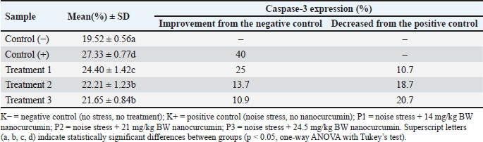
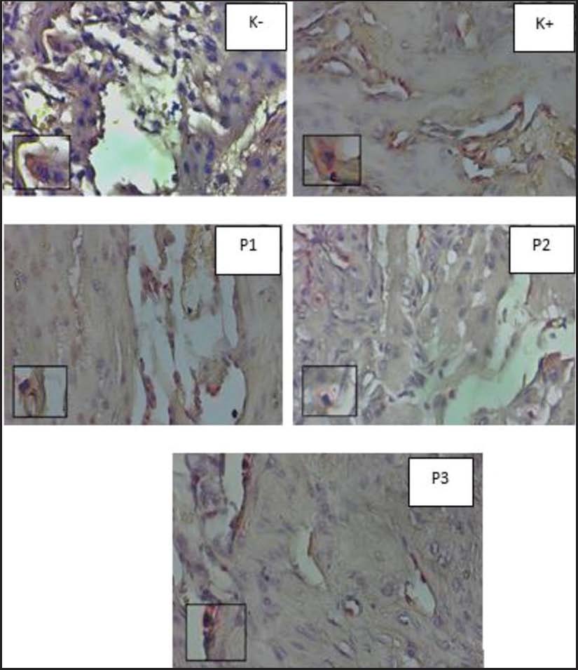
Fig. 1. Representative immunohistochemical staining of caspase-3 in the uterine epithelium of BALB/c mice. K−=negative control; K+=positive control; P1=noise + 14 mg/kg nanocurcumin; P2=noise + 21 mg/kg nanocurcumin; P3=noise + 24.5 mg/kg nanocurcumin. The positive nuclei appear brown (DAB); the negative nuclei appear blue (hematoxylin). Magnification: 400×.
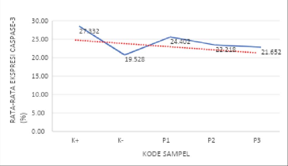
Fig. 2. Linear regression analysis showing the relationship between nanocurcumin dose and uterine Caspase-3 expression (%). Experimental groups: K−, K+, P1, P2, and P3.
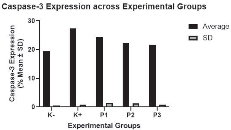
Fig. 3. Caspase-3 expression (%) across the experimental groups. Data are presented as mean ± SD. Administration of nanocurcumin reduced expression in a dose-dependent manner.
Similarly, treatment markedly influenced the expression of interleukin-10 (IL-10). The positive control group (K+: 17.21 ± 0.98%) exhibited significantly reduced IL-10 expression compared with the negative control (K-: 21.20% ± 2.00%), consistent with suppressed anti-inflammatory signaling under acoustic stress. Nanocurcumin administration resulted in enhanced IL-10 expression, with mean values of 23.87% ± 1.81% in P1, 22.91% ± 1.00% in P2, and 24.40% ± 0.94% in P3. These values correspond to increases of 20.04%, 32.3%, and 41.7% compared to K+, respectively. The highest expression level was observed in P3, and Tukey’s post hoc test indicated significant differences between the groups (p < 0.05). Regression analysis confirmed a positive linear relationship between nanocurcumin dose and IL-10 expression (β=0.980, adjusted R²=0.158, p=0.049), indicating a dose-dependent immunomodulatory effect (Fig. 6). The morphological visualization of IL-10 staining is presented in Figure 5, while the quantitative results are summarized in Fig. 4 and Table 2.
Table 2. (IL-10 expression examination results) Mean ± SD values of uterine IL-10 expression (%) in BALB/c mice exposed to gestational noise stress and treated with nanocurcumin.
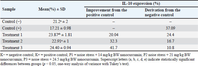
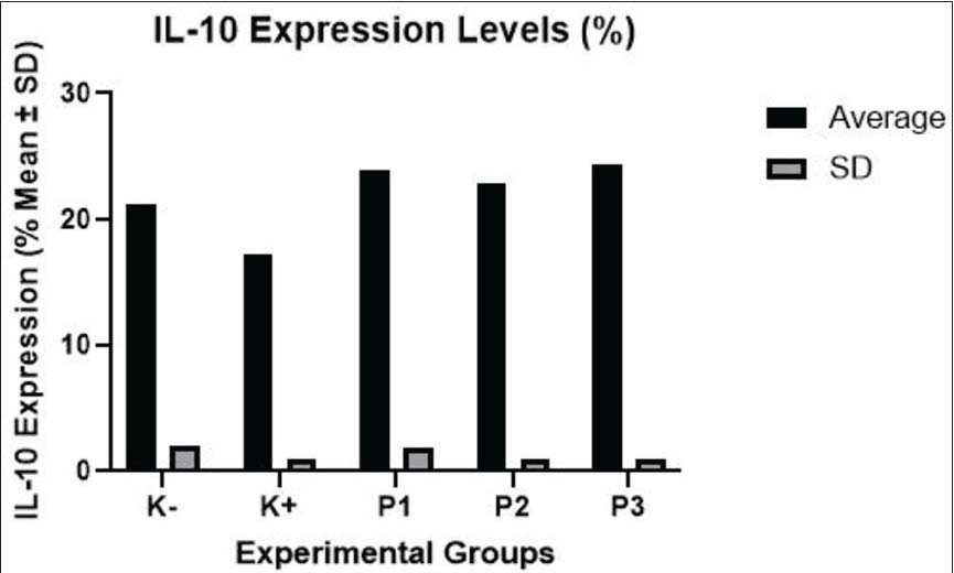
Fig. 4. IL-10 expression (%) across the experimental groups. Data are presented as mean ± SD. Nanocurcumin treatment restored IL-10 expression in a dose-dependent manner.
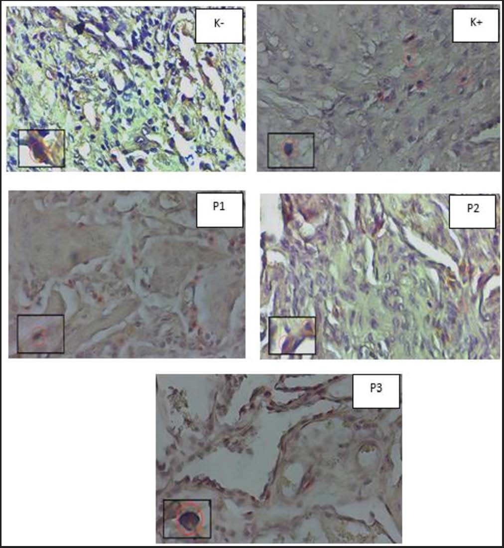
Fig. 5. Representative immunohistochemical staining of IL-10 in the uterine mucosa of mice with BALB/c. Group definitions are shown in Figure 1. The positive nuclei appear brown (DAB); the negative nuclei appear blue (hematoxylin). Magnification: 400×.
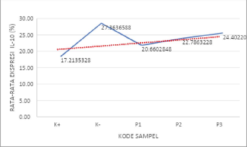
Fig. 6. Linear regression analysis showing the relationship between the dose of nanocurcumin and uterine IL-10 expression (%). Experimental groups: K−, K+, P1, P2, and P3.
Discussion
This study provides novel insights into the gravid uterus’ molecular response to high-decibel noise stress and the potential protective role of nanocurcumin. Repeated exposure to 135 dB white noise during mid to late gestation significantly disrupted uterine immune homeostasis, as indicated by increased Casp-3 and decreased IL-10 expression. These findings confirm that environmental acoustic stress can induce uterine apoptosis and attenuate anti-inflammatory signaling, which is consistent with previous reports on the reproductive toxicity of noise stress (Ristovska et al., 2014; Jafari et al., 2017; Clancy et al., 2023).
Mechanistically, the observed effects may be attributed to the activation of the hypothalamic–pituitary–adrenal (HPA) axis and sympathetic nervous system under noise stress, resulting in elevated glucocorticoid levels and downstream mitochondrial damage (Sherwood, 2016; Wirtz and von Känel, 2017). Elevated Caspase-3 expression in the K+ group confirms activation of the cytochrome c-mediated intrinsic apoptotic pathway, while reduced IL-10 levels indicate compromised immunosuppressive signaling. The therapeutic efficacy of nanocurcumin observed here likely stems from its superior bioavailability and combined antioxidant, anti-apoptotic, and immunomodulatory properties (Gera et al., 2017; Nikdad et al., 2020). Nanocurcumin inhibits oxidative stress, modulates NF-κB activity, and enhances IL-10 production through regulatory T-cell activation (Cui et al., 2021; Bafirman et al., 2023). The activation of the HPA axis leads to increased corticosterone, which can cross the placenta and disrupt uterine function via glucocorticoid receptor signaling. This cascade promotes mitochondrial dysfunction and pro-apoptotic gene activation (e.g., Bax), while downregulating anti-inflammatory cytokines, such as IL-10.
The quantitative findings support these mechanisms. Casp-3 expression was highest in the noise-stressed control group (K+) and significantly reduced in all treatment groups, especially in P3 (Fig. 3). The linear regression plot (Fig. 2) confirms a strong inverse correlation between the nanocurcumin dose and the expression of apoptotic markers. In contrast, IL-10 expression was lowest in K+ but elevated beyond baseline in the nanocurcumin groups, with P3 showing the greatest increase (Fig. 4). The corresponding dose–response trend (Fig. 6) demonstrates a robust upregulation of anti-inflammatory signaling with increasing nanocurcumin concentration. Maintaining a balance between apoptosis and immune regulation is essential for successful pregnancy, as physiological homeostasis at the maternal–fetal interface supports implantation and placental development.
Maintaining a balance between apoptosis and immune regulation is vital for successful pregnancy from a physiological standpoint. Disruptions to this balance may compromise uterine receptivity, placental development, and fetal growth. In particular, IL-10 plays a crucial role in maternal-fetal tolerance, and its suppression is linked to adverse pregnancy outcomes, such as preterm birth and miscarriage (Azizieh and Raghupathy, 2017). Nanocurcumin reduces apoptotic signaling and restores anti-inflammatory balance, supporting its therapeutic relevance in stress-induced reproductive dysfunction.
Although the 135 dB sound intensity used in this model exceeds the average community noise exposure, it is relevant for occupational or military scenarios. Thus, these findings provide translational relevance in evaluating nutraceutical strategies for populations exposed to chronic or extreme environmental stressors (Bronzaft, 2017; Clancy et al., 2023).
The strengths of this study include a well-defined gestational intervention window (GD6–18), the use of validated immunohistochemical markers, and rigorous quantitative analysis. However, several limitations warrant consideration. The small sample size (n=5 per group) may limit the generalizability of the findings. In addition, a lack of gene expression data (e.g., quantitative polymerase chain reaction) and systemic biomarker analysis (e.g., cortisol and oxidative stress indicators) restrict mechanistic interpretation. Furthermore, reproductive endpoints, such as litter size and fetal viability, were not assessed.
Future research should aim to evaluate the long-term outcomes of nanocurcumin supplementation, including fetal development, hormonal profiles, and placental structure. Studies involving chronic exposure to lower-level noise, the inclusion of systemic markers, and the exploration of combined antioxidant therapies could enrich the understanding of protective mechanisms. Investigating pharmacokinetics and safety during gestation is also important for translational application.
In conclusion, this study highlights the adverse effects of GNS on uterine homeostasis and supports the therapeutic potential of nanocurcumin. Nanocurcumin is a promising nonhormonal intervention to preserve reproductive function in acoustically stressed pregnancies by attenuating apoptosis and restoring anti-inflammatory responses.
While IHC provided spatial and semi-quantitative localization of Caspase-3 and IL-10, further studies incorporating gene expression (qPCR), protein-level confirmation (Western blot), and upstream pathway indicators such as NF-κB, Bax/Bcl-2, and oxidative stress biomarkers (e.g., MDA, ROS) would strengthen mechanistic insight. Our findings thus serve as a preliminary indication of nanocurcumin’s effects on apoptotic and immunomodulatory balance, pending further mechanistic validation. The present study focused on molecular markers of apoptosis and inflammation. However, key reproductive outcomes such as fetal viability, resorption rates, litter size, and placental histology were not assessed. Future work should incorporate these parameters to evaluate whether molecular protection translates into physiological reproductive benefit. Curcumin’s antibacterial and anti-inflammatory properties have been extensively reviewed, including its effect on bacterial membranes and oxidative signaling (Zheng et al., 2020). Previous studies have reported that oral nanocurcumin at doses up to 50 mg/kg BW in rodents is safe and does not affect fetal development or maternal behavior (Nainggolan et al., 2012). Our dose range (14–24.5 mg/kg) thus lies within the acceptable margin of safety.
Conclusion
Exposure to high-intensity noise during pregnancy disrupts uterine homeostasis by promoting apoptotic activity and suppressing anti-inflammatory signaling, as evidenced by elevated Caspase-3 and reduced IL-10 expression. Oral nanocurcumin effectively mitigated these alterations in a dose-dependent manner and demonstrated protective effects on the gravid uterus under environmental stress. The observed reduction in Caspase-3 and restoration of IL-10 expression highlight the dual anti-apoptotic and immunomodulatory actions of nanocurcumin. These findings strengthen the evidence supporting bioactive nutraceuticals as strategies to enhance reproductive resilience in adverse environmental conditions. Further studies incorporating chronic noise models, systemic biomarker evaluation, and fetal outcome assessment are necessary to validate the translational relevance of nanocurcumin in reproductive medicine.
Acknowledgments
The authors extend their sincere gratitude to the Faculty of Veterinary Medicine, Universitas Brawijaya, for providing research facilities and technical support. Special thanks are also due to the Biomedical Sciences Laboratory staff for their assistance with histological preparation and immunohistochemistry procedures. The authors appreciate the collaborative efforts and contributions of colleagues, technical personnel, and all team members who supported the experimental workflow and manuscript development.
Funding
This research was funded by the Internal Grant Program (DPP-SPP) of the Faculty of Veterinary Medicine, Universitas Brawijaya (FKH UB), Indonesia. The funding body played no role in the study design, data collection, analysis, interpretation, or preparation of the manuscript.
Authors’ contributions
V.F.H. conceptualized and supervised the study, designed the methodology, and revised the manuscript critically. R.F. performed animal experiments sample processing, and data collection. D.K. performed the statistical analysis and figure preparation. H.U. performed histopathological evaluation and assisted with immunohistochemistry. All authors have read and approved the final version of the manuscript and are responsible for its content. D.R. contributed to the refinement of the experimental design and reviewed the histological protocols. EFL. supported manuscript editing and coordinated animal welfare compliance.
Conflict of interest
The authors declare no conflict of interest regarding the publication of this paper.
Data availability
All data supporting this study’s findings are available within the manuscript. Additional raw datasets generated during the current study are available upon reasonable request from the corresponding author.
References
Azizieh, F.Y. and Raghupathy, R. 2017. IL-10 and pregnancy complications. Clin. Exp. Obstet. Gynecol. 44(2), 252–258.
Bafirman, B., Yulfadinata, A., Agus, A. and Ayubi, N. 2023. Curcumin: compound in turmeric that has the potential to increase serum interleukin-10 (IL-10) levels after high intensity exercise. Retos 52, 37–41; doi:10.47197/retos.v52.101895
Bronzaft, A.L. 2017. Impact of noise on health: the divide between policy and science. Open J. Soc. Sci. 5(5), 108–120; doi:10.4236/jss.2017.55008
Clancy, B.M., Theriault, B.R., Turcios, R., Langan, G.P. and Luchins, K.R. 2023. The effect of noise, vibration, and light disturbances from daily health checks on breeding performance, nest building, and corticosterone in mice. J. Am. Assoc. Lab. Anim. Sci. 62(4), 291–302; doi:10.30802/AALAS_JAALAS-23-000002
Cui, X., Lin, L., Sun, X., Wang, L. and Shen, R. 2021. Curcumin protects against renal ischemia/reperfusion injury by regulating oxidative stress and inflammatory response. Evid. Based. Complement. Alternat. Med. 2021, 1–8.
Farahbakht, E., Alsinani, Y., Safari, M., Hofmeister, M., Rezaie, R., Sharifabadi, A. and Jahromi, M.K. 2023. Immunoinflammatory response to acute noise stress in male rats adapted with different exercise training. Noise Health 25(119), 226–235.
Gera, M., Sharma, N., Ghosh, M., Huynh, D.L., Lee, S.J., Min, T., Kwon, T. and Jeong, D.K. 2017. Nanoformulations of curcumin: an emerging paradigm for improved remedial application. Oncotarget 8(39), 66680–66698; doi:10.18632/oncotarget.19164
Hewlings, S. and Kalman, D. 2017. Curcumin: a review of its effects on human health. Foods 6, 92; doi:10.3390/foods6100092
Jafari, Z., Faraji, J., Mirza Agha, B., Metz, G.A.S., Kolb, B.E. and Mohajerani, M.H. 2017. The adverse effects of auditory stress on mouse uterus receptivity and behavior. Sci. Rep. 7(1), 1063; doi:10.1038/s41598-017-04943-8
Jia, G., Wang, Q., Wang, R., Deng, D. and Xue, L. 2015. Tubeimoside I induces glioma apoptosis through regulation of Bax/Bcl-2 and the ROS/cytochrome c/caspase-3 pathway. Onco. Targets. Ther. 8, 313–311; doi:10.2147/OTT.S76063
Kia, S.J., Basirat, M., Mortezaie, T. and Moosavi, M.S. 2020. Comparison of oral nano-curcumin with oral prednisolone on oral lichen planus: a randomized double-blinded clinical trial. BMC Complement. Med. Ther. 20(1), 328; doi:10.1186/s12906-020-03128-7
Mehrizi, A., Rasoulzadeh, Y., Kazemi, T. and Mesgari Abbasi, M. 2022. Inflammatory and immunological changes caused by noise exposure: a systematic review. J. Environ. Sci. Health C 38(1), 61–90.
Naderyan Fe’Li, S., Monazzam Esmaielpour, M.R., Hokmabadi, R. and Rezaei-Hachesu, V. 2022. Effect of occupational noise exposure on cortisol hormone level: a systematic review. Noise Vib. Worldw. 53(11), 579–588; doi:10.1016/j.noisevw.2022.01.08
Nainggolan, H., Saragih, H., Ricky, D.R. and Manggading, A. 2012. Nanocurcumin sebagai penurun kolesterol pada tikus diabetes. Pros Simp Fisika Nasional XXV 1411:4771. [Indonesian].
National Research Council. 2011. Guide for the Care and Use of Laboratory Animals: Eighth Edition. Washington, DC: National Academies Press.
Nikdad, S., Valizadeh, R., Jafari, S. and Ghorbani, M. 2020. Antioxidative effects of nanocurcumin on liver mitochondria in paraquat-induced oxidative stress. Res. Mol. Med. 8(1), 37–42; doi:10.32598/rmm.8.1.37
Rahiman, N., Markina, Y.V., Kesharwani, P., Johnston, T.P. and Sahebkar, A. 2022. Curcumin-based nanotechnology approaches in restoration of autoimmune diseases. J. Control. Release 348, 264–286; doi:10.1016/j.jconrel.2022.05.046
Ristovska, G., Laszlo, H.E. and Hansell, A.L. 2014. Reproductive outcomes associated with noise exposure: a systematic review. Int. J. Environ. Res. Public Health 11(8), 7931–7952; doi:10.3390/ijerph110807931
Sherwood, L. 2016. Human physiology: from cells to systems. 9th ed. Boston: Cengage Learning.
Syarifah, A.S., Sudjarwo, S.A., Hendarto, H. and I’tishom, R. 2021. Nanocurcumin effect on SOD and caspase-3 in lead acetate-induced rats’ granulosa cells. Indian J. Forensic Med. Toxicol. 15(2), 1961–1969.
Wirtz, P.H. and Von Känel, R. 2017. Psychological stress, inflammation, and coronary heart disease. Curr. Cardiol. Rep. 19(11), 111; doi:10.1007/s11886-017-0919-x
Yates, A.L.G. and Cidlowski, J.A. 2013. Tissue-specific actions of glucocorticoids on apoptosis. Cells 2(2), 202–223.
Zheng, D., Liwinski, T. and Elinav, E. 2020. Antibacterial mechanism of curcumin: a review. Chem. Biodivers. 17(8), e2000171.








