





| Case Report | ||
Open Vet. J.. 2026; 16(8): 5563-5571 !
Open Veterinary Journal, (2026), Vol. 16(8): 5563–5571 Case Report Surgical management of massive hepatic cholangiocarcinoma with suspected pulmonary metastasis in a Dachshund: A palliative approachAlexandra Moura Fraga1*, Leticia Lopes Carrijo1, Mariana Moreira Lopes1, Washington Cleyber de Oliveira Júnior1, Hellen Nayara Silva Oliveira1, Luiz Augusto de Souza1, Vitor Eduardo Arantes de Barros1 and Iago Martins Oliveira21Escola de Veterinária e Zootecnia, Universidade Federal de Goiás (UFG), Goiânia, Brazil 2Escola de Ciências Médicas e da Vida, Pontifícia Universidade Católica de Goiás, Goiânia, Brazil *Corresponding Author: Alexandra Moura Fraga. Escola de Veterinária e Zootecnia, Universidade Federal de Goiás (UFG), Goiânia, Brazil. Email: alexandramf [at] icloud.com Submitted: 09/12/2025 Revised: 11/06/2026 Accepted: 29/06/2026 Published: 08/08/2026 © 2025 Open Veterinary Journal
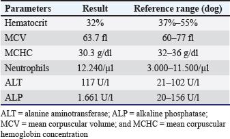
AbstractBackground: Hepatic cholangiocarcinoma is a malignant neoplasm originating from the epithelium of the bile ducts, characterized by infiltrative growth and high metastatic potential. It presents with nonspecific clinical signs, such as abdominal distension, weight loss, and lethargy, which makes early diagnosis difficult. Aim: To describe the clinical, imaging, surgical, and histopathological findings of a massive hepatic cholangiocarcinoma in a dog, emphasizing palliative surgical management. Case Description: A 13-year-old spayed female Dachshund dog was presented with abdominal distension. Physical examination revealed marked abdominal pain and abdominal enlargement. Ultrasonography identified a large cavitary structure in the liver, and thoracic radiography revealed a pulmonary mass. Computed tomography confirmed a massive hepatic neoplastic process without invasion of the caudal vena cava or portal vein. An exploratory celiotomy followed by hepatic lobectomy of the left lateral lobe was performed with palliative intent, aiming to achieve significant clinical improvement and enhanced quality of life. Histopathological examination confirmed a massive-pattern hepatic cholangiocarcinoma. Three months after surgery, follow-up imaging examinations revealed a new hepatic mass, without progression of the pulmonary mass. After 6 months, the patient developed clinical deterioration compatible with paraneoplastic syndrome and died under palliative care. Conclusion: Diagnostic imaging and histopathological findings contributed to the characterization of massive cystic hepatic cholangiocarcinoma in this dog. However, the advanced stage of the disease at diagnosis and the lack of characterization of the pulmonary lesion limited complete disease assessment. Further studies and additional case reports are warranted. Keywords: Bile duct, Dog, Liver, Neoplasm. IntroductionCholangiocarcinoma is the second most frequently diagnosed type of hepatic tumor in dogs (Mshelbwala et al., 2024). This malignant tumor originates from the epithelium of the bile ducts (Maemura et al., 2014) and is characterized by infiltrative growth, guarded prognosis, and high metastatic potential (Tanaka et al., 2022). In dogs, the most common presentation consists of multiple nodules distributed throughout the hepatic lobes (Flores et al., 2013). On the contrary, the massive form is characterized by the presence of a large solitary mass that completely replaces a hepatic lobe and is considered a less common presentation of canine cholangiocarcinoma (Flores et al., 2013). Clinical signs are generally nonspecific, including lethargy, anorexia, weight loss, abdominal distension, and, in some cases, jaundice (Cardoso and Pereira, 2024). Due to these characteristics, early identification is uncommon, and post-diagnosis survival depends on the extent of the disease and the feasibility of complete surgical resection (Reist et al., 2023). Diagnosis is established through laboratory tests and diagnostic imaging, particularly abdominal ultrasonography and computed tomography, which allow evaluation of tumor extent and possible metastases, thereby assisting in therapeutic decision-making (Tanaka et al., 2022). Metastases may occur in regional lymph nodes, lungs, spleen, and other abdominal organs, contributing to the complexity of clinical management (Mshelbwala et al., 2024). Although cholangiocarcinoma is the second most common primary hepatic tumor in dogs, its presentation as a massive cavitary lesion remains poorly described, especially regarding imaging characteristics and therapeutic decision-making in advanced cases. This study aims to describe a case of hepatic cholangiocarcinoma with a massive cavitary presentation in a female dog, emphasizing the imaging findings, the clinical reasoning for palliative surgical management, and its implications for the patient’s quality of life. Case DetailsA 13-year-old spayed female Dachshund dog, weighing 11.7 kg, with updated vaccination and ectoparasite control protocols, was presented to the Veterinary Teaching Hospital of the Federal University of Goiás (UFG, GO, Brazil) with a history of abdominal distension of undetermined duration. Physical examination revealed a grade II/VI systolic murmur at the mitral valve area, nodules in the left mammary chain, abdominal pain, a distended and firm abdomen, and abdominal cutaneous hyperpigmentation. Table 1. Hematological and biochemical test results of a 13-year-old female Dachshund dog with hepatic cholangiocarcinoma.
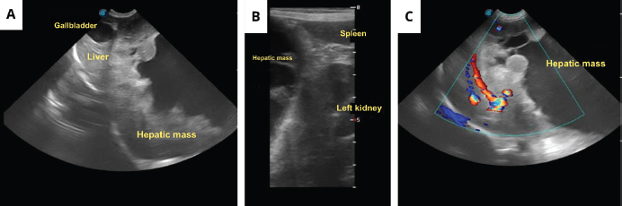
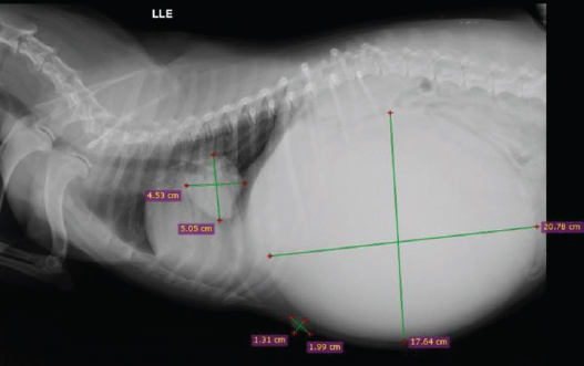
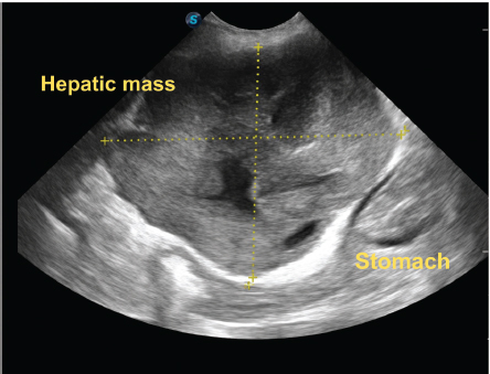
Based on the clinical findings, differential diagnoses included hepatic neoplasia (hepatocellular carcinoma or cholangiocarcinoma), metastatic mammary neoplasia, hepatic cysts, and abscesses. To further investigate the case, laboratory tests, complete abdominal ultrasonography, and thoracic radiography were requested. The coagulation profile was not assessed before surgery, representing a limitation in the preoperative evaluation. Laboratory findings, presented in Table 1, revealed normocytic normochromic anemia, leukocytosis due to neutrophilia without left shift, mild elevation of alanine aminotransferase (ALT), and marked elevation of alkaline phosphatase (ALP). These laboratory abnormalities were consistent with chronic inflammatory disease and cholestasis, which are commonly described in cases of cholangiocarcinoma. Abdominal ultrasonography demonstrated a large cavitary structure containing heterogeneous anechoic particulate material, hyperechoic septations, and peripheral vascularization, without central Doppler flow (Fig. 1A–C). The lesion overlapped the left lateral, caudate, and right lateral hepatic lobes, extending into the mid/caudal abdomen and displacing adjacent organs such as the spleen, left kidney, stomach, and intestinal loops (Fig. 1B). The gallbladder exhibited a thin hyperechoic wall with anechoic content and biliary sludge. Thoracic radiography identified a pulmonary mass (4.53 × 5.05 cm) in the right caudal lung lobe (Fig. 2). Due to the high risk of rupture and hemorrhage associated with severe abdominal pain, urgent hepatic lobectomy was elected without prior cytology or biopsy because of the elevated risk of bleeding and lesion rupture, considering its large size and cavitary nature.
Fig. 1. Abdominal ultrasonography of a 13-year-old female Dachshund dog with hepatic cholangiocarcinoma. (A) Liver with markedly increased dimensions, extending beyond the costal arch due to the presence of a cavitary structure exceeding the technical limits of ultrasonographic scanning, containing heterogeneous particulate anechoic content, interspersed hyperechoic septations, and well-defined, irregular, heterogeneous echogenic margins. (B) Hepatic mass visualized overlapping the topography of the left lateral, caudate, and right lateral hepatic lobes, extending into the mid/caudal abdominal region and causing displacement of adjacent abdominal structures, such as the spleen and left kidney. (C) Hepatic mass showing mild peripheral vascularization on color Doppler imaging.
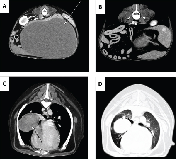
Fig. 2. Thoracic radiograph of a 13-year-old female Dachshund dog with hepatic cholangiocarcinoma. Showing a pulmonary mass in the right caudal lung lobe measuring 4.53 × 5.05 cm. In addition, an oval, homogeneous abdominal structure with regular contours measuring 17.64 × 20.78 cm was observed, as well as an oval, heterogeneous enlargement with partially defined contours in the left cranioventral extra-abdominal region measuring 1.31 × 1.99 cm, suggestive of a mammary neoplasm. For surgical planning, thoracic and abdominal computed tomography was performed using a GE BRIVO 385 scanner (16-slice). A helical acquisition technique was employed, and multiplanar reconstructions with 1.2 mm slice thickness were obtained using both soft and sharp reconstruction filters before and after intravenous contrast administration. The procedure was conducted under general anesthesia, using propofol for anesthetic induction in a dose-to-effect protocol at an approximate dose of 5 mg/kg, until loss of reflexes and successful endotracheal intubation were achieved. Subsequently, for anesthetic maintenance and monitoring during the examination, the patient was maintained in a semi-closed circuit with isoflurane administered to effect. Nonionic iodinated contrast medium (Omnipaque® 300 mg/ml) was administered intravenously via the cephalic vein at a dose of 1.5 ml/kg over 20 seconds. Thoracic images revealed a rounded, heterogeneous, well-defined mass with regular margins and contrast enhancement located in the right middle lung lobe. The lesion exhibited close contact with the right pulmonary artery and caudal vena cava, and adhesions to these structures could not be excluded. The mass measured 4.7 × 4.3 × 3.4 cm along its greatest axes (Fig. 3C and D). Images of the abdominal cavity demonstrated hepatomegaly associated with a massive hepatic mass (19.3 × 12.4 × 18.5 cm), without involvement of the intrahepatic caudal vena cava or portal vein, although indirect compressive effects were observed. Peripheral contrast enhancement involved the left medial and left lateral hepatic lobes, with loss of cleavage planes between the lesion, the quadrate lobe, and the papillary process of the caudate lobe, making it impossible to exclude invasion of these structures (Fig. 3A and B). After evaluation of the computed tomography images, the suspicion of expansive hepatic neoplasia and pulmonary mass was reinforced. However, imaging examinations alone did not allow determination of whether the pulmonary lesion corresponded to a primary or metastatic neoplasm. Nevertheless, due to the patient’s age and clinical condition, as well as the complexity and risks associated with exploratory thoracotomy, surgical intervention of the pulmonary mass for histopathological sampling was not performed. The patient underwent exploratory xipho-pubic celiotomy followed by hepatic lobectomy of the left lateral lobe, with hilar ligation performed using size 0 polydioxanone suture in a Miller’s knot pattern. The tumor was restricted to the left lateral hepatic lobe, and gross evaluation revealed preservation of the remaining hepatic lobes. Furthermore, no evidence of involvement of adjacent blood vessels was identified. The procedure was completed without anesthetic or surgical complications, and the patient remained hospitalized for postoperative monitoring and analgesia. Postoperatively, marked clinical improvement was observed, with a reduction of abdominal distension and improved comfort according to the owner’s report.
Fig. 3. Thoracic and abdominal computed tomography of a 13-year-old female Dachshund dog with hepatic cholangiocarcinoma. (A) Liver presenting increased dimensions, thin borders, regular contours, and heterogeneous texture due to the presence of an extensive expansive lesion (large cavitation), rounded, hypoattenuating, and partially delineated. (B) Liver showing peripheral contrast enhancement involving the left medial and left lateral hepatic lobes. Loss of cleavage planes with the quadrate hepatic lobe and the papillary process of the caudate lobe was observed. (C) Rounded, heterogeneous, well-defined mass with regular margins located in the right middle lung lobe. The lesion exhibited intimate contact with the right pulmonary artery and the caudal vena cava, and adhesions to these structures could not be excluded. (D) Pulmonary mass showing contrast enhancement. The lesion caused the collapse of the right main bronchus and some lobar bronchi. The day after surgery, the patient was discharged. Postoperative supportive therapy included analgesia with dipyrone (500 mg/mL), administered at a dose of 8 drops orally every 8 hours for 5 days, and tramadol (100 mg/mL), 12 drops orally every 8 hours for the same period. Meloxicam (0.5 mg), 1 tablet orally every 24 hours for 3 days, was prescribed as an anti-inflammatory drug, in addition to antibiotic therapy with amoxicillin associated with potassium clavulanate (50 mg), 2 tablets orally every 12 hours for 10 days. The left lateral hepatic lobe resected during surgery was fixed in 10% formaldehyde solution and submitted to the hospital’s Animal Pathology Service for histopathological evaluation. Gross examination demonstrated a massive neoplasm with a whitish surface (Fig. 4A). On longitudinal section, a large volume of brownish fluid compatible with cystic content was observed. The inner cavity ranged from whitish to brownish, with hemorrhagic foci and firm walls (Fig. 4B). Microscopic examination revealed neoplastic proliferation of biliary epithelial cells infiltrating and compressing the hepatic parenchyma, associated with extensive multifocal necrosis, mild neutrophilic inflammatory infiltrate, and marked degeneration of the adjacent tissue, with areas of congestion and hemorrhage. Fibrous connective tissue septa separating multiple lobules of the hepatic mass were also observed (Fig. 4C), in addition to cells ranging from polyhedral to cuboidal morphology and the presence of mitotic figures (Fig. 4D).
Fig. 4. Anatomopathological evaluation of hepatic cholangiocarcinoma in a 13-year-old female Dachshund dog. (A) Neoplastic left hepatic lobe, massive, congested, and cystic. (B) On longitudinal section of the hepatic lobe, the mass presents an irregular surface, brownish-red coloration, multifocal areas of necrosis and hemorrhage, areas of fibrosis (black arrow), with lobulated architecture and marked heterogeneity. (C) Photomicrograph of cholangiocarcinoma at 4× magnification, stained with hematoxylin and eosin, showing thin fibrous connective tissue septa (black arrow) separating multiple lobules of the neoplasm. (D) Photomicrograph of cholangiocarcinoma at 40× magnification, stained with hematoxylin and eosin, showing cells with polyhedral to cuboidal morphology, moderate pale eosinophilic, and the presence of mitotic figures (black arrow). Due to the unavailability of immunohistochemistry services at the hospital and the owner’s financial limitations, the submitted material was not evaluated by immunohistochemistry. For monitoring of possible metastases, follow-up imaging examinations were performed 3 months after surgery. Abdominal ultrasonography identified, in the hepatic topography, the presence of a structure tending toward a circular shape, with well-defined and irregular contours, measuring approximately 67.54 × 52.05 mm, presenting mixed echogenicity and heterogeneous echotexture, with interspersed anechoic areas and peripheral vascularization, without signs of central vascularization on color Doppler evaluation (Fig. 5). Thoracic radiography did not demonstrate enlargement of the previously identified pulmonary mass, and no new lesions were detected. Six months after a left lateral hepatic lobectomy, the patient developed clinical deterioration characterized by weight loss, hyporexia, and muscle weakness, changes suggestive of paraneoplastic syndrome. Considering the clinical condition and guarded prognosis, the owner elected palliative care. For pain management, pregabalin (25 mg), 1 tablet orally every 12 hours, was instituted as continuous therapy. Omega-3 supplementation (500 mg), 1 capsule orally every 24 hours, was also prescribed for continuous use. The patient died approximately 180 days after surgery, corresponding to a survival time of 6 months.
Fig. 5. Abdominal ultrasonography of a 13-year-old female Dachshund dog with hepatic cholangiocarcinoma, 3 months after left lateral hepatic lobectomy. Liver with increased dimensions, extending beyond the costal arch due to the presence of a structure tending toward a circular shape, with well-defined and irregular contours, measuring approximately 67.54 × 52.05 mm, showing mixed echogenicity and heterogeneous echotexture, with interspersed anechoic areas, located in the mid/caudal abdominal region and causing caudal displacement of the stomach to the left. DiscussionIn the present report, cholangiocarcinoma was identified in a 13-year-old spayed female Dachshund dog. The age group and species are consistent with the literature, which describes a greater predisposition in elderly dogs for the development of malignant hepatic neoplasms, although there is no consensus regarding breed or sex predisposition (Mshelbwala et al., 2024). The main clinical sign observed in this patient was abdominal distension secondary to hepatomegaly, a finding described in approximately 42% of dogs with hepatic neoplasms (Patnaik et al., 1980). Similar to the present case, cholangiocarcinoma frequently presents with nonspecific clinical signs, which may or may not be associated with jaundice (Cardoso and Pereira, 2024). The absence of signs of hepatic failure in this dog was likely related to the preserved functional integrity of the remaining hepatic lobes, since manifestations of liver failure generally occur only when more than 70% of the functional hepatic parenchyma is compromised (Fossum, 2021). The hepatic alterations observed on abdominal ultrasonography were essential in supporting the differential diagnosis of neoplasia. However, the sensitivity of ultrasonography for hepatic diseases is considered limited due to the high variability of lesion patterns (Assawarachan et al., 2019). In addition, the pulmonary mass identified on thoracic radiography required further evaluation using more advanced imaging modalities for better case elucidation. Therefore, computed tomography was requested as a complementary examination for better lesion characterization, assessment of possible vascular involvement, and surgical planning. Computed tomography is commonly used to evaluate hepatic masses because it allows the determination of lesion extent and detection of distant metastases. Recent studies report that this examination, when performed with a triphasic contrast protocol, may be useful in distinguishing pathological characteristics of hepatic masses through evaluation of contrast enhancement patterns (Goda et al., 2025). In this report, computed tomographic evaluation demonstrated an extensive lesion occupying the left lateral hepatic lobe, with central cavitation and expansion into the hypogastric region, characteristics compatible with the macroscopic pattern classified as a massive tumor (Fukushima et al., 2012). The location was also consistent with that expected for massive lesions, which preferentially affect the left lateral hepatic lobe in up to 56% of cases (Tanaka et al., 2022). Furthermore, the peripheral contrast enhancement observed in this clinical case may reflect the histological characteristics of cholangiocarcinoma, indicating viable tumor cells at the periphery and central fibrosis or fibrotic tumor cells (Goda et al., 2025). Previous reports generally describe hepatic cholangiocarcinoma in dogs as multiple or diffuse lesions, with massive presentation being less frequently reported (Flores et al., 2013; Mshelbwala et al., 2024). In the present case, the presence of extensive cavitation associated with a marked compressive effect differs from most reports, suggesting potentially more aggressive biological behavior of the neoplasm. The thoracic computed tomography findings were consistent with the alterations previously observed on thoracic radiography, reinforcing the suspicion of pulmonary neoplasia, whether primary or metastatic. However, due to the greater sensitivity and anatomical detail provided by computed tomography, it was possible to identify that the mass was located in the right middle lung lobe rather than in the right caudal lung lobe, as initially suggested by radiography. In addition, the lesion was observed to be in close contact with the right pulmonary artery and the caudal vena cava, and adhesions to these structures could not be ruled out. The patient’s advanced age, associated with the risk of rupture and hemorrhage of the hepatic neoplasm, as well as the possibility of adhesion of the pulmonary mass to the right pulmonary artery and the caudal vena cava, was the determining factor in the decision to prioritize exploratory laparotomy rather than perform surgical intervention on the pulmonary mass. Laboratory tests revealed moderate normocytic normochromic anemia, a pattern frequently associated with chronic inflammatory processes and commonly observed in hepatic cholangiocarcinoma (Tantary et al., 2014). In the biochemical profile, elevated ALT and ALP activities were identified. ALT, a cytosolic hepatocellular enzyme, tends to increase more markedly in neoplasms directly affecting the hepatic parenchyma, such as hepatocellular carcinoma (Liptak et al., 2004). On the contrary, ALP is predominantly associated with the biliary epithelium and shows marked elevation in conditions involving ductal proliferation or cholestasis, alterations frequently observed in cholangiocarcinoma (Mshelbwala et al., 2024; Loan et al., 2025). Therefore, the marked increase in FA observed in this case is compatible with the biochemical pattern reported for neoplasms of biliary origin and supports the diagnosis of cholangiocarcinoma. Complete resection of the affected lobe is recommended for primary malignant hepatic tumors whenever technically feasible (Fossum, 2021). In the present report, surgical excision of the left hepatic lobe was elected despite the presence of the pulmonary mass because of the large tumor volume, the risk of rupture, and the significant pain associated with the lesion (Reist et al., 2023). Given the systemic involvement and the impossibility of curative treatment, the procedure had a strictly palliative purpose, aiming to relieve pain and improve the patient’s quality of life. The pulmonary lesion was not definitively characterized and should, therefore, be interpreted cautiously during tumor staging. Furthermore, the location in the left hepatic lobe favored the surgical decision, since neoplasms in the right hepatic lobe are associated with a higher risk of intraoperative mortality, whereas left lobe lesions present better technical feasibility for safe excision (Trindade and Gerardi, 2018). After the surgical procedure, adjuvant chemotherapy was not instituted. Although conventional chemotherapeutic agents have been used in the treatment of cholangiocarcinoma, their efficacy is still considered variable and poorly established in the veterinary literature (Nakamura et al., 2015). In this case, the absence of complementary therapy was related to the owner’s financial limitations and the advanced stage of the disease at the time of diagnosis. Nevertheless, recent studies have investigated more individualized therapeutic approaches based on the molecular characteristics of the tumor, moving toward precision medicine (Capuozzo et al., 2024). Macroscopically, the neoplasm was well-delimited, voluminous, firm, whitish, and spherical in appearance. This pattern differs from the most common presentation of cholangiocarcinoma, which generally consists of multiple nodules distributed throughout several hepatic lobes, with the massive form being less frequently described in dogs (Flores et al., 2013), representing approximately 22%–46% of cases (Goda et al., 2025). The presence of a cystic cavity filled with serohemorrhagic content may have contributed to the expansive growth of the lesion and its massive presentation (Terai et al., 2022). The whitish coloration and firm consistency observed at the margins are compatible with the abundant fibrous stroma characteristic of cholangiocarcinoma (Flores et al., 2013). However, brownish-green pigmentation observed in this case is uncommon, since neoplastic biliary cells generally do not produce bile. The presence of this pigment may result from passive retention or degenerative tissue changes, although it is not considered a typical finding of the disease (Flores et al., 2013). Histopathological examination demonstrated neoplastic proliferation of biliary epithelial origin, with acinar, tubular, and occasional papillary arrangements, in addition to polyhedral to cuboidal cells intermingling with the hepatic parenchyma. These findings are compatible with the morphological pattern described for hepatic cholangiocarcinoma, including diffuse fibrous stroma and tubules lined by anaplastic cuboidal or columnar cells (Mshelbwala et al., 2024). Treatment consisted of complete resection of the affected hepatic lobe and supportive therapy based on analgesia and omega-3 supplementation, aiming to provide greater comfort and maintain the patient’s quality of life during the postoperative period. The survival time observed in this case, 180 days after surgery, is consistent with findings reported in the literature for dogs with cholangiocarcinoma managed exclusively with surgical treatment (Liptak et al., 2019). Despite the guarded prognosis associated with this neoplasm, the palliative approach adopted contributed to temporary clinical improvement and reduction of the signs associated with the expansive effect of the hepatic mass. The limitations of this study include the diagnosis of the disease at an advanced stage, the absence of definitive characterization of the pulmonary lesion, and the lack of well-established chemotherapy protocols in veterinary medicine for the treatment of cholangiocarcinoma. ConclusionIn this report, hepatic cholangiocarcinoma presented as a massive cystic lesion associated with a marked abdominal compressive effect. Diagnostic imaging examinations contributed to lesion characterization, assessment of its extent, and surgical planning, while histopathological evaluation allowed definitive diagnosis. The main limitations of this case were the advanced stage of the disease at diagnosis and the lack of characterization of the concomitant pulmonary mass, which limited complete disease staging. Considering the still-limited number of reports describing massive cystic presentations of hepatic cholangiocarcinoma in dogs, additional studies are needed to expand knowledge regarding its clinical, imaging, and prognostic characteristics. AcknowledgmentsTo the Veterinary Hospital of the Veterinary and Zootechnical School of the Federal University of Goiás, for supporting us in this case. FundingThis research received no specific grant. Authors' contributionsConceptualization A.M.F, V.E.A.B., I.M.O., and L.A.D.S., methodology A.M.F, formal analysis A.M.F, V.E.A.B., and L.A.D.S., data curation A.M.F, V.E.A.B., and L.A.D.S., conducted the clinical case; A.M.F, drafted the manuscript; A.M.F, revised the manuscript and provided guidance on the clinical case; A.M.F, V.E.A.B., and L.A.D.S translated the manuscript. A.M.F., V.E.A.B., L.A.D.S., L.L.C., M.M.L., W.C.D.O.J., H.N.S.O., I.M.O., and L.G.S.D.O. All authors have read and agreed with the published version of the manuscript. Conflict of interestThe authors declare that there is no conflict of interest. Data availabilityAll data supporting the findings of this study are available within the manuscript. ReferencesCapuozzo, M., Santorsola, M., Ferrara, F., Cinque, C., Farace, S., Patrone, R., Granata, V., Zovi, A., Nasti, G. and Ottaiano, A. 2024. Intrahepatic cholangiocarcinoma biomarkers: towards early detection and personalized pharmacological treatments. Mol. Cell. Probes. 73, 101951; doi: 10.1016/j.mcp.2024.101951 Cardoso, L.M., Pereira, G.Q. and Pereira, G.Q. 2024. Colangiocarcinoma em cão: relato de caso. Rev. Bras. Hig. Sanid. Anim. 18(6), 6; doi: 10.5935/1981-2965.20240026 Flores, M.M., Bianchi, R.M., Kommers, G.D., Irigoyen, L.F., Barros, C.S.L. and Fighera, R.A. 2013. Prevalência e achados epidemiológicos, anatomopatológicos e imuno-histoquímicos dos tumores hepáticos malignos primários de cães da Região Central do Rio Grande do Sul (1965–2012). Pesq. Vet. Bras. 33, 497–511; doi: 10.1590/S0100-736X2013000400014 Fossum, W.T. 2021. Hepatobiliary neoplasia.In Small animal surgery. 5 ed. Rio de Janeiro: Elsevier, pp: 1725–32. Fukushima, K., Kanemoto, H., Ohno, K., Takahashi, M., Nakashima, K., Fujino, Y., Uchida, K., Fujiwara, R., Nishimura, R. and Tsujimoto, H. 2012. CT characteristics of primary hepatic mass lesions in dogs. Vet. Radiol. Ultrasound 53, 252–257; doi: 10.1111/j.1740-8261.2011.01917.x Goda, Y., Yamamoto, S., Miyoshi, K., Hanazono, K., Sakai, M.M.T., Tamura, M., Deguchi, T., Endo, Y., Kadosawa, T., Fujita, M., Itami, T. and Torisu, S. 2025. Triple-phase contrast-enhanced computed tomography findings in dogs with cholangiocarcinoma and combined hepatocellular-cholangiocarcinoma: pilot study. Open. Vet. J. 15, 5739–5747; doi: 10.5455/OVJ.2025.v15.i11.29 Liptak, J.M., Dernell, W.S., Monnet, E., Powers, B.E., Bachand, A.M., Kenney, J.G. and Withrow, S.J. 2004. Massive hepatocellular carcinoma in dogs: 48 cases (1992–2002). J. Am. Vet. Med. Assoc. 225, 1225–1230; doi: 10.2460/javma.2004.225.1225 Loan, N.V.T.H., Chao, N.V. and Nhung, T.T. 2025. Clinical features and hematological and biochemical blood parameters of dogs with hepatobiliary disorders. Vet. World 18, 986–993; doi10.14202/vetworld.2025.986-993 Maemura, K., Natsugoe, S. and Takao, S. 2014. Molecular mechanism of cholangiocarcinoma carcinogenesis. J. Hepatobiliary. Pancreat. Sci. 21, 754–760; doi10.1002/jhbp.126 Mshelbwala, F.M., Ajayi, O.L., Adebiyi, A.A., Olaniyi, M.O., Oladipo, T.M., Okpe, E.F., Rahman, S.A., Makinde, A.F., Kadiri, A.K.F., Abakpa, S.A.V. and Olasoju, M.I. 2024. Cholangiocarcinoma: consistent clinical, cytological, hematological, and biochemical findings and pathomorphology of the liver and kidney in five exotic dog breeds in Abeokuta, Nigeria. Vet. World 17, 2053–2061; doi: 10.14202/vetworld.2024.2053-2061 Nakamura, H., Arai, Y., Totoki, Y., Shirota, T., Elzawahry, A., Kato, M., Hama, N., Hosoda, F., Urushidate, T., Ohashi, S., Hiraoka, N., Ojima, H., Shimada, K., Okusaka, T., Kosuge, T., Miyagawa, S. and Shibata, T. 2015. Genomic spectra of biliary tract cancer. Nat. Genet. 47, 1003–1010; doi: 10.1038/ng.3375 Patnaik, A.K., Hurvitz, A.I. and Lieberman, P.H. 1980. Canine hepatic neoplasms: a clinicopathologic study. Vet. Pathol. 17, 553–564; doi: 10.1177/030098588001700504 Reist, A.M., Reagan, J.K., Fujita, S.K. and Walny, A.M. 2023. Histopathologic findings and survival outcomes of dogs undergoing liver lobectomy as treatment for spontaneous hemoabdomen secondary to a ruptured liver mass: retrospective analysis of 200 cases (2012–2020). J. Am. Vet. Med. Assoc. 261, 237–245; doi: 10.2460/javma.22.03.0130 Tanaka, T., Noguchi, S., Wada, Y., Nishida, H. and Akiyoshi, H. 2022. Computed tomography findings in canine cholangiocellular carcinoma. Vet. Rec. Case Rep. 10, 303; doi: 10.1002/vrc2.303 Tantary, H.A., Soodan, J.S., Chirag, S., Ansari, M.M., Kumar, S. and Imtiyaz, T. 2014. Diagnostic studies in dogs with hepatic disorders. Int. J. Vet. Sci. 3(4), 210–215. Terai, K., Ishigaki, K., Kagawa, Y., Okada, K., Yoshida, O., Sakurai, N., Heishima, T. and Asano, K. 2022. Clinical, diagnostic, and pathologic features and surgical outcomes of combined hepatocellular-cholangiocarcinoma in dogs: 14 cases (2009–2021). J. Am. Vet. Med. Assoc. 260, 1668–1674; doi: 10.2460/javma.21.08.0379 Trindade, A.B. and Gerardi, D.G. 2018. Manejo clínico e cirúrgico de um cão com colangiocarcinoma difuso. Acta Sci. Vet. 46(Suppl. 1), 290; doi: 10.22456/1679-9216.86822 | ||

| How to Cite this Article |
| Pubmed Style Fraga AM, Carrijo LL, Lopes MM, Júnior WCDO, Oliveira HNS, Souza LAD, Barros VEAD, Oliveira IM. Surgical management of massive hepatic cholangiocarcinoma with suspected pulmonary metastasis in a Dachshund: A palliative approach. Open Vet. J.. 2026; 16(8): 5563-5571. doi:10.5455/OVJ.2026.v16.i8.50 Web Style Fraga AM, Carrijo LL, Lopes MM, Júnior WCDO, Oliveira HNS, Souza LAD, Barros VEAD, Oliveira IM. Surgical management of massive hepatic cholangiocarcinoma with suspected pulmonary metastasis in a Dachshund: A palliative approach. https://www.openveterinaryjournal.com/?mno=302562 [Access: August 08, 2026]. doi:10.5455/OVJ.2026.v16.i8.50 AMA (American Medical Association) Style Fraga AM, Carrijo LL, Lopes MM, Júnior WCDO, Oliveira HNS, Souza LAD, Barros VEAD, Oliveira IM. Surgical management of massive hepatic cholangiocarcinoma with suspected pulmonary metastasis in a Dachshund: A palliative approach. Open Vet. J.. 2026; 16(8): 5563-5571. doi:10.5455/OVJ.2026.v16.i8.50 Vancouver/ICMJE Style Fraga AM, Carrijo LL, Lopes MM, Júnior WCDO, Oliveira HNS, Souza LAD, Barros VEAD, Oliveira IM. Surgical management of massive hepatic cholangiocarcinoma with suspected pulmonary metastasis in a Dachshund: A palliative approach. Open Vet. J.. (2026), [cited August 08, 2026]; 16(8): 5563-5571. doi:10.5455/OVJ.2026.v16.i8.50 Harvard Style Fraga, A. M., Carrijo, . L. L., Lopes, . M. M., Júnior, . W. C. D. O., Oliveira, . H. N. S., Souza, . L. A. D., Barros, . V. E. A. D. & Oliveira, . I. M. (2026) Surgical management of massive hepatic cholangiocarcinoma with suspected pulmonary metastasis in a Dachshund: A palliative approach. Open Vet. J., 16 (8), 5563-5571. doi:10.5455/OVJ.2026.v16.i8.50 Turabian Style Fraga, Alexandra Moura, Leticia Lopes Carrijo, Mariana Moreira Lopes, Washington Cleyber De Oliveira Júnior, Hellen Nayara Silva Oliveira, Luiz Augusto De Souza, Vitor Eduardo Arantes De Barros, and Iago Martins Oliveira. 2026. Surgical management of massive hepatic cholangiocarcinoma with suspected pulmonary metastasis in a Dachshund: A palliative approach. Open Veterinary Journal, 16 (8), 5563-5571. doi:10.5455/OVJ.2026.v16.i8.50 Chicago Style Fraga, Alexandra Moura, Leticia Lopes Carrijo, Mariana Moreira Lopes, Washington Cleyber De Oliveira Júnior, Hellen Nayara Silva Oliveira, Luiz Augusto De Souza, Vitor Eduardo Arantes De Barros, and Iago Martins Oliveira. "Surgical management of massive hepatic cholangiocarcinoma with suspected pulmonary metastasis in a Dachshund: A palliative approach." Open Veterinary Journal 16 (2026), 5563-5571. doi:10.5455/OVJ.2026.v16.i8.50 MLA (The Modern Language Association) Style Fraga, Alexandra Moura, Leticia Lopes Carrijo, Mariana Moreira Lopes, Washington Cleyber De Oliveira Júnior, Hellen Nayara Silva Oliveira, Luiz Augusto De Souza, Vitor Eduardo Arantes De Barros, and Iago Martins Oliveira. "Surgical management of massive hepatic cholangiocarcinoma with suspected pulmonary metastasis in a Dachshund: A palliative approach." Open Veterinary Journal 16.8 (2026), 5563-5571. Print. doi:10.5455/OVJ.2026.v16.i8.50 APA (American Psychological Association) Style Fraga, A. M., Carrijo, . L. L., Lopes, . M. M., Júnior, . W. C. D. O., Oliveira, . H. N. S., Souza, . L. A. D., Barros, . V. E. A. D. & Oliveira, . I. M. (2026) Surgical management of massive hepatic cholangiocarcinoma with suspected pulmonary metastasis in a Dachshund: A palliative approach. Open Veterinary Journal, 16 (8), 5563-5571. doi:10.5455/OVJ.2026.v16.i8.50 |


