





| Case Report | ||
Open Vet. J.. 2026; 16(8): 5538-5543
Open Veterinary Journal, (2026), Vol. 16(8): 5538–5543 Case Report Ascaridia dissimilis infestation in organic turkeys with high daily mortalityPhilipp Kern1*, Benjamin Schade2, Brigitte Böhm2 and Ferdinand Schmitt21Geflügelpraxis Philipp Kern, Neumünster, Germany 2Bavarian Animal Health Service, Poing, Germany *Corresponding Author: Philipp Kern. Geflügelpraxis Philipp Kern, Neumünster, Germany. Email: philipp.kern [at] gefluegelpraxis-kern.de Submitted: 12/08/2025 Revised: 06/06/2026 Accepted: 25/06/2026 Published: 08/08/2026 © 2025 Open Veterinary Journal
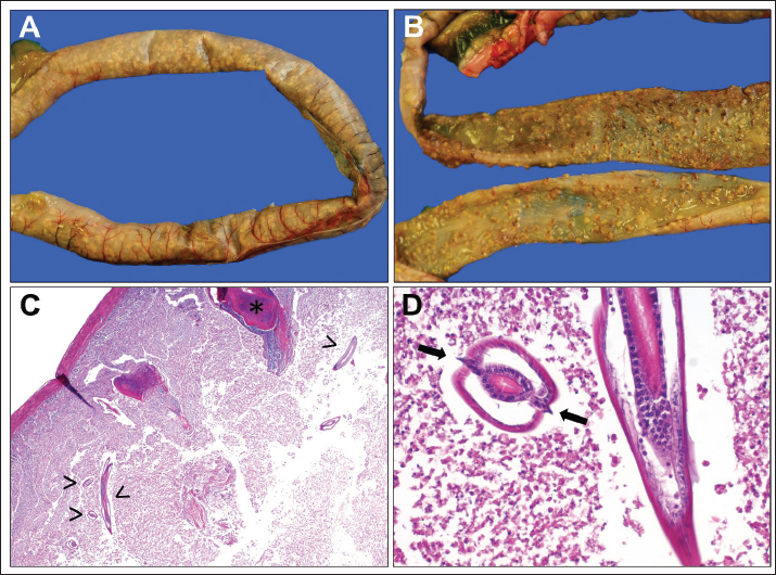
AbstractBackground: Parasite infestations of poultry kept in organic production systems are re-emerging, but case reports of Ascaridia dissimilis infections in turkeys reared under organic conditions are limited. Case Description: A flock of organic B.U.T. six turkeys at the age of 12 weeks experienced an acute increase in mortality in spring. Necropsy of affected turkeys revealed severe intestinal lesions with necrotic enteritis. Histopathology confirmed the presence of ascarid larvae within intestinal lesions, establishing the definitive diagnosis: ascaridiasis caused by A. dissimilis, the turkey roundworm. The remaining turkeys in the flock were immediately treated with fenbendazole in the drinking water. Within a week after treatment, the daily mortality rate dropped dramatically and returned to normal. The warm and humid spring weather, wet litter, and the use of gas heaters likely accelerated parasite egg development and led to an early, overwhelming infestation. Conclusion: This outbreak points out that A. dissimilis, typically a subclinical parasite, can cause fatal enteritis in young turkeys under certain conditions. Prompt diagnosis via necropsy and histology was critical, as routine fecal exams can miss prepatent infestations. A targeted parasite monitoring strategy is crucial to detect and manage Ascaridia infestations before losses occur. Effective control of A. dissimilis in turkeys requires vigilant monitoring and timely intervention, alongside good husbandry practices, to prevent severe disease and economic loss in poultry production. Keywords: Ascarid infestation, Enteritis, Mortality, Organic farming, Turkeys. IntroductionAscaridia spp. are large nematode parasites (roundworms) that inhabit the intestines of birds. Among these, Ascaridia galli predominantly infects chickens (and other fowl), whereas Ascaridia dissimilis is the chief ascarid of turkey. Ascaridia dissimilis is considered the most prevalent intestinal helminth of domestic turkeys, with some surveys indicating nearly 100% of flocks harboring the parasite (Yazwinski et al., 2009). In most cases, these infestations (termed ascaridiasis) are subclinical, causing only mild weight depression or reduced feed conversion. However, heavy infestations can induce clinical disease characterized by diarrhea, intestinal inflammation or blockage, and even mortality. The impact on production can be significant, as parasitic burdens within the gut impair nutrient absorption and growth, hence prompting the need for effective control measures in commercial poultry operations. Adult Ascaridia worms are large, whitish roundworms that reside in the lumen of the small intestine of poultry. Adult Ascaridia are the largest nematodes found in poultry, reaching up to approx 10–12 cm in length, with females being larger than males. These worms have a direct life cycle: eggs laid by females are shed in the feces and embryonate in the environment without an intermediate host. Under warm and humid conditions, eggs can develop into the infective stage in about 1–2 weeks. For instance, at optimal temperatures (~33°C), infective larvae form within ~12 days. The embryonated eggs are extremely resilient, capable of surviving for months to a year in poultry litter, which leads to continuous re-infestation cycles in houses where litter is reused or not thoroughly cleaned. Turkeys become infested by ingesting these hardy eggs (via contaminated feed, water, or litter). Upon ingestion, larvae hatch in the upper digestive tract (crop/proventriculus) and are carried to the small intestine, where they penetrate the mucosal lining. The larvae may cause local tissue damage during this migratory phase, occasionally even entering the hepatic portal circulation and causing liver granulomas. Within ~4–6 weeks post-infection, larvae mature into adult worms and begin producing eggs (the prepatent period) (Höglund and Jansson, 2011). Especially, because A. dissimilis larvae tend to remain in the intestinal wall for an extended time, and many may die before maturing, even moderate egg outputs can coincide with substantial tissue larval burdens. Turkeys often develop some age-related resistance to high worm loads, but immunity is incomplete and may not prevent reinfection entirely (Norton et al., 1994). This article presents a case report of fatal A. dissimilis infestation in young turkeys, followed by a discussion of diagnostic approaches and management strategies to prevent such severe ascaridiasis. Case DetailsA flock of organic B.U.T. six turkeys at the age of 12 weeks experienced an acute increase in mortality in spring. The turkeys had been raised on a reused litter base in an indoor barn. The spring season was unusually warm and humid, and propane gas heaters were used in the barn to maintain brooding temperatures for the young poults. Over a span of a week, the farm recorded a sharp rise in daily deaths up to 0.8%. Affected turkeys were found dead or moribund, with a few showing non-specific signs such as dullness and inappetence shortly before death. In a test slaughter, numerous small button-like, yellowish dots protruding from the intestinal mucosa were found in the jejunum and ileum, so flock veterinarians suspected coccidiosis and treated the birds with toltrazuril for 3 days at a dosage of 7 mg/kg body weight. On gross post-mortem examination of two turkeys, the predominant finding was severe enteritis centered in the mid-intestinal tract. The small intestines (especially the jejunum) were distended and inflamed. Both birds showed numerous multifocal necrotic lesions in the mucosa of the duodenum and jejunum and scattered necrosis in the caeca (Fig. 1A and B). This necrosis had a diameter of approximately 1 mm and was covered by dense fibrin. The gizzards were filled with litter and grit. The carcasses were in suboptimal condition with poor muscle tone and mild dehydration, indicating the disease’s impact on the physical development of the birds. Histological sections of the affected intestine revealed cross and longitudinal sections of nematode larvae in the mucosa and lumen, as well as multifocal fibrinoid mucosal necrosis (Fig. 1C and D). The larvae were surrounded by inflammatory infiltrates. They had a single lateral alae and a diameter of approximately 100–110 micrometers, confirming tissue invasion by A. dissimilis during its developmental stages. In addition, a mild acute multifocal necrotizing splenitis was detected. Bacteriological cultures of the intestinal content were also performed. Gallibacterium anatis was obtained from the intestinal lesions. Escherichia coli was detected in the liver, indicating a possible secondary bacterial infection overlaying the parasitic damage. No primary viral pathogens were detected in routine virology screenings. The definitive diagnosis for the flock’s problem was severe ascaridiasis due to A. dissimilis infection, leading to necrotic enteritis and high mortality. This conclusion was based on the histological demonstration of A. dissimilis larvae within affected tissues, which together constitute the diagnostic gold standard for this parasitic disease. Before necropsy, a fecal examination from the flock had been inconclusive—at the time of initial losses, no parasite eggs were found in pooled fecal samples. This is explained by the prepatent period of A. dissimilis: the young worms had not yet matured to egg-laying stages when the mortalities began. Thus, reliance on fecal egg counts alone would have failed to promptly identify the outbreak. Only post-mortem examination could confirm the presence of the parasite in this early phase. Once the diagnosis was made, the remaining turkeys in the flock were immediately treated with an anthelmintic (fenbendazole in the drinking water, 2,5 mg/kg for 5 days). Within a week after treatment, the daily mortality rate dropped dramatically and eventually returned to normal. Worm burden reduction was confirmed by finding far fewer worms in a sample of treated birds necropsied a week later, and subsequent fecal checks showed significantly reduced egg counts. The case thus illustrates the importance of thorough diagnostic investigations (necropsy and histopathology) in uncovering A. dissimilis as the hidden cause of an otherwise perplexing high-mortality episode. DiscussionThis case highlights that A. dissimilis, often considered a minor or subclinical parasite in turkeys, can, under certain circumstances, cause severe disease outbreaks. The turkeys in this flock suffered necrotic enteritis as a direct result of massive A. dissimilis larval infestation in the intestinal wall. Previous reports have similarly documented high mortality in young turkeys heavily parasitized by A. dissimilis, sometimes accompanied by necrotic jejunitis and overgrowth of Clostridium perfringens and E. coli. In those instances, removal of the worms via anthelmintic treatment led to a prompt decline in deaths, underscoring the worms’ role as the inciting cause. Most A. dissimilis infestations in older turkeys remain subclinical and only impair performance (growth rate and feed efficiency), but low to moderate worm burdens do not impair weight at slaughter (Collins et al., 2024). However, in young growing birds with overwhelming exposure, the parasite can be lethal. The pathophysiology involves larval worms damaging the intestinal mucosa (causing hemorrhage and necrosis), which not only directly disturbs digestion but also creates a favorable environment for opportunistic pathogens. This outcome is preventable with better parasite control, recognizing that even “invisible” parasites can wreak havoc if ignored. The warm and humid conditions in the barn played a pivotal role in this outbreak. Ascaridia eggs thrive in such conditions; adequate moisture and heat greatly speed up egg embryonation. In environment A. dissimilis eggs normally take 2–3 weeks (or longer under cool conditions) to become infective. In this case, an unusually warm spring and the use of gas brooders likely created ideal microclimatic conditions on the litter surface, allowing eggs to embryonate in as little as ~10–14 days. Moreover, the litter was noted to be wet in many areas. Moisture is essential for egg development and survival. Combined with reused litter from a previous flock, these factors meant that the young poults were probably exposed to a large reservoir of infective eggs very early in life. Such a heavy initial exposure would lead to a high larval burden all at once, overwhelming the birds before any immunity could develop. A field study by Norton et al. (Norton et al., 1994) also found that A. dissimilis larvae appeared in the livers of turkeys within 2–3 weeks of placement on contaminated litter, coinciding with the onset of liver granulomas. In that study, despite multiple piperazine treatments, larvae persisted, and liver lesions continued, pointing to how quickly the life cycle can start and the difficulty of interrupting it once eggs are in the environment. It underlines that environmental management is as important as treatment. Litter management should aim to break the cycle: thorough cleaning and disinfection between flocks (or using fresh litter) can significantly delay or reduce infection (Collins et al., 2024). In chickens, thorough barn sanitation has been shown to delay the appearance of Ascaridia eggs in feces from 6 weeks by more than 11 weeks. In flocks raised on old litter, ascarid eggs can be detected already 6–7 weeks after chicks are placed (Höglund and Jansson, 2011; Tarbiat et al., 2022). Although similar data for turkeys is limited, it is reasonable to expect parallel dynamics with A. dissimilis. Therefore, to prevent early heavy infestations, producers should maintain dry litter (to hinder egg development) and consider periodic removal or treatment of litter (e.g., windrowing, composting, or chemical disinfectants) to reduce carry-over of infective eggs.
Fig. 1. A & B: Gross examination of the closed (A) and dissected (B) duodenum and jejunum, showing multifocal necrosis in the mucosa; the necrotic lesion is about 1 mm in diameter. C & D: Histopathological examination of the lesions showed numerous nematode larvae (arrow heads) with single lateral alae (arrows) visible in the mucosa of the jejunum and scattered necrosis in the caeca (Asterix). Hematoxylin-eosin (HE) stain; C: 20×, D: 200×, respectively; Scale bars C: 200 μm, D: 50 μm. The definitive identification of A. dissimilis infection in live birds can be challenging due to the parasite’s prepatent period. Routine fecal examination is a common diagnostic tool; finding the characteristic thick-shelled, oval eggs in droppings confirms ascarid infection. However, as this case demonstrated, sole reliance on fecal exams can be misleading early in an outbreak. Young birds may carry substantial numbers of immature worms before any eggs are shed (Tarbiat et al., 2022). In such scenarios, necropsy and histopathology are indispensable diagnostic methods. By directly visualizing the parasites, necropsy provides an immediate answer that fecal screening might miss. Intestinal scrapings followed by microscopy enable the rapid detection of ascarid larvae in the necrotic lesions. These avoid time spent on tissue processing for histopathology. However, severe autolysis and solid fibrin exudates sometimes complicate a definitive detection. In addition, histological detection of larvae in tissues not only confirms the presence of A. dissimilis during prepatency but also distinguishes the damage caused by the worms from other possible etiologies. In our case, the larval identification in gut sections clinched the diagnosis. This approach should be considered the gold standard, especially in episodes of unexplained weight loss, enteritis, or mortality in young flocks where parasitism is on the differential list. It is worth noting that A. dissimilis is generally host-specific to turkeys (with rare crossover to chickens), so finding large ascarids in turkeys virtually always points to this species. Other roundworms of poultry (such as Heterakis sp. in the ceca) are much smaller and occupy different gut regions, aiding in visual differentiation at necropsy. From a flock health perspective, recognizing a lethal A. dissimilis outbreak has important implications: it calls for immediate anthelmintic intervention and a reevaluation of parasite control protocols on the farm. Once A. dissimilis infestation is confirmed, treatment should be administered promptly to reduce worm burdens. In some countries, fenbendazole (a benzimidazole anthelmintic) is the only approved drug for ascarid control in turkeys. It is highly effective when used correctly; studies report >99% efficacy in clearing A. dissimilis with recommended dosages (Collins et al., 2021). In the present case, the use of fenbendazole likely saved the remaining flock from further losses. Alternatives such as levamisole have also been used in the past with success, though they may not have formal approval in poultry. While deworming is essential, it should be coupled with management changes to address the source of infestation (egg-laden litter). Anthelmintic resistance is an emerging concern in A. dissimilis. The frequent, long-term use of fenbendazole in commercial turkey operations, sometimes given as a preventative every 4 weeks, i.e., even before worms reach egg-laying age, exerts considerable selection pressure on the parasite. Recent reports have confirmed fenbendazole-resistant isolates of A. dissimilis on farms where this drug was used intensively (Kaplan et al., 2004; Howell, 2008; Kaplan and Vidyashankar, 2012; Collins et al., 2019). To mitigate this risk, farmers should avoid under-dosing (ensure correct delivery of the full dose to all birds) and ideally incorporate parasite monitoring to inform treatment need, rather than treating blindly on a fixed schedule (Smith et al., 1999; Jackson and Coop, 2000; Collins et al., 2021). Rotation with other anthelmintic classes is limited by the lack of alternatives in turkeys, making prudent use of the existing drug all the more important. A proactive worm monitoring program can help balance effective control with judicious drug use (Tarbiat et al., 2022). Because A. dissimilis has a relatively long prepatent period (comparable to A. galli in chickens), timing of monitoring is crucial. The following strategy is recommended for turkey flocks, especially those raised on litter floors: Initial Fecal Examination (Week 6): Collect pooled fecal samples from the flock starting around 5–6 weeks of age (or approximately 5–6 weeks after placement on contaminated ground). This timing aligns with the earliest expected shedding of Ascaridia eggs post-infection (Höglund and Jansson, 2011). In a heavily contaminated environment, some turkeys may begin passing eggs by this age. A simple flotation microscopy exam can detect oval, thick-shelled eggs if present. If eggs are detected at this first check, it indicates an established infection; anthelmintic treatment should be administered without delay to prevent escalation. Regular Follow-up Sampling: If the initial sample is negative (no eggs seen), do not assume the flock is parasite-free. Latent infection could still be in the prepatent stage. Continue to monitor fecal samples weekly or biweekly from 6 weeks onward up to about 12–14 weeks of age. It often takes up to 7–8 weeks for lighter infections to become patent. So monitoring increases the chances of catching a later-developing infection. In practical terms, many veterinarians will sample at 6, 8, 10, and 12 weeks, which covers two parasite life cycles. Any appearance of eggs during this period should prompt treatment and possibly an intensification of cleaning measures. Extended Monitoring (as needed): In operations with a history of persistent ascarid issues, it may be prudent to perform additional fecal checks beyond 12 weeks (e.g., at 16 or 18 weeks for turkeys grown to 20+ weeks). While mature turkeys develop some resistance and are less likely to suffer mortality from worms, they can harbor parasites that contaminate the environment. Periodic monitoring throughout the grow-out, and even testing a sample of intestines at slaughter, can inform the farm’s parasite status and control efficacy. If all fecal examinations remain negative up to the mid-grow-out, one can be reasonably confident that no significant A. dissimilis cycle is ongoing in that flock. The presence of any Ascaridia eggs in the flock feces should be taken seriously. Even a low egg count suggests that some birds have adult worms. Treat the flock promptly when eggs are first detected, rather than waiting for clinical signs. Repeat a fecal exam in ~1–2 weeks to check that egg counts have dropped (which indicates successful worm reduction). If eggs persist post-treatment, it could signal drug resistance or reinfection from the environment, and further action will be needed (e.g., changing the drug or more aggressive sanitation). Vice versa, if no eggs are detected in the monitoring period, routine deworming might be skipped for that flock, thus reducing unnecessary drug use. Monitoring reports over time also help evaluate whether control programs (including cleaning between flocks and treatments) are effectively minimizing parasite exposure. By adhering to a monitoring schedule based on the parasite’s biology, farmers can catch an A. dissimilis infestation early—before it causes losses—and intervene in a timely manner. It effectively creates an alarm system that goes off when the parasite begins to circulate in the flock. This targeted approach enhances bird welfare and farm profitability, and it can prolong the useful life of anthelmintics by avoiding their overuse. ConclusionAscaridia dissimilis infestation in turkeys should not be underestimated as a potential cause of disease. This case report of a fatal ascaridiasis outbreak in B.U.T. six turkeys demonstrates that under conducive environmental conditions, even young birds can acquire life-threatening worm burdens. The gold standard for diagnosis in such scenarios is thorough necropsy examination and histopathology, which can reveal the parasites during their prepatent stages when fecal tests are still negative. Early detection and diagnosis enable prompt treatment (e.g., with fenbendazole), thereby curtailing mortality and preventing further spread. Going forward, an integrated parasite control program is essential for at-risk flocks. Such a program includes stringent litter hygiene (to reduce the reservoir of infective eggs) and regular parasitological monitoring to inform targeted anthelmintic treatments. By implementing these measures, turkey producers can prevent severe losses due to A. dissimilis, improve the overall health and productivity of their flocks, and ensure sustainable management of this common intestinal parasite. AcknowledgmentsThe authors thank Astrid Nagel, Marion Segl, Friedrich Faßler, and Johann Gschlössl for excellent technical assistance. Furthermore, we are grateful for the broad information provided by the farmer and the valuable input of Prof. em. Dr. Christian Bauer. Conflict of interestThe authors declare that there is no conflict of interest. FundingParts of this work were financially supported by the Free State of Bavaria and the Bavarian Joint Founding Scheme for the Control and Eradication of Contagious Livestock Diseases. Author’s contributionsP.K. and F.S. investigated, treated the affected flock, and drafted the manuscript. B.B. and B.S. performed the pathological examinations, revised, and edited the manuscript. All authors read and approved the final version of the manuscript. Data availabilityAll data supporting the findings of this case report are available within the manuscript. ReferencesCollins, J.B., Jordan, B., Baldwin, L., Hebron, C., Paras, K., Vidyashankar, A.N. and Kaplan, R.M. 2019. Resistance to fenbendazole in Ascaridia dissimilis, an important nematode parasite of turkeys. Poultry Sci. 98(11), 5412–5415; doi: 10.3382/ps/pez379 Collins, J.B., Jordan, B., Vidyashankar, A.N., Castro, P.J., Fowler, J. and Kaplan, R.M. 2021. Impact of fenbendazole resistance in Ascaridia dissimilis on the economics of production in turkeys. Poultry Sci. 100(11), 101435; doi:10.1016/j.psj.2021.101435 Collins, J.B., Shaver, A.O., Schaye, E.S., Volpe, T., Nunn, L.R., Zamanian, M. and Andersen, E.C. 2024. Lack of correlation between parasite burden and key weight metrics in poultry infected with intestinal ascarids. Micro. Publ. Biol; doi:10.17912/micropub.biology.001197 Höglund, J. and Jansson, D.S. 2011. Infection dynamics of Ascaridia galli in non-caged laying hens. Vet. Parasitol. 180(3–4), 267–273; doi:10.1016/j.vetpar.2011.03.031 Howell, J. 2008. What's new in parasitology?. Vet. Nursing. J. 23(8), 19–24; doi:10.1080/17415349.2008.11013710 Jackson, F. and Coop, R.L. 2000. The development of anthelmintic resistance in sheep nematodes. Parasitology 120(7), 95–107; doi:10.1017/S0031182099005740 Kaplan, R.M. and Vidyashankar, A.N. 2012. An inconvenient truth: global worming and anthelmintic resistance. Vet. Parasitol. 186(1), 70–78; doi:10.1016/j.vetpar.2011.11.048 Kaplan, R.M., Klei, T.R., Lyons, E.T., Lester, G., Courtney, C.H., French, D.D., Tolliver, S.C., Vidyashankar, A.N. and Zhao, Y. 2004. Prevalence of anthelmintic resistant cyathostomes on horse farms. J. Am. Vet. Med. Assoc. 225(6), 903–910; doi:10.2460/javma.2004.225.903 Norton, R.A., Bayyari, G.R,, Skeeles, J.K., Huff, W.E. and Beasley, J.N. 1994. A survey of two commercial turkey farms experiencing high levels of liver foci. Avian. Dis. 38(4), 887–894. Smith, G., Grenfell, B.T., Isham, V. and Cornell, S. 1999. Anthelmintic resistance revisited: under-dosing, chemoprophylactic strategies, and mating probabilities. Int. J. For Parasitol. 29(1), 77–91; doi:10.1016/S0020-7519(98)00186-6 Tarbiat, B., Jansson, D.S. and Höglund, J. 2022. Implementation of a targeted treatment strategy for the sustainable control of Ascaridia galli infections in laying hens. Vet. Rec. Open 9(1), e37; doi:10.1002/vro2.37 Yazwinski, T.A., Tucker, C.A., Reynolds, J., Johnson, Z. and Pyle, D. 2009. Efficacies of fenbendazole and levamisole in the treatment of commercial turkeys for Ascaridia dissimilis infections. J. Appl. Poultry. Res. 18(2), 318–324. Available via https://www.sciencedirect.com/science/article/pii/S1056617119305458 | ||
| How to Cite this Article |
| Pubmed Style Kern P, Schade B, Bohm B, Schmitt F. Ascaridia dissimilis infestation in organic turkeys with high daily mortality. Open Vet. J.. 2026; 16(8): 5538-5543. doi:10.5455/OVJ.2026.v16.i8.47 Web Style Kern P, Schade B, Bohm B, Schmitt F. Ascaridia dissimilis infestation in organic turkeys with high daily mortality. https://www.openveterinaryjournal.com/?mno=302137 [Access: August 08, 2026]. doi:10.5455/OVJ.2026.v16.i8.47 AMA (American Medical Association) Style Kern P, Schade B, Bohm B, Schmitt F. Ascaridia dissimilis infestation in organic turkeys with high daily mortality. Open Vet. J.. 2026; 16(8): 5538-5543. doi:10.5455/OVJ.2026.v16.i8.47 Vancouver/ICMJE Style Kern P, Schade B, Bohm B, Schmitt F. Ascaridia dissimilis infestation in organic turkeys with high daily mortality. Open Vet. J.. (2026), [cited August 08, 2026]; 16(8): 5538-5543. doi:10.5455/OVJ.2026.v16.i8.47 Harvard Style Kern, P., Schade, . B., Bohm, . B. & Schmitt, . F. (2026) Ascaridia dissimilis infestation in organic turkeys with high daily mortality. Open Vet. J., 16 (8), 5538-5543. doi:10.5455/OVJ.2026.v16.i8.47 Turabian Style Kern, Philipp, Benjamin Schade, Brigitte Bohm, and Ferdinand Schmitt. 2026. Ascaridia dissimilis infestation in organic turkeys with high daily mortality. Open Veterinary Journal, 16 (8), 5538-5543. doi:10.5455/OVJ.2026.v16.i8.47 Chicago Style Kern, Philipp, Benjamin Schade, Brigitte Bohm, and Ferdinand Schmitt. "Ascaridia dissimilis infestation in organic turkeys with high daily mortality." Open Veterinary Journal 16 (2026), 5538-5543. doi:10.5455/OVJ.2026.v16.i8.47 MLA (The Modern Language Association) Style Kern, Philipp, Benjamin Schade, Brigitte Bohm, and Ferdinand Schmitt. "Ascaridia dissimilis infestation in organic turkeys with high daily mortality." Open Veterinary Journal 16.8 (2026), 5538-5543. Print. doi:10.5455/OVJ.2026.v16.i8.47 APA (American Psychological Association) Style Kern, P., Schade, . B., Bohm, . B. & Schmitt, . F. (2026) Ascaridia dissimilis infestation in organic turkeys with high daily mortality. Open Veterinary Journal, 16 (8), 5538-5543. doi:10.5455/OVJ.2026.v16.i8.47 |


