





| Review Article | ||
Open Vet. J.. 2026; 16(8): 5014-5039
Open Veterinary Journal, (2026), Vol. 16(8): 5014–5039 Review Article Integrating intestinal morphology, cytokine, and organ indices in relation to gut health and physiological adaptation in pigs: Narrative reviewAdivhaho Khavhadi, Peter Ayodeji Idowu, Mamokoma Cathrine Modiba and Takalani Judas Mpofu*Department of Animal Sciences, Faculty of Science, Tshwane University of Technology, Pretoria, South Africa *Corresponding Author: Takalani Judas Mpofu. Department of Animal Sciences, Faculty of Science, Tshwane University of Technology, Pretoria, South Africa. Email: MpofuTJ [at] tut.ac.za Submitted: 29/11/2025 Revised: 30/06/2026 Accepted: 09/07/2026 Published: 08/08/2026 © 2025 Open Veterinary Journal
AbstractThe interplay between gut morphology, cytokine activity, and systemic physiological responses regulates pig intestinal health. In this narrative review, 137 studies were used to illustrate the interrelationship between the villus–crypt structure, inflammatory and regulatory cytokines, and organ indices under different nutritional, environmental, and pathogenic stressors. Key findings demonstrate stressor-induced villus shrinkage, crypt hyperplasia, and cytokine-mediated inflammation compromising nutrient absorption/barrier integrity, with organ indices signaling systemic adaptations. Therefore, this review validates the need for multi-omics, standardized morphometric methods, and targeted nutritional interventions to support the development of intestinal resilience for sustainable pig production. Keywords: Gastrointestinal tract, Immune system, Pro- and anti-inflammatory cytokines, Sustainable pig production, Swine physiology. IntroductionIntestinal health is a central determinant of pig growth, immunity, and productivity (Gardiner et al., 2020; Yakout and Eckhardt, 2022). The gastrointestinal tract (GIT) facilitates nutrient digestion, absorption, and host complex mucosal immune system functions (Yakout and Eckhardt, 2022). A healthy gut requires effective digestion and absorption, a stable and diverse microbial community, and a strong mucosal barrier (Idowu et al., 2025a). It also maintains balance and responds appropriately to challenges such as dietary changes, weaning, and pathogen exposure (Szabó et al., 2023). This situation leads to the efficient utilization of feed, disease resilience, and improved performance (Zou et al., 2016). However, the intestine responds to nutritional shifts, weaning stress, pathogens, and environmental challenges, all of which can compromise its structural and immunological ability (Zheng et al., 2021). Several studies have identified three indicators to assess gut health: intestinal morphology (Yi et al., 2018), cytokine activity (Mathew et al., 2016; Nordgreen et al., 2020), and organ indices (Zhao et al., 2019; Li et al., 2020). Villus height (VH), crypt depth (CD), and villus-to-crypt ratio provide histological insights into absorptive capacity and epithelial turnover (Zou et al., 2016). Cytokines include tumor necrosis factor-alpha (TNF-α), interleukin-1 beta (IL-1β), IL-6, IL-10, and transforming growth factor-β (TGF-β), which mediate inflammatory and regulatory pathways that control barrier function and cellular renewal (De Groot et al., 2024). Lastly, organ indices, such as the relative weight of the liver, spleen, or intestine, can indicate adaptation or physiological stress and reflect systemic metabolic or immunological load (Pluske et al., 2018; Ferreira et al., 2019). In most cases, these parameters are often studied independently; nevertheless, their biological functions are interconnected. Studies have shown that alterations in the villus–crypt structure can stimulate the release of pro- and anti-inflammatory cytokines (Liu et al., 2022), which can lead to cytokine dysregulation. Gut dysfunction and immune activation can modify organ indices through metabolic and inflammatory pathways (Eming et al., 2017). While significant studies have focused on each factor, an integrated framework of how each factor exerts a cumulative effect on gut function and pig performance is limited. Therefore, this review synthesizes studies relating to intestinal morphology with cytokine responses and organ indices through a unified perspective. It examines evidence across nutrition, development, stress physiology, and disease and how these biomarkers influence intestinal resilience and gut health. The review also highlights inconsistencies in research findings and possible future research, which will be critical to ensure sustainable pig production. MethodsFollowing a narrative approach, this review was conducted to summarize the findings of relevant studies published in peer-reviewed journals from January 2004 to August 2025. The electronic search was performed using PubMed, Web of Science, Scopus, and Google Scholar. The search strategy adopted Boolean combinations of appropriate keywords and phrases, including “intestinal morphology,” “gut histology,” “villus height,” “crypt depth,” “organ indices,” “cytokines,” “inflammatory response,” “immune modulation,” and “gastrointestinal tract.” Only published articles examining intestinal morphology, internal organ weights or indices, and cytokine expression in pigs were retained for qualitative synthesis after screening in accordance with the above recommendations. Studies using in vivo and in vitro methods to describe the measurable alterations in intestinal architecture or immunologic parameters were added to the literature. A search was conducted for convergent evidence, emerging trends, and discrepancies regarding morphological adaptations, organ development, and cytokine regulation, as well as their physiological or dietary variability. Using this integrative narrative style, the literature review served to draw attention to consensus, show knowledge gaps, and recommend future research directions relevant to pig gut health and systemic immune modulation. Overview of pig intestinal morphology, cytokines, and organ indicesThe GIT is the structural basis that facilitates nutrient absorption, digestion, and assimilation (Pluske et al., 2018; Idowu et al., 2025b), thereby having a central influence on growth performance, metabolic efficiency, and health in pigs (Stanifer et al., 2020; Sun and Zhang, 2022). The intestinal mucosal layer (mucus layer, epithelial monolayer, and extensive enteric immune system) is a highly dynamic interface that continuously adapts to dietary content, microbial activity, and physiological stressors (Roura et al., 2016; Genova et al., 2022). Within this context, intestinal morphology, cytokine activity, and systemic organ indices represent interdependent indicators of gut function and host resilience (Wang et al., 2020a; Liu et al., 2021). Morphological parameters of the small intestine (VH, CD, and villus-to-crypt ratio) are indicators of epithelial turnover and absorptive potential (De Groot et al., 2024). The duodenum, jejunum, and ileum naturally differ in their villus structure and physiological roles. Nevertheless, all the segments exhibited structural flexibility in response to diet, age, and stress (Wang et al., 2020a). Enhanced villus architecture, characterized by increased VH with moderate CD, is generally associated with improved enzyme activity and nutrient utilization, particularly in young pigs (Cheng et al., 2023). The intake of diets rich or partially rich in fermentable fiber, such as wheat bran, increases VH to CD (VH:CD) ratios (Itza-Ortiz et al., 2019). In contrast, evidence linking villus metrics to growth performance in finishing pigs remains inconsistent, suggesting that mature gastrointestinal systems rely more heavily on microbial equilibrium, functional efficiency, and systemic metabolic integration than structural remodeling alone (Elefson et al., 2021; Dou et al., 2023; Piles et al., 2025). The dynamic nature of the intestinal epithelium explains why morphological alterations often originate from changes in immune signaling (Wang et al., 2025). Proinflammatory cytokines such as TNF-α, IL-1β, and IL-6 are rapidly induced during dysbiosis, pathogenic exposure, or nutritional imbalance, which disrupts the mucosal integrity through increased epithelial apoptosis, tight-junction disruption, and accelerated crypt hyperplasia (Hu et al., 2023; Wang et al., 2025). These processes contribute to villus atrophy, which is commonly observed during weaning and inflammatory stress. Conversely, regulatory cytokines (IL-10 and TGF-β) facilitate epithelial remodeling, maintain immune tolerance toward commensal microbiota, and support mucosal stability (Wang et al., 2020b). The balance between inflammatory activation and regulatory control determines whether epithelial remodeling results in functional regeneration or compromised absorptive capacity. Dietary composition and microbial fermentation further shape these responses (Święch et al., 2016; De Groot et al., 2024). Although lacking villi, the hindgut contributes significantly to intestinal health by producing short-chain fatty acids (SCFAs) such as butyrate, which fuel colonocyte metabolism, promote epithelial proliferation, and strengthen barrier integrity (Patil et al., 2020; Hu et al., 2023; Fei et al., 2025). Adequate fiber enhances microbial diversity and promotes SCFA production. On the other hand, excessive or poorly fermentable fiber can generate mucosal irritation, increase epithelial turnover, and induce local inflammation that ultimately disrupts villus architecture in the small intestine (Patil et al., 2020; Fei et al., 2025). In the ileum, where Peyer’s patches coordinate mucosal immune activities, nutrient availability, and micronutrients, such as vitamin A and zinc, critically influence lymphoid development and epithelial barrier maintenance (Da Silva et al., 2017; De Groot et al., 2024). Physiological consequences of intestinal disturbances extend beyond the GIT and are reflected in systemic organ indices. Altered intestinal morphology and cytokine imbalance increase the metabolic and immunological load on peripheral organs such as the liver, spleen, and kidneys (Lewis et al., 2019). Enlargement of the colon or small intestine could reflect increased epithelial turnover or inflammation associated with dysbiosis or dietary stress (Elefson et al., 2021). Similarly, elevated liver indices often indicate the need for detoxification by microbial or inflammatory metabolites, whereas spleen enlargement is a marker of improved antigenic stimulation and chronic immune activation (Guo et al., 2024). These organ-level responses capture the downstream effects of gut perturbations and illustrate how intestinal disturbances influence whole-body homeostasis through immune and metabolic pathways (Lewis et al., 2019; Elefson et al., 2021). Intestinal morphology, cytokine dynamics, and organ indices constitute an integrated biological framework linking luminal conditions (dietary composition, microbial activity, and intestinal chemical environment) to systemic physiology. Diet shapes microbial fermentation, microbial metabolites influence epithelial structure, cytokines regulate the balance between injury and repair, and organ indices reflect these interactions’ cumulative metabolic and immunological consequences. Therefore, the gut health and productivity of pigs are best evaluated through the integrated interpretation of these indicators rather than an isolated assessment. This will provide a mechanistic basis for understanding pigs’ intestinal resilience and growth performance (Fig. 1). Measurement techniques for intestinal morphologyA comprehensive evaluation of pigs’ gut health requires measurement approaches that capture structural integrity, immune-mediated remodeling, and systemic physiological responses. Since intestinal morphology, cytokine signaling, and organ indices represent interconnected biological layers, no single technique is sufficient to characterize gut function in isolation. An integrative framework relies on complementary methodologies that operate across cellular, tissue, and whole-organ scales. This allows mechanistic interpretation of how dietary and environmental factors influence intestinal resilience, productivity, and physiological adaptation of pigs. Structural assessment of intestinal morphologyHistological staining and histomorphometry are key techniques for assessing intestinal morphology (Röhe et al., 2018). Histological staining and histomorphometry remain foundational tools for quantifying intestinal architecture, particularly VH, CD, epithelial thickness, and VH:CD ratios (Huang et al., 2025). These parameters provide insight into epithelial turnover, absorptive capacity, and tissue remodeling, which are frequently influenced by immune activation and nutritional status (Chen et al., 2021). Changes in villus and crypt morphology often reflect cytokine-driven processes, inflammation-induced apoptosis, or compensatory crypt hyperplasia, thereby linking structural observations to immune signaling dynamics. Electron microscopy further enhances morphological assessment by resolving ultrastructural features within enterocytes, such as tight junction integrity, microvillus organization, and mitochondrial density. These features, which are not always detectable using conventional light microscopy, are particularly relevant for interpreting barrier function and epithelial stress responses (Guo et al., 2024).
Fig. 1. Relationship between pig intestinal morphology, cytokines, and organ indices. Whole-organ and functional imaging techniquesNoninvasive imaging modalities, including endosonography, computed tomography, magnetic resonance imaging, and scintigraphy, complement histological approaches by enabling the assessment of intestinal structure and function at the organ and system levels (Gao et al., 2013). These techniques are particularly valuable for evaluating intestinal wall thickness, organ enlargement, luminal diameter, and regional motility, which reflect cumulative structural and inflammatory adaptations. Functional imaging techniques, such as scintigraphy, provide additional insight into gastrointestinal transit and absorptive efficiency, thereby linking morphological alterations to functional outcomes (Roura et al., 2022). Imaging-derived measures help distinguish adaptive intestinal remodeling from pathological dysfunction when combined with cytokine profiles and histomorphometric data (Chen et al., 2021). In vitro and other methodsIn vitro procedures, such as organ baths and barostat techniques, are used to investigate intestinal motility and drug reactions. Organ baths enable the controlled observation of smooth muscle contractility and relaxation, shedding light on motility diseases (Gao et al., 2013). The barostat method is used to evaluate intestinal wall tension and compliance, which is useful for understanding diseases such as hemorrhagic bowel syndrome. Other measurement approaches include pH measurements, which can reveal information regarding intestinal luminal conditions, and stereological methods, which quantify certain morphological characteristics by reconstructing tissue samples in three dimensions (Chen et al., 2021). Autoradiography, which uses radioactively labeled chemicals, enables the analysis of metabolic processes within the intestinal mucosa and is critical for establishing the drug absorption capacities (Tan et al., 2019; Chen et al., 2021). These approaches complement one another by providing a more thorough picture of intestinal morphology and function, particularly in studies evaluating treatment responses and the impact of nutrition on gut health. Organ indices as systemic indicators of gut–immune–metabolic integrationOrgan indices measure the size of internal organs, such as the heart, liver, spleen, kidney, and lungs, relative to body weight (Stanifer et al., 2020). In growing pigs, visceral organs account for less than 15% of total body mass, yet they require a large amount of energy (Lewis et al., 2019). Diets that enlarge these organs increase their energy demands, potentially limiting the energy available for total growth (Tables 2 and 3). Visceral organ weight in maturing pigs correlates with body weight (Elefson et al., 2021). Nevertheless, organ indicators are context-dependent and may represent physiological adaptations rather than a direct assessment of improved function (Guo et al., 2024). Changes in liver index may reflect adaptive metabolic responses to diet, such as enlargement in response to high-fiber feeding to promote enhanced breakdown and detoxification activities (Lewis et al., 2019). In toxicological assessments, enlargements of liver or kidney indices may indicate stress or toxicity caused by dietary components (Lewis et al., 2019). Emerging feed additives that modify organ indices must be carefully evaluated to determine safe inclusion levels to avoid deleterious consequences (Elefson et al., 2021). The organ index is calculated using the following formula:
The organ index is derived as the ratio of organ weight to body weight, which provides a normalized approximation to account for differences in body size between animals or treatments. This quantitative method is frequently utilized in biological and medical research to analyze changes in organ size in response to diverse physiological, toxicological, or developmental situations (Stanifer et al., 2020). The study by López-Ruiz et al. (2023) observed that the small intestine organ index is reduced by 10%–15% (baseline ~55 to 47 g/kg) in weaner pigs (7–15 kg BW, 28-day trial) on high insoluble fiber (14% from wheat bran + sugar beet pulp, vs. 4% control). This reduction is caused by 20%–30% shorter villi and thinner mucosa from accelerated transit (DM retention time 24 hours vs. 36 hours), which reduces liver organ index by 7%–12% (32 to 28 g/kg) due to decreased glycogen deposition and bile acid synthesis amid 15%–25% reduced metabolizable energy intake. Nutrition has a major impact on pig organ growth and function via many physiological routes (Elefson et al., 2021). It provides essential substrates for cellular proliferation, differentiation, and tissue development in diverse organs (Guo et al., 2024). Nutritional status can influence gastrointestinal shape, enzyme activity, and barrier integrity, all of which affect nutrient absorption and immunological function (Table 1). These dietary impacts go beyond the stomach; food availability influences organ size, metabolic activity, and developmental trajectories in relation to overall body growth (Xiong et al., 2019). Nutritional factors further influence cytokine expression with regard to organ indices (Table 2). Early-life nutrition is especially important for organs such as the brain and kidneys, where inadequate food can cause long-term functional problems. Furthermore, nutrition influences oxidative stress and inflammatory responses, which impact organ health and susceptibility to environmental stresses (Rodrigues et al., 2022a). Macronutrient and micronutrient balance can also influence gut microbiota composition and systemic immunological activity, which affects organ homeostasis and function (Szabó et al., 2023). Table 1 summarizes the dietary components that affect organ growth and function, and Table 3 shows feed ingredients affecting organ size and function. Table 1. Dietary components influencing organ development and function.
Factors influencing pigs’ gut healthThe complex interaction of nutritional inputs, developmental processes, environmental pressures, and pathogenic and toxic exposures affects pigs’ gut health. As the GIT integrates digestion, absorption, immunity, and microbial metabolism, its structural and functional state reflects a dynamic equilibrium that can either support high productivity or predispose pigs to disease, inefficiency, and systemic dysfunction (Wang et al., 2025). NutritionThe feed ingredients and compositions of plants, insects, and plants affect the intestinal morphology of pigs (Grela et al., 2012; Cheng et al., 2023; Guo et al., 2024; Fei et al., 2025). Dietary supplements, such as prebiotics and probiotics, have also been reported to regulate intestinal structure and function (Enaud et al., 2020; Almeida et al., 2023; Idowu et al., 2025b). Table 4 presents the effects of different feed ingredients on pig intestinal morphology. Notably, high-fiber diets can alter the intestinal structure by increasing the VH and CD, thereby improving nutritional absorption (Araújo et al., 2016; Enaud et al., 2020). This is attributed to the fact that fibers regulate microbial fermentation in the hindgut, resulting in SCFAs, such as butyrate, directly stimulating enterocyte proliferation and differentiation. Butyrate increases the expression of tight junction proteins and intestinal alkaline phosphatase, thereby improving mucosal integrity (Almeida et al., 2023). However, the type and solubility of fibers determine the feed’s beneficial level. Soluble fibers are more fermentable and likely to activate beneficial bacteria, but large amounts of insoluble or nonfermentable fibers may cause abrasion and excessive mucosal turnover (Hassani, 2022). De Groot et al. (2024) reported that increasing fiber inclusion levels deepens crypts while decreasing VH. This finding establishes how excessive fiber, particularly if it is poorly digested, can cause structural regression in the gut. Deepened crypts frequently indicate compensatory hyperproliferation of immature enterocytes, which seek to replace damaged or undeveloped villi. This could indicate gut irritation, increased epithelium turnover, and inflammation, especially under stress conditions such as early weaning, which already impair intestinal morphology (Almeida et al., 2023). Thus while moderate fiber intake can be healthy, excessive or poorly chosen fiber sources may damage gut growth. Table 2. Nutritional modulation of cytokine expression and activity and its association with intestinal morphology, organ indices, and productivity outcomes.
Increased gastrointestinal motility in pigs reduces digesta retention time, which may affect nutrient absorption efficiency (Schop et al., 2019; Alagbe et al., 2022). Microbial activity, particularly fiber fermentation, results in the production of bioactive compounds that promote gut immunity and morphology (Liu et al., 2022). However, if microbial fermentation produces an excessive amount of lactic acid or branched-chain fatty acids, mucosal inflammation or acidification may occur, which can harm gut epithelial cells (Chen et al., 2021). Protein concentrations and amino acid composition affect intestinal development (Araújo et al., 2016). Glutamine, threonine, and arginine are essential for intestinal cell proliferation and barrier integrity (Iskandar et al., 2023). Functional amino acid supplementation significantly improves intestinal development and health in weaned piglets, with effects varying by dose and age. In the study by Jiang et al. (2024), the dipeptide Gly-Gln, fed at 0.125%–0.50%, enhanced gut barrier integrity and immune function, with ≥ 0.25% improving VH and tight-junction gene expression (ZO-1, occludin, and claudin-1) while reducing IL-1β and IFN-γ. Similarly, Iskandar et al. (2023) showed that supplementing 1% Gln + 0.5% Glu + 0.1% Asp improved intestinal integrity by increasing occludin and claudin abundance and promoting epithelial proliferation early after weaning (day 5). By day 21, this blend enhanced amino acid transporter expression and activated the mTOR–S6K1–4EBP1 pathway, supporting protein synthesis and metabolic maturation. Enterocytes prefer glutamine as an energy substrate, whereas threonine is required for mucin formation, which covers the gut and acts as a barrier against pathogens (Almeida et al., 2023). Furthermore, protein malnutrition or indigestible protein diets shorten villi due to a decrease in nutrient supply and an increase in the production of harmful metabolites, such as ammonia or phenols, during protein fermentation in the colon (Zhang et al., 2020). Normal protein levels promote villi development, whereas low-protein or low-quality feed is likely to lead to the formation of shorter villi and reduce nutritional absorption efficiency (Zhang et al., 2020; Iskander et al., 2023). This emphasizes the necessity of ensuring a balanced amino acid composition to support epithelial turnover, tight junction integrity, and mucosal immune responses, particularly during the early phase of maturation of the intestinal barrier (Herisson et al., 2025). Table 3. Effects of plant-, insect-, and animal-based feed ingredients on pig organ size and function.
Table 4. Effects of plant-based feed ingredients on intestinal morphology and function in pigs.
Dietary lipids, especially unsaturated fatty acids, enhance VH and crypt structure, thereby improving nutritional absorption. These lipids provide dense energy, which alters membrane fluidity and signal transduction pathways in gut epithelial cells (Covantev et al., 2021; Awuchi et al., 2022). Omega-3 fatty acids regulate inflammation and cell proliferation, whereas CLA promotes tight junction integrity and mucosal development (Liu, 2015). Dietary fatty acid supplementation in sows and weaned piglets influences postnatal intestinal health. In sows, combining coated sodium butyrate (1 g/kg), medium-chain fatty acids (7.75 g/kg), and n-3 PUFAs (68.2 g/kg) lowers the jejunal expression of TNF-α, IL-1β, and TLR4 and promotes colonic microbiota shifts (You et al., 2023). Complementary findings in weaned pigs demonstrated that lowering the dietary n-6:n-3 PUFA ratio from 20:1 to 4:1 improved intestinal tight-junction integrity and reduced diarrhea and circulating inflammatory mediators, including TNF-α, COX-2, PGE2, and LTB4, particularly under poor sanitary conditions (Shin et al., 2017). Collectively, both studies indicate that targeted fatty acid nutrition, either through sow diets or post-weaning PUFA balance, enhances gut barrier function, attenuates inflammation, and supports early-life growth, even when piglets are exposed to dietary or environmental stressors. Developmental stageAge and developmental stage of pigs influence the intestinal morphology and function during the weaning phase (Pluske et al., 2018). Development begins in utero and continues throughout early postnatal life, affecting nutrient digestion, immunological development, and pathogen susceptibility (Cheng et al., 2023). GIT is mechanically underdeveloped at birth and dependent on colostrum. The colon contains immunoglobulins, growth factors, and bioactive substances that promote epithelial cell differentiation and proliferation (Galli et al., 2024). In commercial pig production, early weaning occurs between 14 and 30 days of age, before full intestinal and immune maturity (St-Pierre et al., 2023). This transition introduces dietary, environmental, and social stresses that disrupt GIT maturation (Nordgreen et al., 2020). It can lead to villus atrophy, characterized by shorter villi and deepened crypt hyperplasia (Liu et al., 2022). These morphological changes reduce the available surface area for nutrient absorption, adversely affecting growth and health. As piglets become older (1–21 days), intestinal enzymes and nutrient transporters are formed, although they may not be uniformly developed. Cheng et al. (2023) observed that Taoyuan Black and Xiangcun Black piglets had an increase in jejunal lactase and maltase activities at 1–21 days. Duroc piglets exhibited increased ileal digestive enzymes and amino acid, glucose, and fatty-acid transporters at 1 day old, with Xiangcun Black piglets showing higher transporter expression by 24 days. The weaning period is the most important age-related challenge. Initial weaning at 14–21 days often leads to villus atrophy, crypt hyperplasia, and decreased brush-border enzyme activity, indicative of acute epithelial loss (Pluske et al., 2018; Cheng et al., 2023). Al Masri et al. (2015) observed that weaning before 28 days leads to severe and rapid mucosal disruption with a decrease in VH within the first 3–5 days post-weaning. Less-than-optimal feeding in the first few days after early weaning contributes to villus collapse. Ming et al. (2021) compared piglets weaned at 28 days to those weaned at 21 days and observed significantly improved duodenal, jejunal, and ileal morphology by day 14 after the first feeding. Taken together, these findings suggest that intestinal morphology is subject to age-dependent transitions with rapid development from 1 to 24 days. At 28–35 days post-weaning, enhanced intestinal structure, enzyme expression, and barrier stability at 28–35 days were observed (Ming et al., 2021). These age-dependent patterns point to the necessity for selective nutrition and management for intestinal development and protection from weaning-related health effects. BreedPig breeds exhibit substantial genetic variation in intestinal morphology and digestive function, including VH, CD, and overall intestinal length (Barea et al., 2011; Cheng et al., 2023). Liu et al. (2022) found that Taoyuan pigs outperformed Duroc × Landrace × Yorkshire hybrids in terms of ADG, feed intake, and nutrient digestibility (GE: +3.2%, CP: +4.1%; p < 0.05). This validates the functional linkage between increased villus architecture and greater nutrient assimilation, resulting in significant productivity benefits and establishing intestinal morphology as a reliable growth predictor in native breeds (Liu et al., 2022). Taoyuan pigs had significantly greater jejunal VH and a higher villus height-to-crypt depth (V/C) ratio. This indicates a more developed absorptive surface and enhanced mucosal maturity at the age of 60 days. The high-fiber diet further emphasized these breed differences by improving VH and V/C ratio in both breeds, but the response was more pronounced in Taoyuan pigs. Collectively, these results demonstrate that Taoyuan pigs possess a more developed intestinal mucosal structure than Duroc pigs, which likely contributes to their superior digestive capacity when exposed to fiber-rich diets. At the molecular level, insulin-like growth factor (IGF) and growth hormone (GH) signaling pathways play crucial roles in epithelial cell proliferation and crypt–villus homeostasis. Dou et al. (2023) observed that IGF-1 mRNA expression and circulating IGF-binding proteins vary significantly among breeds during weaning; however, these transcriptional differences do not translate to proportional protein expression or morphological outcomes. Furthermore, IGF-1 and IGFBP-3 abundance was linked to breed-specific resilience to weaning stress rather than direct structural remodeling of the intestine. This underscores that mRNA abundance should be interpreted as a potential indicator rather than a definitive growth or intestinal morphology predictor. Furthermore, genes associated with mucosal immunity and barrier integrity, such as toll-like receptors (TLRs) and tight-junction components such as ZO-1 and occludin, influence epithelial maintenance under microbial and inflammatory stress (Guo et al., 2024). However, as shown by Dou et al. (2023), increased ZO-1 and occludin transcription in oregano-supplemented pigs was accompanied by post-transcriptional regulation at the protein level, mediated by phosphorylation and protein–protein interactions that stabilized the epithelial barrier. Metabolomic and proteomic research (Ding et al., 2024) explains how these molecular responses are mitigated by translation efficiency, protein folding, and post-translational modifications (phosphorylation, ubiquitination). Hence, although elevated IGF-1 protein levels are often associated with enhanced villus regeneration, the actual physiological outcome depends on downstream receptor sensitivity and intracellular signaling capacity. Stressors (weaning and environmental)Given their substantial effects on gastrointestinal health and general animal welfare, weaning and environmental stresses pose serious challenges in pig production (Zheng et al., 2021). During the weaning process, piglets experience a variety of stressors, including sudden nutritional changes, psychological separation from the sow, and environmental adaptation. These disruptions cause significant physiological changes in the GIT, such as decreased gastric motility and elevated gastric pH due to decreased acid secretion. These changes increase the risk of diarrhea and enteric infections after weaning (Fan et al., 2025). Crypt hyperplasia and villous atrophy in the small intestine are examples of structural changes that impair absorption and digestive effectiveness. These changes have been linked to weaning-induced anorexia, stress-related hormonal fluctuations, and inflammatory responses, with blood glucagon levels significantly rising in the days following weaning (Basuroy et al., 2006; Rao, 2008). Moreover, at weaning, the immune system and gut microbiota of piglets are immature. This underdeveloped state renders piglets vulnerable to oxidative stress and epithelial damage upon exposure to dietary antigens and environmental factors. In most cases, these can further impair intestinal integrity and performance (Moeser et al., 2017). Environmental stressors, such as thermal extremes (heat or cold), disrupt the gut microbiome, induce oxidative damage, and compromise the mucosal barrier (Roura et al., 2016). Thermal extremities, such as heat or cold stress, significantly affect the gut microbiota balance, leading to dysbiosis, fewer beneficial commensals, and more opportunistic infections. This imbalance reduces microbial fermentation and SCFA production, both of which are necessary for the energy supply and epithelial integrity of enterocytes (Fan et al., 2025). The resulting microbial imbalance causes excessive reactive oxygen species formation, exceeding antioxidant defenses, which generate oxidative damage to lipids, proteins, and DNA within intestinal epithelial cells (Li et al., 2025). Oxidative stress impairs mitochondrial activity, causing enterocyte death and decreased cellular proliferation. As a result of diminished epithelial renewal, the VH decreases while the CD increases as a compensatory hyperplasia response, resulting in young epithelial cells that are less effective in barrier and absorptive tasks. Furthermore, oxidative stress impairs the expression and function of tight junction proteins, such as occludin, claudins, and zonula occludens-1 (ZO-1), leading to increased intestinal permeability (Panwar et al., 2021). This breakdown allows toxins and germs to leave the mucosa, leading to local immune activation and persistent inflammation and, in some cases, the release of proinflammatory cytokines, including TNF-α and IL-6, leading to epithelial injury (Fan et al., 2025). The cascade of action of microbiome disruption, oxidative injury, and the inflammatory response significantly reduces nutrient absorption efficiency. Phenotypically, this activity lowers feed conversion ratios and growth rates in pigs (Lauridsen, 2020). Furthermore, poor hygiene in pigs is another major stressor that impairs gut health and morphology, which constantly affects the immune system (Fan et al., 2025). Exposure to high quantities of environmental pathogens and noxious gases such as ammonia causes microbial dysbiosis and jeopardizes the epithelial barrier integrity of the GIT (Li et al., 2025). This breakdown causes the translocation of lipopolysaccharides and other pathogen-associated molecular patterns, which trigger immune responses via toll-like receptor signaling. Proinflammatory cytokines, including TNF-α, IL-1β, and IL-6, continue to be produced, hindering intestinal epithelial turnover and repair mechanisms (Lanspa et al., 2022). These cytokines cause morphological changes, such as villus atrophy, crypt hyperplasia, and decreased villus-to-crypt ratios, which ultimately limit the absorption of nutrients (Bischoff, 2011). These stresses work together to cause systemic inflammatory responses that might affect behavior and nutrient utilization and predispose piglets to gastrointestinal illnesses (Table 5). Pathogens and mycotoxinsDifferent fungi produce secondary metabolites called mycotoxins (Szabó et al., 2023). They are the most common natural pollutants found in the diets of both humans and animals, and some of them can cause inflammation (Jiang et al., 2024). Aspergillus, Fusarium, and Penicillium are the primary fungi that produce mycotoxins (Nordgreen et al., 2020). Their presence in pig feed is a serious problem for the health and well-being of the animals due to their numerous detrimental consequences (Maes et al., 2020). Mycotoxins directly compromise the anatomy and function of the pig gastrointestinal system (Pierron et al., 2016). Reduced VH and increased CD, which are indicators of compromised intestinal morphology, might result from damage to the intestinal epithelium caused by exposure to mycotoxins such as deoxynivalenol (DON) and fumonisins (Wan et al., 2022). If the gut barrier is compromised, nutritional absorption is reduced, and individuals are vulnerable to enteric infections due to structural alterations. Mycotoxins can also alter the gut microbiota and cause systemic and local immunological reactions that intensify intestinal dysfunction and inflammation (Pluske et al., 2018). Pigs exposed to mycotoxins thus frequently show decreased feed intake, poorer growth, and increased susceptibility to illness (Szabó et al., 2023), which could be a result of compromised intestinal morphology. Pigs that consume mycotoxin-contaminated feed release proinflammatory cytokines such as TNF-α, IL-1β, and IL-6 (Wan et al., 2022). In addition to mediating physiological reactions such as fever and decreased appetite, these cytokines affect behavior and brain function (Nordgreen et al., 2020). In modern intensive pig farming systems, pig exposure to diseases is a serious concern (Maes et al., 2020). Intestinal pathogens are major causes of gastrointestinal disorders and productivity loss. The porcine epidemic diarrhea virus (PEDv), rotaviruses, Lawsonia intracellularis, and enterotoxigenic Escherichia coli (ETEC) are among the pathogens that cause direct harm to the intestinal mucosa through infection and toxin production (Jiang et al., 2024). Villous atrophy, crypt hyperplasia, mucosal inflammation, and barrier disruption are among the distinctive morphological changes that result from this, endangering the ability of pigs to absorb nutrients, defend themselves, and maintain their general health (Kumar et al., 2022). Table 6 shows the effects of intestinal pathogens on the morphology and function of the pig gut. Table 5. Effects of major stressors on pig gut parameters.
The gut health axisThe gut microbiota relates to various organs in pigs via a complex and bidirectional pathway referred to as the gut–organ axis (Wang et al., 2025). These interactions operate through coordinated gut–organ axes that link the intestinal microbiota, immune signaling, and systemic organ function (Fig. 2). The gut microbiota produces metabolites and signaling molecules that influence the function, metabolic activity, and immunological responses of the liver, lungs, brain, heart, kidneys, and spleen. Changes in gut health or microbial balance can impact the development, susceptibility to illness, and systemic equilibrium of these organs. The gut microbiota profoundly affect the function of extraintestinal organs in pigs through biochemical, metabolic, and immunological pathways Microbial metabolites, such as SCFAs, bile acids, and amino acid derivatives, are key signaling molecules that regulate systemic metabolism, immune function, and barrier integrity (Patil et al., 2020; Hu et al., 2023; Fei et al., 2025). SCFAs from porcine colon microbiota (butyrate 15–25 mM) directly feed colonocyte metabolism in pigs (Tong et al., 2025). As the liver is known as the main recipient of blood from the intestine, compounds migrating from the gut have a significant impact on it. In weaning pigs, gut-derived LPS increases liver TNF-α levels by 3.2-fold, as evaluated by portal vein sampling (Guo et al., 2024). SCFAs and bile acid metabolites aid in maintaining hepatic barrier integrity and controlling inflammatory signaling, both of which are essential for a healthy liver (Wang et al., 2025). The gut microbiota modulates pulmonary immunity by maintaining cytokine balance and epithelial barrier function. Microbiota-produced metabolites contribute to respiratory pathogen resistance, and dysbiosis renders pigs susceptible to PRRS and M. hyopneumoniae infections (Enaud et al., 2020; Hu et al., 2023). The gut–brain axis functions through endocrine and immune mechanisms (Makris et al., 2021). SCFAs and microbially derived neurotransmitters, such as serotonin, influence stress responses, cognition, and neuroinflammation (De Wouw et al., 2018). In return, the central nervous system (CNS) modulates gut motility, permeability, and secretory activity via autonomic and hormonal pathways, thereby sculpting a bidirectional regulatory loop (Carabotti et al., 2015; Hu et al., 2023). The microbial community stimulates the development of immune organs and modulates immune cell functions by promoting immune factor production, thereby strengthening the mucosal barrier against pathogens (Patil et al., 2020). The host contributes to this defense by secreting digestive enzymes, mucus, defensins, and secretory IgA, which contribute to physical and chemical gut barriers (Wen et al., 2024). Notably, antigen-presenting cells, such as the dendritic cells in Peyer’s patches, produce regulatory cytokines at the cellular level. This initiates defenses against infections and preserves immune tolerance to commensal microorganisms (Patil et al., 2020). Piglets raised without any gut microbes (germ-free pigs) have immature immune systems, as immune cells, such as dendritic cells, macrophages, and T cells, do not fully develop in their intestines (Patil et al., 2020; Jiang et al., 2024). The intestinal barrier comprises physical (epithelial cells and tight junctions), chemical (mucus and antimicrobial peptides), biological (commensal microorganisms), and immunological components that work together to inhibit pathogen spread and preserve homeostasis (Wang et al., 2025). Beneficial microorganisms convert dietary fiber into SCFAs, which act as an energy source for intestinal cells and have anti-inflammatory properties, supporting gut barrier integrity (Wen et al., 2024). Stressors, such as weaning, illnesses, or antibiotic use, can disturb these regulated relationships, leading to dysbiosis, enhanced gut permeability, and inflammation that threatens growth and health (Jiang et al., 2024). Table 6. Effects of intestinal pathogens on pig gut morphology and function.
The gut–heart axis explains the mechanistic link between intestinal integrity and cardiovascular function. Gut-derived systemic inflammation impairs cardiac performance by activating inflammatory signaling pathways that drive pathological cardiac restructuring (Violi et al., 2023). Compromise of the intestinal barrier permits the translocation of proinflammatory cytokines and microbial-derived toxins into the systemic circulation, promoting cardiomyocyte hypertrophy, myocardial fibrosis, and reduced contractile function (Herisson et al., 2025). Gut dysbiosis was associated with loss of barrier integrity, increased bacterial translocation, and sustained systemic inflammation in a clinically relevant porcine model of metabolic syndrome and heart failure with preserved ejection fraction, underscoring the functional relevance of the gut–heart axis (Herisson et al., 2025). Consistently, gut-derived signals have been shown to upregulate TNF-α and TLR4 expression in cardiac tissue, further linking intestinal dysfunction to inflammatory-driven structural remodeling of the heart (Salomon et al., 2023). Several gut-derived metabolites also influence renal metabolism, with citrulline being metabolized to arginine in the kidney and active in renal metabolism and immunity (Khan et al., 2021). Conversely, dysbiosis promotes the production and translocation of uremic toxins, such as indoxyl sulfate, which promote kidney injury via oxidative stress and inflammatory activation (Hu et al., 2023; Fei et al., 2025). The spleen, a central immunological organ, dynamically responds to gut-derived signals. Microbial metabolites and cytokines modulate splenic immune responses, influencing systemic immune homeostasis, pathogen resistance, and vaccine efficacy (Khan et al., 2021).
Fig. 2.Diagram showing the bidirectional interaction between gut–liver axis, gut–lung axis, gut–brain axis, gut–heart-axis, gut–kidney axis, and gut–spleen axis in pigs via gut–organ axes. The gut microbiota communicates bidirectionally with the liver, lungs, brain, heart, kidneys, and spleen through microbial metabolites (short-chain fatty acids and bile acids), immune mediators (cytokines), and microbial-associated molecular patterns (lipopolysaccharide). These signals regulate epithelial integrity, immune balance, and metabolic function under homeostatic conditions. Disruption of the intestinal barrier or microbial dysbiosis facilitates the systemic translocation of inflammatory mediators, increasing the metabolic and immune load on peripheral organs and contributing to impaired growth performance and health. This framework highlights the intestine as a central regulator of pigs’ systemic physiological adaptation. Integrated effects on systemic physiology and growth performanceIntestinal morphology as a functional biomarkerIntestinal morphology, such as VH, CD, and the villus-to-crypt ratio, remains a widely used alternative for assessing pigs’ gut function (Xie et al., 2025). However, morphological traits do not independently determine growth; rather, they reflect the cumulative influence of diet, microbial activity, immune signaling, and environmental stress. Villus elongation generally enhances absorptive capacity, increases brush-border enzyme activity, and improves nutrient digestibility (Wang et al., 2025). In contrast, villus atrophy and crypt hyperplasia typically signify increased epithelial turnover during stress, inflammation, or pathogen exposure (Kvidera et al., 2024). These changes represent a physiological response rather than a direct causal mechanism of growth loss. This means that reduced performance cannot be explained solely by intestinal structural changes. Intestinal morphology should be viewed as a functional biomarker that reflects the nutritional status, microbial balance, and immune activity of the pig. It represents the downstream outcome of these interacting factors rather than a primary cause of productivity differences. For instance, decreases in VH after weaning or mycotoxin exposure occur in parallel with reduced feed intake and elevated cytokine activity. This masks the explanation of reduced performance as purely a structural change (Holanda and Kim, 2022). Therefore, intestinal morphology should be regarded as a functional biomarker, a downstream indicator of nutritional adequacy, microbial stability, and immune balance and should not be considered a primary productivity determinant (Fig. 1 and Table 2). Cytokine signatures as gut stress indicatorsCytokine profiles provide a sensitive profile of the physiological status of the gut and are central to linking intestinal disturbances with systemic metabolic status (Rodrigues et al., 2022). A balanced cytokine environment characterized by the controlled expression of proinflammatory mediators (TNF-α, IL-1β, IL-6) and adequate anti-inflammatory signals (IL-10, TGF-β) supports epithelial renewal, barrier maintenance, and mucosal tolerance. The disruption of this balance serves as an early indicator of stress (Carta et al., 2021). Proinflammatory cytokines increase rapidly during weaning stress, mycotoxin exposure, microbial dysbiosis, or pathogen challenge (Nordgreen et al., 2020; Holanda and Kim, 2022). They impair tight-junction proteins, increase epithelial permeability, and shift crypt–villus dynamics toward hyperproliferation and villus shortening when sustained (Nordgreen et al., 2020). This inflammatory state diverts nutrients away from growth toward immune activation, resulting in measurable reductions in ADG and feed efficiency (Niu et al., 2022). Therefore, cytokines transit from being defensive signals to being a predictor of growth loss when their expression remains elevated beyond the immediate challenge. In contrast, robust IL-10 and TGF-β responses resolve inflammation, promote epithelial healing, and restore barrier function (Tang et al., 2022). Their relative abundance moderates villus damage severity and systemic inflammatory load magnitude. Thus, cytokine signatures offer a mechanistic bridge linking intestinal insult to structural change, metabolic expenditure, and ultimately growth outcomes. Organ indices as systemic response indicatorsThe liverThe livers of pigs are critical organs that perform various physiological activities necessary for growth, metabolism, and overall health. The liver is the largest internal organ and is responsible for metabolism, detoxification, immunological control, and nutrient storage (Hassani, 2022). It neutralizes toxins, medications, and toxic chemicals, ensuring their safe removal. Its filtering system, assisted by Kupffer cells, removes pathogens and detritus from the bloodstream. This reservoir ensures a consistent supply of these essential nutrients throughout the physiological demand. The liver contributes to immune protection by creating Kupffer cells, which destroy infections and stimulate immune responses (Hassani, 2022). Kupffer cell immune surveillance is important for supporting mucosal immunity because it controls systemic bacterial translocation from the intestines, which regulates immune responses at the intestinal level and prevents systemic inflammation (Mahadevan, 2020). The close relationship of the liver with the GIT via the hepatic portal system places it at the forefront of filtering microbial products and metabolites derived from the gut microbiome, thereby regulating mucosal immunity and tolerance (Fei et al., 2025). One of the liver’s most impressive properties is its ability to regenerate. The liver can regain its mass and function within days or weeks, even after significant tissue loss, depending on the species. This is critical for recovery from damage or partial hepatectomy (Hassani, 2022; Mahadevan, 2020). The liver works closely with other organs; its bile duct communicates with the gallbladder, which stores bile for digestion. Its metabolic and detoxifying functions affect the kidneys, whereas its protein production benefits the circulatory system. The incorporation of the liver into these systems demonstrates its systemic relevance (Fei et al., 2025). Its interaction with the gut includes direct immunological crosstalk, as cytokines such as IL-1 and TNF-α released in the gut during inflammation can activate hepatic immune responses, which further modulate intestinal immune homeostasis (Palomo et al., 2015). Gut cytokines, such as IL-1 and TNF-α, can trigger hepatic immune responses, thereby affecting intestinal immune homeostasis (Ferreira et al., 2019). This leads to direct immunological crosstalk. The spleenThe spleen is a secondary lymphoid organ in developing pigs that aids in immunological function, blood filtration, and the removal of old or damaged red blood cells. Although it is not required for survival, its absence can increase vulnerability to infections and hematologic disorders (Hassani, 2022). The white pulp, which is rich in lymphocytes and distributed around the main arteries, helps with adaptive immunity. The red pulp, which contains the splenic cords and sinuses, helps filter blood and eliminate senescent erythrocytes (Lewis et al., 2019). The spleen performs a range of crucial functions, including hematological regulation and immunological response (Lewis et al., 2019). Culling is the removal of aging or dysfunctional erythrocytes (Khan et al., 2021). Red pulp macrophages play a crucial role in identifying and phagocytosing these cells, thereby preserving blood integrity (Khan et al., 2021). The spleen collects intraerythrocytic inclusions to keep structurally intact red blood cells. This process is essential for appropriate oxygen transport in the bloodstream (Khan et al., 2021). The spleen is a lymphoid organ that presents circulating antigens in the white pulp to lymphocytes. This connection enhances antigen-specific antibody production and immune response activation (Covantev et al., 2021). These immune processes are critical for gut integrity because systemic immune responses help with intestinal immune surveillance, particularly when pathogens or bacterial components from the intestine, which is common in dysbiosis or inflammation, cross the mucosal barrier and enter the bloodstream (Lewis et al., 2019). The spleen serves as a hematopoietic center during fetal development. In growing pigs, extramedullary hematopoiesis can lead to pathological conditions such as myelofibrosis (Khan et al., 2021). Immune responses in the spleen modulate intestinal inflammation via cytokine signaling, and spleen-derived T and B lymphocytes influence mucosal immunity by migrating to the intestinal lamina propria, particularly during infections or inflammatory bowel disease (Covantev et al., 2021). The kidneyThe kidneys of growing pigs are important organs that maintain homeostasis by filtering blood, regulating fluid and electrolyte balance, and excreting metabolic waste products (Meijers et al., 2018). These organs also play an important role in the metabolism of minerals, such as calcium and phosphorus, which are required for skeletal development in developing pigs (Judd et al., 2024). The renal function of pigs is intimately related to intestinal absorption processes, as the intestine regulates food acquisition, which influences renal excretory demands (Wang et al., 2019). Furthermore, the kidneys interact with the immune system by filtering circulating immune complexes and controlling inflammatory responses, which are important throughout growth when pigs are exposed to environmental antigens (Chi et al., 2021). Pig kidneys undergo significant morphological and functional maturation during early development, which is required for adaptation to postnatal life and dietary changes. The nephrogenesis process continues postnatally, increasing the number of nephrons and renal filtration capacity in relation to the growth rate and metabolic requirements of pigs (Judd et al., 2024). Systemic variables, such as hormones and immunological mediators, as well as intestinal barrier function and immune competence, influence this developmental trajectory (Meijers et al., 2018). Thus, the intestine and kidney constitute a physiological axis that coordinates nutrient absorption and waste output to maintain internal balance (Liu et al., 2021). The intestine in developing pigs acts as the main interface for immune surveillance and nutrient absorption with a complex mucosal barrier that inhibits pathogen translocation while permitting selective nutritional uptake (Meijers et al., 2018). The production of mucins and antimicrobial peptides by intestinal epithelial cells supports innate immunity, which is crucial for shielding the kidney from systemic infections (Cheng et al., 2023). Immunoglobulin A (IgA), which is produced by gut-associated lymphoid tissue (GALT), contributes to mucosal immunity and systemic immunological control, thus indirectly affecting the renal immune state (Judd et al., 2024). The prevention of inflammatory kidney injury during hormonal signals, such as parathyroid hormone (PTH) and vitamin D metabolites, tightly regulates the kidney processing of minerals such as calcium and phosphorus, which are also affected by intestinal absorption efficiency (Olsen et al., 2020). A feedback loop between the kidney and the gut is demonstrated by the kidney converting vitamin D into its active form, calcitriol, which increases intestinal calcium uptake (Judd et al., 2024). The interconnected nature of these organs in growing pigs is highlighted by the fact that disruptions in this axis can result in mineral imbalances that impact immune function and bone growth (Meijers et al., 2018). Renal inflammation can also affect vitamin D metabolism, thus jeopardizing the integrity of the intestinal barrier and the immunological response (Chi et al., 2021). Renal inflammation in chronic kidney disease (CKD) reduces the kidney’s ability to convert 25-hydroxyvitamin D to its active form, 1,25-dihydroxyvitamin D, due to decreased renal mass, decreased 1α-hydroxylase enzyme activity, and inflammatory cytokine-mediated suppression of vitamin D receptor expression (Chi et al., 2021). This deficit affects intestinal barrier integrity by downregulating tight junction proteins, such as occludin and claudins, which are transcriptionally controlled by the VDR, resulting in increased intestinal permeability. Vitamin D deficiency affects immune regulation by lowering the expression of antimicrobial peptide (AMP) and increasing the expression of proinflammatory cytokines, including TNF-α and IL-6, leading to mucosal inflammation (Olsen et al., 2020). Vitamin D deficiency caused by renal inflammation affects intestinal epithelial tight junctions, creating increased intestinal permeability and impairing immune defenses through decreased AMP production and increased proinflammatory signaling, in addition to its classical role in calcium and phosphate absorption (Chi et al., 2021). This integrative effect impairs intestinal barrier function and mucosal immune response. Growing pigs’ immune system is actively involved in the kidneys and intestines, where immune cells, including dendritic cells and macrophages, help pigs avoid infections and control inflammation (Meijers et al., 2018). Renal and intestinal epithelial cells express TLRs, which identify microbiological components and release cytokines that regulate both local and systemic immunity (Meijers et al., 2018). Although this innate immune activation is necessary to preserve tissue homeostasis, it must be strictly controlled to avoid persistent inflammation that could harm intestinal and renal tissues (Judd et al., 2024). This defense is further reinforced by adaptive immunity, which includes B and T lymphocytes, which produce reactions to pathogens encountered at mucosal surfaces (Patil et al., 2020). In growing pigs, the kidney–intestine–immune axis is strongly influenced by nutritional status and gut microbiota composition (Olsen et al., 2020). Dietary components modulate the gut microbial communities that generate metabolites such as SCFAs, which have systemic anti-inflammatory effects affecting kidney function (Wang et al., 2020b). On the other hand, kidney disease can change the profiles of systemic metabolites, which can impact immunological responses and intestinal barrier function (Meijers et al., 2018). Kidney disease largely affects systemic metabolites by inducing metabolite accumulation that the kidneys are normally responsible for clearing. Protein-bound metabolites, such as phenylacetylglutamine, hippurate, indoxyl sulfate, and p-cresol sulfate, can accumulate more than urea and creatinine due to reduced renal filtration and secretion functions (Rhee, 2018). Furthermore, kidney illness impacts systemic metabolism by affecting insulin resistance, protein energy wasting, and gut microbiome composition, all of which might influence the generation of specific metabolites (Olsen et al., 2020). Kidney disease causes complicated immunological dysregulation. In AKI, innate immune cells such as neutrophils and macrophages are activated quickly, releasing proinflammatory cytokines and aggravating tissue damage (Chi et al., 2021). CKD is characterized by chronic inflammation caused by innate immune cells and adaptive immunological responses, including T and B cells, resulting in long-term tissue damage. This immune dysregulation involves the activation of pattern recognition receptors (such as TLRs), inflammasomes (such as NLRP3), and the release of proinflammatory cytokines (Meijers et al., 2018). Intestinal barrier function is disrupted in CKD, resulting in intestinal dysbiosis, increased permeability, and bacterial endotoxin translocation, all of which trigger systemic inflammation and immunological activation (Meijers et al., 2018). Persistent innate immunological activation may result in immune paralysis or acquired immunosuppression, which contributes to poor host defense and systemic inflammation. This two-way communication emphasizes the importance of the coordinated management of nutrition, gut health, and renal function to maximize development and disease resistance in pigs (Judd et al., 2024). The heartThe function of the heart is closely linked to that of other organ systems, particularly the intestine and immune system, which work together to maintain homeostasis and support growth (Wang et al., 2020a). Hypertrophic cardiomyocyte development and improved vascularization, which are controlled by systemic factors, including hormones and nutritional status, affect how the pig heart adjusts to increase workload throughout growth (Herisson et al., 2025). In addition, the gut microbiota affects heart function by producing metabolites, such as trimethylamine N-oxide and SCFAs, that modulate inflammation and vascular tone (Xing et al., 2024). Therefore, the intestine–heart axis is essential for maximizing cardiovascular development and function in pigs. Systemic immune activation, which is frequently caused by microbial translocation or intestinal barrier breaches, can promote inflammation and oxidative stress and can affect cardiac function and possibly hinder heart growth and function (Elefson et al., 2021). Therefore, gut integrity is crucial for preserving heart health by regulating immunological responses. GALT coordinates mucosal immunity and systemic immune education in the colon of pigs, making it a vital immunological organ (Herisson et al., 2025). Immunoglobulin A (IgA) and antimicrobial peptides are secreted by intestinal epithelial cells and prevent pathogen invasion and systemic inflammation, which can harm the heart (Chen et al., 2021). Furthermore, cytokines and chemokines generated in the gut during immune responses can reach the bloodstream and affect cardiac function by altering endothelial cell and cardiomyocyte signaling pathways (Olsen et al., 2020). This gut-immune-heart communication demonstrates the importance of coordinated organ system function during pig growth. These micronutrient deficiencies can reduce immune responses and affect myocardial energy generation, increasing the heart’s vulnerability to infections that could harm its function. Inflammation serves a dual purpose in the heart of developing pigs: it promotes healing but can also be dangerous if it persists or becomes severe (Herisson et al., 2025). When intestinal and cardiac cells express TLRs, they identify microorganisms and trigger nuclear factor kappa B (NF-κB) signaling, which can influence cardiac remodeling by coordinating immunological responses (Xing et al., 2024). Systemic inflammation that hinders heart function and growth might result from dysregulated immunological activation, which frequently starts in the gut due to increased permeability or dysbiosis (Olsen et al., 2020). Therefore, preserving the integrity of the intestinal barrier is essential for avoiding immune-mediated cardiac injury. The impact of the gut microbiome on the immune system extends to cardiovascular health through the regulation of metabolic pathways and systemic inflammation (Wang et al., 2020a). Microbial metabolites such as butyrate shield heart tissue from immune-mediated damage by enhancing regulatory T-cell function and lowering proinflammatory cytokines (Wang et al., 2020a). Microbial dysbiosis may promote the synthesis of toxic compounds that worsen oxidative stress and vascular inflammation, which can impair the development of pig hearts (Xing et al., 2024). These results highlight the importance of a balanced gut microbiota for the overall health of the heart, intestines, and immune system. The lungsTo maintain homeostasis and protect against environmental pathogens, lung function is strongly linked with that of other organ systems, especially the immune system and intestine (Wang et al., 2019). This development is impacted by systemic elements, including vitamins A and D, which control immunological responses and epithelial integrity in lung tissue, as well as nutritional conditions (Enaud et al., 2020). Furthermore, in growing pigs, the gut–lung axis, which is regulated by immunological signaling and microbial metabolites, is crucial in determining inflammation and lung immunity (Wang et al., 2019). Gut health has a direct effect on lung defense and development. Frontline defense is provided by pulmonary immune cells, such as dendritic cells and alveolar macrophages, which phagocytose pathogens and coordinate adaptive immune responses (Enaud et al., 2020). According to Liu et al. (2021), intestinal immune activation, i.e., through GALT, affects circulating lymphocyte populations and cytokine profiles, which in turn affects systemic immunity and pulmonary immunological preparedness. This two-way connection highlights the significance of the synchronized immunological function of the intestine and lungs. The intestinal barrier is crucial to prevent infections and endotoxins from spreading throughout the body and causing pulmonary inflammation (Chen et al., 2021). Intestinal permeability is increased when this barrier is disrupted, such as during weaning stress or enteric infections. This permits microbial products to enter the bloodstream and intensifies lung inflammation by activating TLRs on immune cells and pulmonary epithelial cells (Liu et al., 2021). Preventing immune-mediated injury to lung tissue during growth depends on gut integrity. The lung development and immune competence of pigs depend on the intestinal absorption of nutritional components such as zinc, selenium, and omega-3 fatty acids (Olsen et al., 2020). Reduced antioxidant capacity, impaired mucociliary clearance, and a weakened immune system increase the vulnerability of people to respiratory infections (Vu et al., 2021). Lung-derived surfactant proteins, whose production is affected by systemic nutritional and inflammatory states, also regulate local immune responses and preserve airway patency (Enaud et al., 2020). These findings demonstrate how immunity, lung physiology, and nutrition are interrelated. During growth, lung inflammation has two sides: it can help remove pathogens, but if left unchecked, it can cause tissue damage (Liu et al., 2021). Although chronic activation, which is frequently associated with gut-derived endotoxemia, might hinder alveolar development and function, activation of the TLR and nuclear factor kappa B (NF-κB) signaling pathways in lung cells initiates the production of cytokines that attract immune cells (Wang et al., 2019). To balance defense and tissue preservation in developing pigs, regulatory systems involving immunological tolerance and anti-inflammatory cytokines are crucial (Xing et al., 2024). The gut–lung axis, where microbial metabolites such as SCFAs affect systemic inflammation and improve regulatory T-cell activity, has a significant effect on lung immunity (Liu et al., 2021). Dysbiosis of the gut microbiome can increase vulnerability to respiratory disorders by encouraging proinflammatory states and compromising lung mucosal immunity (Wang et al., 2019). Therefore, interventions that focus on the gut microbiota composition have the potential to enhance pig development and lung health. Therefore, genes, environment, and early-life conditions are important contributors to organ indices in pigs. Sex-biased gene expression shapes the metabolic load and immune organ size, where the lymphoid organs are generally larger and more active in females (Wang et al., 2020). Heat, weaning, and pathogens disrupt liver, kidney, and lung development but amplify immune organ enlargement through lymphoid activation (Nordgreen et al., 2020; Elefson et al., 2021). Increased stocking density suppresses hepatic and renal growth, and such a high stocking density can contribute to splenic hypertrophy due to chronic stress and increased disease pressure (Pluske et al., 2018; Elefson et al., 2021). Intrauterine growth restriction redirects nutrients toward vital organs, such as the heart and brain, at the expense of the liver, kidneys, and digestive tissues, resulting in long-term functional deficits. These factors indicate that organ development is strongly responsive to physiological stressors, management practices, and developmental programming. Organs as systemic response indicatorsIn pigs, greater liver weight indicates increased detoxification from gut-derived endotoxins and inflammatory cytokines such as TNF-α and IL-6 after stresses such as weaning or mycotoxin exposure (Reiner et al., 2021). Kupffer cells in the liver are activated to clear pathogens, linking intestinal barrier dysfunction to metabolic overload. Dietary nutrients such as protein, selenium, and zinc increase their size and antioxidant capacity to support these adaptations (Plaza-Díaz et al., 2020). Spleen enlargement indicates chronic immunological activation, with lymphoid hyperplasia filtering translocated microorganisms from the weakened gut mucosa during proinflammatory surges (Barrea et al., 2018). Relative spleen weight correlates with antigenic stimulation caused by weaning-induced dysbiosis, redirecting energy from growth to adaptive immunity, while selenium and antioxidants keep it functioning against oxidative stress (Upadhaya and Kim, 2021). Spleen indices in growing pigs provide context-dependent information about immune load, which is frequently correlated with cytokine dysregulation. Endotoxin leakage across the gut–heart axis causes cardiac hypertrophy by decreasing contractility through inflammatory cascades and oxidative damage (Guo et al., 2025). Visceral heart growth follows trends in overall body weight, demonstrating dietary influences on metabolic partitioning. Lung indices indicate gut–lung axis abnormalities, where SCFA deficiency from dysbiosis reduces epithelial barrier integrity and increases respiratory pathogen sensitivity (Verma et al., 2024). Antioxidant minerals, such as selenium, improve lung protection and fibrosis resistance, demonstrating how gut stresses spread to other organs (Arigliani et al., 2018). Lung relative weights stay steady throughout healthy growth but diverge under inflammatory conditions, allowing for better assessment of environmental or nutritional impacts. Oxidative stress from filtering dysbiosis-derived uremic toxins causes kidney enlargement, which is intensified by phosphorus or methionine abnormalities that hinder detoxification (Elefson et al., 2021). This index works with cytokine profiles to track renal metabolic changes, with methionine and selenium promoting growth and stress resistance. In longitudinal investigations, kidney weights are normalized to body size, allowing the detection of aberrations caused by toxicological or developmental problems (Kapp and Tyl, 2016). Cytokines in pigs growingCytokines act as communication messengers, influencing immune cell behavior and steering responses to infections and trauma. They are essential for the induction and control of immunological responses (Ferreira et al., 2018). Proinflammatory cytokines, such as TNF and IL-1, are essential for combating pathogens, whereas anti-inflammatory cytokines regulate immune responses to prevent tissue damage and autoimmune reactions (Ray, 2016). The main cytokines, their roles, and typical patterns under common stressors in pigs are summarized in Table 7. The cytokine profiles of pigs are important indicators of inflammation and intestinal health, particularly during times of physiological stress such as weaning, food changes, or infection exposure. Immunological and epithelial cells emit proinflammatory cytokines, including TNF-α, IL-1β, and IL-6, which are frequently upregulated when the gut is challenged (Celi et al., 2017; Jayaraman and Nyachoti, 2017). Although necessary for the removal of pathogens, this inflammatory response can weaken the gut barrier by causing the rupture of tight connections between epithelial cells, increasing intestinal permeability, and changing the architecture of the villus–crypt (Nordgreen et al., 2020). Pigs with prolonged or excessive cytokine production are therefore more prone to malabsorption, gastrointestinal illnesses, and poorer growth performance. Gut homeostasis is largely dependent on the ratio of proinflammatory to anti-inflammatory cytokines (Szabó et al., 2023). Anti-inflammatory cytokines, such as IL-10, facilitate the restoration of barrier integrity and normal gut function by reducing inflammation and promoting tissue repair (Bischoff, 2011). Therefore, a healthy cytokine profile is defined by a controlled and balanced response that permits efficient defense without resulting in collateral tissue damage, rather than by the absence of immune activity. Chronic imbalance further hampered pig performance and nutrient uptake, which can result in crypt hyperplasia, villus atrophy, and a reduction in absorptive surface area due to a persistent increase in proinflammatory cytokines (Rodrigues et al., 2022b). Furthermore, the gut microbiota has a major effect on the alteration of cytokine patterns. A stable and varied microbiome strengthens the integrity of the gut barrier, which encourages the creation of compounds that can reduce needless inflammation and promote the release of cytokines (Celi et al., 2017). Dysbiosis or microbial imbalance can induce a cytokine milieu that promotes inflammation and compromises gut health, which can also cause inappropriate immune activation. Interactions among intestinal morphology, organ indices, and cytokinesIntestinal morphology, cytokine signaling, and organ indices represent interconnected components of a coordinated physiological response that governs pig health and performance. Cytokines function as upstream regulators linking nutritional, microbial, and environmental stimuli to structural and functional changes in the intestine (Ray, 2016; Kvidera et al., 2017). Proinflammatory cytokines such as TNF-α, IL-1β, and IL-6 stimulate crypt cell proliferation, shorten enterocyte lifespan, and disrupt tight-junction integrity, leading to villus atrophy and crypt hyperplasia (Pluske et al., 2018; Cheng et al., 2023). These morphological changes reduce absorptive surface area and digestive enzyme activity, thereby impairing nutrient use and growth performance (Flohr et al., 2018; Stanifer et al., 2020). In contrast, regulatory cytokines, such as IL-10 and TGF-β, promote epithelial repair, stabilize tight-junction proteins (occludin and claudins), and support balanced epithelial turnover, resulting in higher villus-to-crypt ratios and improved intestinal function (Song et al., 2014; Vinicius Ferreira et al., 2018). Table 7. Main cytokines, their roles, and typical patterns under common stressors.
Intestinal morphology alterations impose systemic metabolic and immune demands that are reflected in organ indices. Loss of epithelial integrity increases intestinal permeability, allowing microbial products and inflammatory mediators to translocate into the circulation, thereby elevating hepatic detoxification demands and often increasing liver indices (Guo et al., 2024; Kvidera et al., 2024). Concurrently, sustained antigenic exposure enhances immune activation in secondary lymphoid organs, particularly the spleen, leading to splenic enlargement and altered spleen indices (Elefson et al., 2021; Szabó et al., 2023). Similarly, increases in intestinal weight or length frequently reflect compensatory epithelial hyperplasia during chronic inflammatory or nutritional stress rather than improved functional capacity (Al Masri et al., 2015; Gardiner et al., 2020). Thus, organ indices serve as downstream indicators of prolonged gut-derived immune and metabolic load. These integrated responses have direct implications for pig performance and health. Pigs exhibit efficient digestion, stable immune regulation, and improved growth efficiency when cytokine balance, intestinal architecture, and organ indices remain aligned with physiological homeostasis (Flohr et al., 2018; Wang et al., 2020c). Conversely, persistent inflammatory cytokine signaling drives maladaptive intestinal remodeling and secondary organ stress, diverting energy from growth toward immune activation and tissue maintenance (Kvidera et al., 2017; Nordgreen et al., 2020). Intestinal morphology should be interpreted as a functional biomarker reflecting underlying cytokine activity and systemic adaptation rather than as a primary productivity determinant. Integrating intestinal morphometry with cytokine profiles and organ indices provides a mechanistically grounded framework for understanding how nutritional and environmental stressors affect pig health and performance. Future research directionsAdvancing pigs’ gut health research requires methodological harmonization and deeper mechanistic integration to strengthen causal inference and translational relevance. Most existing studies have examined intestinal morphology, cytokines, microbiota, or organ indices in isolation, limiting the ability to distinguish the primary drivers of gut stress from downstream adaptive responses. Future research should prioritize integrative designs that directly link gut-derived biomarkers to growth performance metrics, such as feed efficiency and ADG, under standardized grower–finisher conditions representative of commercial systems. Multi-omics approaches, such as metagenomics, transcriptomics, proteomics, and metabolomics, are essential for understanding how dietary inputs, microbial metabolites, and immune signaling converge to regulate epithelial turnover, cytokine dynamics, and systemic metabolic adaptation. Integrative analyses will enable the modeling of regulatory networks that connect intestinal structure with immune and organ-level responses to improve the mechanistic interpretation of performance outcomes. To enhance applicability, composite gut health indices combining omics-derived biomarkers with phenotypic traits, such as villus integrity, SCFA profiles, and feed conversion ratio, should be validated across weaning, nursery, and finishing stages. The standardization of the intestinal morphometric methodology remains a priority. Studies have shown variation in sampling location, fixation procedures, section orientation, and analytical thresholds, limiting cross-study comparability and weakening meta-analytical inference. To ensure reproducibility and facilitate integration with immune and metabolic datasets, future studies should adopt harmonized protocols for tissue collection, histological processing, and quantitative image analysis. Moreover, growing evidence highlights the need to move beyond gut-centric analyses toward a systems-level understanding of gut–organ communication. Bidirectional signaling between the intestine and peripheral organs, including the liver, lungs, kidneys, heart, and spleen, mediated by microbial metabolites, cytokines, and immune cell activities, remains underexplored. Targeted investigation of the gut–liver, gut–lung, and gut–kidney axes will clarify how microbial translocation and inflammatory spillover propagate systemic physiological stress. This will provide insight into adaptive organ responses and pathological enlargement. ConclusionGut health in pigs emerges from the integrated interaction between intestinal structure, microbial composition, immune signaling, and systemic organ responses. Villi morphology, cytokine profiles, and organ indices function as complementary indicators of intestinal integrity rather than independent determinants of growth performance across nutritional, environmental, and pathological contexts. Alterations in these biomarkers consistently reflect underlying physiological stressors, such as weaning, dysbiosis, and exposure to mycotoxins or pathogens, rather than direct causal effects on productivity. The maintenance of epithelial barrier integrity, microbial stability, and immune balance supports efficient nutrient absorption while minimizing the metabolic costs associated with chronic inflammation. Conversely, sustained cytokine activation and gut structural disruption are frequently accompanied by coordinated changes in liver, spleen, kidney, lung, and heart indices, indicating broader systemic involvement. These patterns underscore the importance of interpreting gut-related biomarkers within a holistic physiological framework rather than as isolated traits. Future progress will depend on integrative multi-omics strategies, standardized morphometric methodologies, and expanded investigation of gut–organ axes to more precisely define the mechanisms linking intestinal health to pig growth performance and resilience. AcknowledgmentsNone. Conflict of interestThe authors declare that there is no conflict of interest. FundingNone. Authors’ contributionsConceptualization, Adivhaho Khavhadi.; methodology, Adivhaho Khavhadi.; software, Adivhaho Khavhadi.; validation, Adivhaho Khavhadi, Peter Ayodeji Idowu, Mamokoma Cathrine Modiba, and Takalani Judas Mpofu.; formal analysis, Adivhaho Khavhadi.; investigation, Adivhaho Khavhadi.; resources, Adivhaho Khavhadi.; data curation, Adivhaho Khavhadi.; writing—original draft preparation, Adivhaho Khavhadi.; writing—review and editing, Adivhaho Khavhadi, Peter Ayodeji Idowu, Mamokoma Cathrine Modiba, and Takalani Judas Mpofu.; visualization, Adivhaho Khavhadi.; supervision, Peter Ayodeji Idowu, Mamokoma Cathrine Modiba, and Takalani Judas Mpofu.; project administration, Adivhaho Khavhadi, Peter Ayodeji Idowu, Mamokoma Cathrine Modiba, and Takalani Judas Mpofu.; funding acquisition, Adivhaho Khavhadi, Peter Ayodeji Idowu, Mamokoma Cathrine Modiba, and Takalani Judas Mpofu. All authors have read and agreed to the published version of the manuscript. Data availabilityAll data were provided in the manuscript. ReferencesAlagbe, E.O., Aderibigbe, A.S., Schulze, H., Ajuwon, K.M. and Adeola, O. 2022. Gastrointestinal dynamics, immune response, and nutrient digestibility of weanling pigs fed diets supplemented with enzymatically treated yeast. J. Anim. Sci. 100(12), skac377; doi:10.1093/jas/skac377 Al Masri, S., Hunigen, H., Al Aiyan, A., Rieger, J., Zentek, J., Richardson, K. and Plendl, J. 2015. Influence of age at weaning and feeding regimes on the postnatal morphology of the porcine small intestine. J. S. Health Prod. 23(4), 186–203; doi:10.54846/jshap/875 Almeida, C., Gonçalves-Nobre, J.G., Alpuim Costa, D. and Barata, P. 2023. The potential links between human gut microbiota and cardiovascular health and disease—Is there a gut-cardiovascular axis? Front Gastroenterol. 2, 1235126; doi:10.3389/fgstr.2023.1235126 Araújo, D.D., Amorim, A.B., Saleh, M.A.D., Curcelli, F., Perdigón, P.L., Bicudo, S.J. and Berto, D.A. 2016. Nutritional evaluation of integral cassava root silages for growing pigs. Anim. Nutr. 2(3), 149–153; doi:10.1016/j.aninu.2016.04.006 Arigliani, M., Spinelli, A.M., Liguoro, I. and Cogo, P. 2018. Nutrition and lung growth. Nutrients 10(7), 919; doi:10.3390/nu10070919 Awuchi, C.G., Ondari, E.N., Nwozo, S., Odongo, G.A., Eseoghene, I.J., Twinomuhwezi, H., Ogbonna, C.U., Upadhyay, A.K., Adeleye, A.O. and Okpala, C.O.R. 2022. Mycotoxins’ toxicological mechanisms involving humans, livestock and their associated health concerns: a review. Toxins (Basel) 14(3), 167; doi:10.3390/toxins14030167 Barrea, L., Di Somma, C., Muscogiuri, G., Tarantino, G., Tenore, G.C., Orio, F., Colao, A. and Savastano, S. 2018. Nutrition, inflammation and liver-spleen axis. Crit. Rev. Food Sci. Nutr. 58(18), 3141–3158; doi:10.1080/10408398.2017.1353479 Basuroy, S., Seth, A., Elias, B., Naren, A.P. and Rao, R. 2006. MAPK interacts with occludin and mediates EGF-induced prevention of tight junction disruption by hydrogen peroxide. Biochem. J. 393(1), 69–77; doi:10.1042/BJ20050959 Bischoff, S.C. 2011. “Gut health”: a new objective in medicine? BMC Med. 9(1), 24; doi:10.1186/1741-7015-9-24 Boontiam, W., Phaengphairee, P., Hong, J. and Kim, Y.Y. 2022. Full-fatted Hermetia illucens larva as a protein alternative: effects on weaning pig growth performance, gut health, and antioxidant status under poor sanitary conditions. J. Appl. Anim. Res. 50(1), 732–739; doi:10.1080/09712119.2022.2147185 Carabotti, M., Scirocco, A., Maselli, M.A. and Severi, C. 2015. The gut-brain axis: interactions between enteric micro-biota, central and enteric nervous systems. Ann. Gastroententerol. 28(2), 203. https://pmc.ncbi.nlm.nih.gov/articles/PMC4367209/ Celi, P., Cowieson, A.J., Fru-Nji, F., Steinert, R.E., Kluenter, A.M. and Verlhac, V. 2017. Gastrointestinal functionality in animal nutrition and health: new opportunities for sustainable animal production. Anim. Feed Sci. Technol. 234, 88–100; doi:10.1016/j.anifeedsci.2017.09.012 Chen, Y., Xie, Y., Zhong, R., Han, H., Liu, L., Chen, L., Zhang, H., Beckers, Y. and Everaert, N. 2021. Effects of graded levels of xylo-oligosaccharides on growth performance, serum parameters, intestinal morphology, and intestinal barrier function in weaned piglets. J. Anim. Sci. 202199(7), skab183. Cheng, Y., Ding, S., Azad, M.D., Song, B. and Kong, X. 2023. Comparison of the pig breeds in the small intestinal morphology and digestive functions at different ages. Metabolites 13(1), 132; doi:10.3390/metabo13010132 Chi, M., Ma, K., Wang, J., Ding, Z., Li, Y., Zhu, S., Liang, X., Zhang, Q., Song, L. and Liu, C. 2021. The immunomodulatory effect of the gut microbiota in kidney disease. J. Immunol. Res. 2021, 1–16; doi:10.1155/2021/5516035 Chia, S.Y., Tanga, C.M., Osuga, I.M., Alaru, A.O., Mwangi, D.M., Githinji, M., Dubois, T., Ekesi, S., Van Loon, J.J.A. and Dicke, M. 2021. Black soldier fly larval meal in feed enhances growth performance, carcass yield and meat quality of finishing pigs. J. Insects Food Feed 7(4), 433–447; doi:10.3920/JIFF2020.0072 Covantev, S., Uzdenov, R., Turovets, D. and Belic, O. 2021. Spleen: an organ of multiple shapes. Iberoam. J. Med. 3(4), 326–331; doi: 10.53986/ibjm.2021.0052 Da Silva, C., Wagner, C., Bonnardel, J., Gorvel, J.P. and Lelouard, H. 2017. The Peyer’s patch mononuclear phagocyte system at steady state and during infection. Front. Immunol. 8, 1254; doi:10.3389/fimmu.2017.01254 De Groot, N., Fariñas, F., Cabrera-Gómez, C.G., Pallares, F.J. and Ramis, G. 2024. Blend of organic acids improves gut morphology and affects inflammation response in piglets after weaning. Front. Anim. Sci. 5, 1308514; doi:10.3389/fanim.2024.1308514 Ding, S., Cheng, Y., Azad, M.A.K., Zhu, Q., Huang, P. and Kong, X. 2024. Development of small intestinal barrier function and underlying mechanism in Chinese indigenous and Duroc piglets during suckling and weaning periods. Anim. Nutr. 16, 429–442; doi:10.1016/j.aninu.2023.09.005 Dou, M., Azad, M.A.K., Cheng, Y., Ding, S., Liu, Y., Song, B. and Kong, X. 2023. Expressions of insulin-like growth factor system among different breeds impact piglets’ growth during weaning. Animals 13(19), 3011; doi: 10.3390/ani13193011 Elefson, S.K., Lu, N., Chevalier, T., Dierking, S., Wang, D., Monegue, H.J., Matthews, J.C., Jang, Y.D., Chen, J., Rentfrow, G.K., Adedokun, S.A. and Lindemann, M.D. 2021. Assessment of visceral organ growth in pigs from birth through 150 kg. J. Anim. Sci. 99(9), skab249; doi:10.1093/jas/skab249 Eming, S.A., Wynn, T.A. and Martin, P. 2017. Inflammation and metabolism in tissue repair and regeneration. Science 356(6342), 1026–1030; doi:10.1126/science.aam7928 Enaud, R., Prevel, R., Ciarlo, E., Beaufils, F., Wieërs, G., Guery, B. and Delhaes, L. 2020. The gut-lung axis in health and respiratory diseases: a place for inter-organ and inter-kingdom crosstalks. Front. Cell Infect. Microbiol. 10, 9; doi:10.3389/fcimb.2020.00009 Fan, X., Tian, X. and Li, M. 2025. Challenges of heat stress on intestinal health in pig husbandry. Fundam. Res. 5(6), S2667325825003061; doi:10.1016/j.fmre.2025.06.006 Fei, D.L., Liu, Y., Liu, J., Yu, Z. and Dong, J.L. 2025. Gut microbiota and the “gut-liver-kidney axis” theory: from mechanisms to therapeutics. Front. Microbiol. 16, 1554458; doi:10.3389/fmicb.2025.1554458 Ferreira, V.L., Borba, H.H., Bonetti, A.D.F., Leonart, L. and Pontarolo, R. 2018. Cytokines and interferons: types and functions. Autoantibodies Cytokines 13, 65–87; doi:10.5772/intechopen.74550 Ferreira, V., Borba, H., Bonetti, A., Leonart, L. and Pontarolo, R. 2018. Cytokines and Interferons: Types and Functions. doi:10.5772/intechopen Flohr, J.R., Dritz, S.S., Tokach, M.D., Woodworth, J.C., DeRouchey, J.M. and Goodband, R.D. 2018. Development of equations to predict the influence of floor space on average daily gain, average daily feed intake and gain: feed ratio of finishing pigs. Animals 12(5), 1022–1029; doi:10.1017/S1751731117002440 Galli, G.M., Andretta, I., Levesque, C., Stefanello, T., Carvalho, C.L., Perez Pelencia, J.Y., Bueno Martins, G., Souza De Lima Cony, B., Romeiro De Oliveira, C., Franceschi, C.H. and Kipper, M. 2024. Using probiotics to improve nutrient digestibility and gut-health of weaned pigs: A comparison of maternal and nursery supplementation strategies. Front. Vet. Sci. 11, 1356455; doi:10.3389/fvets.2024.1356455 Gao, Y., Han, F., Huang, X., Rong, Y., Yi, H. and Wang, Y. 2013. Changes in gut microbial populations, intestinal morphology, expression of tight junction proteins, and cytokine production between two pig breeds after challenge with Escherichia coli K88: a comparative study1. J. Anim. Sci. 91(12), 5614–5625; doi:10.2527/jas.2013-6528 Gardiner, G.E., Metzler-Zebeli, B.U. and Lawlor, P.G. 2020. Impact of intestinal microbiota on growth and feed efficiency in pigs: # review. Microorganisms 8(12), 1886; doi:10.3390/microorganisms8121886 Genova, J.L., Oliveira, N.T.E., Scherer, C.P., Carvalho, P.L.O., Costa, A.B.S., Artoni, S.M.B., Júnior, A.C.G., Santos, L.B.A., Carvalho, S.T. and Bortoluzzi, J. 2022. Bone, microbiological and intestinal characteristics of piglets fed diets containing Lithothamnium calcareum. S. Afr. J. Anim. Sci. 52(1), 98–110; doi:10.4314/sajas.v52i1.12 Grela, E., Matras, J., Pisarski, R. and Sobolewska, S. 2012. The effect of supplementing organic diets with fish meal and premix on the performance of pigs and some meat and blood characteristics. Pol. J. Vet. Sci. 15(2), 379–386; doi:10.2478/v10181-012-0057-8 Guo, S., Zhang, W., Cui, X. and Yin, B. 2025. The bidirectional regulatory mechanism of gut microbiota metabolites on myocardial injury in heart failure from the perspective of the gut-heart axis: a review. Front. Microbiol. 16, 1710051; doi:10.3389/fmicb.2025.1710051 Guo, Y., Liu, J., Tuo, Q., Zhang, D., Wanapat, M. and Xin, G. 2024. The effect of dietary supplementation of Lycium barbarum leaves on the growth performance, organ indexes and intestinal microflora of rats. Front. Vet. Sci. 11, 1416793; doi:10.3389/fvets.2024.1416793 Hassani, M.K. 2022. “Liver structure, function and its interrelationships with other organs: a review.” Inter. J. Dent. Med. Sci. Res. 4(1), 88–92; doi: 10.%2035629/5252-04018892Huang T, Tang X, Ye Q, Yuan M, Zhang X, He, J. and Xiang, R. 2025. Effect of dietary supplementation with Vitex negundo L. var. cannabifolia extract on the growth performance, blood chemistry, gut morphology, and gut microbiota of broilers. Front. Vet. Sci. 12, 1713722; doi:10.3389/fvets.2025.1713722 Herisson, F.M., Cluzel, G.L., Llopis-Grimalt, M.A., O’Donovan, A.N., Koc, F., Karnik, K., Laurie, I., Canene-Adams, K., Ross, R.P., Stanton, C. and Caplice, N.M. 2025. Targeting the gut-heart axis improves cardiac remodeling in a clinical scale model of cardiometabolic syndrome. JACC Basic Transl. Sci. 10(1), 1–15; doi:10.1016/j.jacbts.2024.09.004 Holanda, D.M. and Kim, S.W. 2022. Impacts of weaning weights and mycotoxin challenges on jejunal mucosa-associated microbiota, intestinal and systemic health, and growth performance of nursery pigs. J. Anim. Sci. Biotechnol. 13(1), 43; doi:10.1186/s40104-022-00691-6 Hong, J., Ndou, S.P., Adams, S., Scaria, J. and Woyengo, T.A. 2020. Canola meal in nursery pig diets: growth performance and gut health. J. Anim. Sci. 98(11), skaa338; doi:10.1093/jas/skaa338 Hu, J., Chen, J., Ma, L., Hou, Q., Zhang, Y., Kong, X., Huang, X., Tang, Z., Wei, H., Wang, X. and Yan, X. 2024. Characterizing core microbiota and regulatory functions of the pig gut microbiome. Inter. Soc. Microbiol. J. 18(1), wrad037; doi:10.1093/ismejo/wrad037 Hu, R., Li, S., Diao, H., Huang, C., Yan, J., Wei, X., Zhou, M., He, P., Wang, T., Fu, H., Zhong, C., Mao, C., Wang, Y., Kuang, S. and Tang, W. 2023. The interaction between dietary fiber and gut microbiota, and its effect on pig intestinal health. Front. Immunol. 14, 1095740; doi:10.3389/fimmu.2023.1095740 Hugman, J.M. 2020. Nutritional value of heat-processed field pea and lentil grains as alternative feedstuffs for pigs. doi:10.7939/R3-S0XT-ES63 Idowu, P.A., Mpofu, T.J., Magoro, A.M., Modiba, M.C., Nephawe, K.A. and Mtileni, B. 2025a. Impact of probiotics on chicken gut microbiota, immunity, behavior, and productive performance—a systematic review. Front. Anim. Sci. 6, 1562527; doi:10.3389/fanim.2025.1562527 Idowu, P.A., Mbambalala, L., Akinmoladun, O.F. and Idowu, A.P. 2025b. Gut microbiome modulation by probiotics: implications for livestock growth performance and health—narrative review. Appl. Microbiol. 5(4), 149; doi:10.3390/applmicrobiol5040149 Iskandar, H., Andersson, G., Sonjaya, H., Arifiantini, R., Said, S., Hasbi, H., Maulana, T. and Baharun, A. 2023. Protein identification of seminal plasma in Bali bull (Bos javanicus). Animals 13(3), 514; doi:10.3390/ani13030514 Itza-Ortiz, M., Segura-Correa, J., Parra-Suescún, J., Aguilar-Urquizo, E. and Escobar-Gordillo, N. 2019. Correlation between body weight and intestinal villi morphology in finishing pigs. Acta Univ. 29, 1–7; doi:10.15174/au.2019.2354 Jayaraman, B. and Nyachoti, C.M. 2017. Husbandry practices and gut health outcomes in weaned piglets: a review. Anim. Nutr. 3(3), 205–211; doi:10.1016/j.aninu.2017.06.002 Jiang, Z., Yang, M., Su, W., Mei, L., Li, Y., Guo, Y., Li, Y., Liang, W., Yang, B., Huang, Z. and Wang, Y. 2024. Probiotics in piglet: from gut health to pathogen defense mechanisms. Front. Immunol. 15, 1468873; doi:10.3389/fimmu.2024.1468873 Judd, E., Kumar, V., Porrett, P.M., Hyndman, K.A., Anderson, D.J., Jones-Carr, M.E., Shunk, A., Epstein, D.R., Fatima, H., Katsurada, A., Satou, R., Navar, L.G. and Locke, J.E. 2024. Physiologic homeostasis after pig-to-human kidney xenotransplantation. Kidney Int. 105(5), 971–979; doi: 10.1016/j.kint.2024.01.016 Kapp, R.W. and Tyl, R.W. 2016. Developmental toxicity of the kidney. In Reproductive toxicology. London, UK: CRC Press, pp: 209–220; doi:10.3109/9781420073447-14 Kemboi, D.C., Antonissen, G., Ochieng, P.E., Croubels, S., Okoth, S., Kangethe, E.K., Faas, J., Lindahl, J.F. and Gathumbi, J.K. 2020. A review of the impact of mycotoxins on dairy cattle health: challenges for food safety and dairy production in Sub-Saharan Africa. Toxins (Basel) 12(4), 222; doi:10.3390/toxins12040222 Ketpanyapong, W. and Marupanthorn, K. 2023. Effect of Moringa oleifera leaf extract on growth performance, blood indices, diarrheal rate, and fecal microbial shedding in weaned pigs. J. Anim. Health Prod. 11(4), 410–419. Khan, N., Kaur, S., Knuth, C.M. and Jeschke, M.G. 2021. CNS-spleen axis– a close interplay in mediating inflammatory responses in burn patients and a key to novel burn therapeutics. Front. Immunol. 12, 720221; doi:10.3389/fimmu.2021.720221 Kumar, D., Shepherd, F.K., Springer, N.L., Mwangi, W. and Marthaler, D.G. 2022. Rotavirus infection in swine: genotypic diversity, immune responses, and role of gut microbiome in rotavirus immunity. Pathogens 11(10), 1078; doi:10.3390/pathogens11101078 Kvidera, S.K., Mayorga, E.J., McCarthy, C.S., Horst, E.A., Abeyta, M.A. and Baumgard, L.H. 2024. Effects of supplemental citrulline on thermal and intestinal morphology parameters during heat stress and feed restriction in growing pigs. J. Anim. Sci. 102, skae120; doi:10.1093/jas/skae120 Lanspa, M., Kothe, B., Pereira, M.R., Kesselman, M.M. and Petrosky, S.N. 2022. A systematic review of nutritional interventions on key cytokine pathways in rheumatoid arthritis and its implications for comorbid depression: Is a more comprehensive approach required?. Cureus, 14(8). Lauridsen, C. 2020. Effects of dietary fatty acids on gut health and function of pigs pre- and post-weaning. J. Anim. Sci. 98(4), skaa086; doi:10.1093/jas/skaa086 Lee, H.J., Choi, I.H., Kim, D.H., Joo, Y.H. and Kim, S.C. 2017. Influence of fermented fish meal supplementation on growth performance, blood metabolites, and fecal microflora of weaning pigs. Rev. Bras. de Zootec. 46(5), 433–437; doi:10.1590/s1806-92902017000500010 Lee, I.K., Kye, Y.C., Kim, G., Kim, H.W., Gu, M.J., Umboh, J., Maaruf, K., Kim, S.W. and Yun, C.H. 2016. Stress, nutrition, and intestinal immune responses in pigs—a review. Asian-Australas. J. Anim. Sci. 29(8), 1075–1082; doi:10.5713/ajas.16.0118 Lewis, S.M., Williams, A. and Eisenbarth, S.C. 2019. Structure and function of the immune system in the spleen. Sci. Immunol. 4(33), eaau6085; doi:10.1126/sciimmunol.aau6085 Li, C.W.D., Herpich, C., Haß, U., Kochlik, B., Weber, D., Grune, T. and Norman, K. 2025. Essential amino acids and branched-chain amino acids are associated with skeletal muscle and inflammatory parameters in older age. Biogerontology 26(2), 66; doi:10.1007/s10522-025-10206-1 Li, X., Xiong, X., Wu, X., Liu, G., Zhou, K. and Yin, Y. 2020. Effects of stocking density on growth performance, blood parameters and immunity of growing pigs. Anim. Nutr. 6(4), 529–534; doi:10.1016/j.aninu.2020.04.001 Li, Y., Guo, Y., Wen, Z., Jiang, X., Ma, X. and Han, X. 2018. Weaning stress perturbs gut microbiome and its metabolic profile in piglets. Sci. Rep. 8(1), 18068; doi:10.1038/s41598-018-33649-8 Li, Y., Song, Z., Kerr, K.A. and Moeser, A.J. 2017. Chronic social stress in pigs impairs intestinal barrier and nutrient transporter function, and alters neuro-immune mediator and receptor expression. PLoS One 12(2), e0171617; doi:10.1371/journal.pone.0171617 Liu, A., Chen, X., Huang, Z., Chen, D., Yu, B., Chen, H., He, J., Yan, H., Zheng, P., Yu, J. and Luo, Y. 2022. Effects of dietary lycopene supplementation on intestinal morphology, antioxidant capability and inflammatory response in finishing pigs. Anim. Biotechnol. 33(3), 563–570; doi:10.1080/10495398.2021.2009490 Liu, Q., Tian, X., Maruyama, D., Arjomandi, M. and Prakash, A. 2021. Lung immune tone via gut-lung axis: gut-derived LPS and short-chain fatty acids’ immunometabolic regulation of lung IL-1β, FFAR2, and FFAR3 expression. Am. J. Physiol. Lung Cell Mol. Physiol. 321(1), L65–L78; doi:10.1152/ajplung.00421.2020 Liu, Y. 2015. Fatty acids, inflammation and intestinal health in pigs. J. Anim. Sci. Biotechnol. 6(1), 41; doi:10.1186/s40104-015-0040-1 López-Ruiz, R., Marin-Saez, J., Cunha, S.C., Fernandes, A., de Freitas, V., Viegas, O. and Ferreira, I.M.P.L.V.O. 2023. Investigating the impact of dietary fibers on mycotoxin bioaccessibility during in vitro biscuit digestion and metabolites identification. Foods (Basel, Switzerland) 12(17), 3175; doi:10.3390/foods12173175 Maes, D.G.D., Dewulf, J., Piñeiro, C., Edwards, S. and Kyriazakis, I. 2020. A critical reflection on intensive pork production with an emphasis on animal health and welfare. J. Anim. Sci. 98(Supplement_1) S15–S26; doi:10.1093/jas/skz362 Mahadevan, V. 2020. Anatomy of the liver. Surgery (Oxford) 38(8), 427–431; doi:10.1016/j.mpsur.2014.10.004 Makkar, H.P.S., Tran, G., Heuzé, V. and Ankers, P. 2014. State-of-the-art on use of insects as animal feed. Anim. Feed Sci. Technol. 197, 1–33; doi:10.1016/j.anifeedsci.2014.07.008 Makris, A.P., Karianaki, M., Tsamis, K.I. and Paschou, S.A. 2021. The role of the gut-brain axis in depression: endocrine, neural, and immune pathways. Hormones 20(1), 1–12; doi:10.1007/s42000-020-00236-4 Mathew, D.J., Lucy, M.C. and Geisert, D. 2016. Interleukins, interferons, and establishment of pregnancy in pigs. Reproduction 151(6), R111–R122; doi:10.1530/REP-16-0047 McClellan, K.A., Fowler, E.C., Perez-Palencia, J.Y., St-Pierre, B., Weaver, E.M., Levesque, C.L., Koch, K., Mueller, S. and Hong, J. 2025. Supplemental effects of acidifier and encapsulated butyrate solely and combined in high canola meal diets for nursery pigs. J. Anim. Sci. 103, skaf111; doi:10.1093/jas/skaf111 Meijers, B., Farré, R., Dejongh, S., Vicario, M. and Evenepoel, P. 2018. Intestinal barrier function in chronic kidney disease. Toxins (Basel) 10(7), 298; doi:10.3390/toxins10070298 Mejicanos, G., Sanjayan, N., Kim, I.H. and Nyachoti, C.M. 2016. Recent advances in canola meal utilization in swine nutrition. J. Anim. Sci. Technol. 58(1), 7; doi:10.1186/s40781-016-0085-5 Ming, D., Wang, W., Huang, C., Wang, Z., Shi, C., Ding, J., Liu, H. and Wang, F. 2021. Effects of weaning age at 21 and 28 days on growth performance, intestinal morphology and redox status in piglets. Animals 11(8), 2169; doi:10.3390/ani11082169 Moeser, A.J., Pohl, C.S. and Rajput, M. 2017. Weaning stress and gastrointestinal barrier development: implications for lifelong gut health in pigs. Anim. Nutr. 3(4), 313–321; doi:10.1016/j.aninu.2017.06.003 Ncobela, C.N., Kanengoni, A.T., Hlatini, V.A., Thomas, R.S. and Chimonyo, M. 2017. A review of the utility of potato by-products as a feed resource for smallholder pig production. Anim. Feed Sci. Technol. 227, 107–117; doi:10.1016/j.anifeedsci.2017.02.008 Niu, X., Ding, Y., Chen, S., Gooneratne, R. and Ju, X. 2022. Effect of immune stress on growth performance and immune functions of livestock: mechanisms and prevention. Animals 12(7), 909; doi:10.3390/ani12070909 Nordgreen, J., Edwards, S.A., Boyle, L.A., Bolhuis, J.E., Veit, C., Sayyari, A., Marin, D.E., Dimitrov, I., Janczak, A.M. and Valros, A. 2020. A proposed role for pro-inflammatory cytokines in damaging behavior in pigs. Front. Vet. Sci. 7, 646; doi:10.3389/fvets.2020.00646 Olsen SC, Boggiatto, P. and Kanipe, C. 2020. Immune responses and clinical effects of experimental challenge of elk with Brucella abortus strain 2308. Vet. Immunol. Immunopathol. 227, 110086. Palomo, J., Dietrich, D., Martin, P., Palmer, G. and Gabay, C. 2015. The interleukin (IL)-1 cytokine family –balance between agonists and antagonists in inflammatory diseases. Cytokine 76(1), 25–37; doi:10.1016/j.cyto.2015.06.017 Panwar, S., Sharma, S. and Tripathi, P. 2021. Role of barrier integrity and dysfunctions in maintaining the healthy gut and their health outcomes. Front. Physiol. 12, 715611; doi:10.3389/fphys.2021.715611 Patil, Y., Gooneratne, R. and Ju, X.-H. 2020. Interactions between host and gut microbiota in domestic pigs: a review. Gut. Microbes 11(3), 310–334; doi:10.1080/19490976.2019.1690363 Piles, M., Tusell, L., Mora, M., Garcia-Baccino, C., Cudrey, D., Hassenfratz, C., Mercat, M.J. and David, I. 2025. Feeding behavior in group-housed growing-finishing pigs and its relationship with growth and feed efficiency. Vet. Sci. 12(2), 168; doi:10.3390/vetsci12020168 Pierron, A., Alassane-Kpembi, I. and Oswald, I.P. 2016. Impact of two mycotoxins deoxynivalenol and fumonisin on pig intestinal health. Porc. Health Manag. 2, 21; doi:10.1186/s40813-016-0041-2 Plaza-Díaz, J., Solís-Urra, P., Rodríguez-Rodríguez, F., Olivares-Arancibia, J., Navarro-Oliveros, M., Abadía-Molina, F. and Álvarez-Mercado, A.I. 2020. The gut barrier, intestinal microbiota, and liver disease: molecular mechanisms and strategies to manage. Int. J. Mol. Sci. 21(21), 8351; doi:10.3390/ijms21218351 Pluske, J.R., Turpin, D.L. and Kim, J.C. 2018. Gastrointestinal tract (gut) health in the young pig. Anim. Nutr. 4(2), 187–196; doi:10.1016/j.aninu.2017.12.004 Rambau, M.Q. 2023. Effects of exogenous enzymes on the nutritive value of macadamia spp. Nut oil cake as a protein source for growing pigs. Dissertation, University of Vázquez. Rao, R. 2008. Oxidative stress-induced disruption of epithelial and endothelial tight junctions. Front. Biosci. 13, 7210; doi:10.2741/3223 Ray, A. 2016. Cytokines and their role in health and disease: a brief overview. MOJ Immunol. 4(2); doi:10.15406/moji.2016.04.00121 Reiner, G., Kuehling, J., Loewenstein, F., Lechner, M. and Becker, S. 2021. Swine inflammation and necrosis syndrome (SINS). Animals 11(6), 1670; doi:10.3390/ani11061670 Rhee, E.P. 2018. A systems-level view of renal metabolomics. Semin Nephrol. 38(2), 142–150; doi:10.1016/j.semnephrol.2018.01.005 Rodrigues, L.A., Koo, B., Nyachoti, M. and Columbus, D.A. 2022a. Formulating diets for improved health status of pigs: current knowledge and perspectives. Animals 12(20), 2877; doi:10.3390/ani12202877 Rodrigues, L.A., Koo, B., Nyachoti, M. and Columbus, D.A. 2022b. Formulating diets for improved health status of pigs: current knowledge and perspectives. Animals 12(20), 2877; doi:10.3390/ani12202877 Roura E, Koopmans S.J and Lallès J.P, et al. 2016. Critical review evaluating the pig as a model for human nutritional physiology. Nutrition Res. Rev. 29(1), 60–90; doi:10.1017/S0954422416000020 Roura, E., Müller, M., Campbell, R.G., Ryoo, M. and Navarro, M. 2022. Digestive physiology and nutrition of swine. Sustain. Swine Nutr. 1–36; doi:10.1002/9781119583998.ch1 Röhe, I., Hüttner, F.J., Plendl, J., Drewes, B. and Zentek, J. 2018. Comparison of different histological protocols for the preservation and quantification of the intestinal mucus layer in pigs. Eur. J. Histochem. 62(1), 2874; doi:10.4081/ejh.2018.2874 Salomon, J.D., Qiu, H., Feng, D., Owens, J., Khailova, L., Osorio Lujan, S., Iguidbashian, J., Chhonker, Y.S., Murry, D.J., Riethoven, J.J., Lindsey, M.L., Singh, A.B. and Davidson, J.A. 2023. Piglet cardiopulmonary bypass induces intestinal dysbiosis and barrier dysfunction associated with systemic inflammation. Dis. Models Mech. 16(5), dmm049742; doi:10.1242/dmm.049742 Schop, M., Jansman, A.J., de Vries, S. and Gerrits, W.J. 2019. Increasing intake of dietary soluble nutrients affects digesta passage rate in the stomach of growing pigs. Br. J. Nutr. 121(5), 529–537; doi:10.1017/S0007114518003756 Serem, J.K., Wahome, R.G., Gakuya, D.W., Kiama, S.G., Gitao, G.C. and Onyango, D.W. 2017. Growth performance, feed conversion efficiency and blood characteristics of growing pigs fed on different levels of Moringa oleifera leaf meal. J. Vet. Med. Anim. Health 9(11), 327–333; doi:10.5897/jvmah2017.0570Shah, A.A., Totakul, P., Matra, M., Cherdthong, A., Harnboonsong, Y. and Wanapat, M. 2022. Nutritional composition of various insects and potential uses as alternative protein sources in animal diets. Anim. Biosci. 35(2), 317–331; doi:10.5713/ab.21.0447 Shin, T.K., Yi, Y.J., Kim, J.C., Pluske, J.R., Cho, H.M., Wickramasuriya, S.S., Kim, E., Lee, S.M. and Heo, J.M. 2017. Reducing the dietary omega-6 to omega-3 polyunsaturated fatty acid ratio attenuated inflammatory indices and sustained epithelial tight junction integrity in weaner pigs housed in a poor sanitation condition. Anim. Feed Sci. Technol. 234, 312–320; doi:10.1016/j.anifeedsci.2017.04.022 Sogari, G., Bellezza Oddon, S., Gasco, L., Van Huis, A., Spranghers, T. and Mancini, S. 2023. Review: Recent advances in insect-based feeds: from animal farming to the acceptance of consumers and stakeholders. Animal 17, 100904; doi:10.1016/j.animal.2023.100904 Song, D., Moon, H. and Kang, B. 2015. Porcine epidemic diarrhea: a review of current epidemiology and available vaccines. Clin. Exp. Vaccine Res. 4(2), 166; doi:10.7774/cevr.2015.4.2.166 Song, J., Xiao, K., Ke, Y.L., Jiao, L.F., Hu, C.H., Diao, Q.Y., Shi, B. and Zou, X.T. 2014. Effect of a probiotic mixture on intestinal microflora, morphology, and barrier integrity of broilers subjected to heat stress. Poult. Sci. 93(3), 581–588; doi:10.3382/ps.2013-03455 Stanifer, M.L., Guo, C., Doldan, P. and Boulant, S. 2020. Importance of type I and III interferons at respiratory and intestinal barrier surfaces. Front. Immunol. 11, 608645; doi:10.3389/fimmu.2020.608645 Stein, H.H., Lagos, L.V. and Casas, G.A. 2016. Nutritional value of feed ingredients of plant origin fed to pigs. Anim. Feed Sci. Technol. 218, 33–69; doi:10.1016/j.anifeedsci.2016.05.003 St-Pierre, B., Perez Palencia, J.Y. and Samuel, R.S. 2023. Impact of early weaning on development of the swine gut microbiome. Microorganisms 11(7), 1753; doi:10.3390/microorganisms11071753 Sun, J. and Zhang, Y.G. 2022. Vitamin D receptor influences intestinal barriers in health and disease. Cells 11(7), 1129; doi:10.3390/cells11071129 Święch, E., Barszcz, M., Tuśnio, A. and Taciak, M. 2016. Gut morphology of young pigs fed diets differing in standardized ileal digestible threonine and wheat gluten used as a source of non-essential amino acids. J. Anim. Feed Sci. 25(3), 226–234; doi:10.22358/jafs/65556/2016 Szabó, C., Kachungwa Lugata, J. and Ortega, A.D. 2023. Gut health and influencing factors in pigs. Animals 13(8), 1350; doi:10.3390/ani13081350 Szczepanik, K., Furgał-Dierżuk, I., Gala, Ł. and Świątkiewicz, M. 2022. Effects of Hermetia illucens larvae meal and astaxanthin as feed additives on health and production indices in weaned pigs. Animals 13(1), 163; doi:10.3390/ani13010163 Tan, H., Pan, Y., Chen, D., Tao, Y., Zhou, K., Liu, Z. 2019. Discovery of the marker residue of olaquindox in pigs, broilers, and carp. J. Agric. Food. Chem. 67(23), 6603–6613; doi: 10.1021/acs.jafc.8b06026 Tang, X., Xiong, K., Fang, R. and Li, M. 2022. Weaning stress and intestinal health of piglets: a review. Front. Immunol. 13, 1042778; doi:10.3389/fimmu.2022.1042778 Tourkochristou, E., Triantos, C. and Mouzaki, A. 2021. The influence of nutritional factors on immunological outcomes. Front. Immunol. 12, 665968; doi:10.3389/fimmu.2021.665968 Tüzün, A.E., Olgun, O., Yıldız, A.Ö. and Şentürk, E.T. 2020. Effect of different dietary inclusion levels of sunflower meal and multi-enzyme supplementation on performance, meat yield, ileum histomorphology, and pancreatic enzyme activities in growing quails. Animals 10(4), 680; doi:10.3390/ani10040680 Upadhaya, S.D. and Kim, I.H. 2021. The impact of weaning stress on gut health and the mechanistic aspects of several feed additives contributing to improved gut health function in weanling piglets—a review. Animals 11(8), 2418; doi:10.3390/ani11082418 Venegas, L., Araya, C., Palomo, R., Galarce, N., Siel, D., Yáñez, J.M., Correa-Fiz, F., Calderón-Amor, J., Cartes, D., Ceballos, M.C., Piña, A., Guzmán-Pino, S. and Luna, D. 2025. The role of human–pig interactions in modulating gut microbiota, stress, and performance. Porcine Health Manag. 11(1), 51; doi:10.1186/s40813-025-00465-2 Verma, A., Bhagchandani, T., Rai, A., Nikita, Sardarni, U. K., Bhavesh, N. S., Gulati, S., Malik, R. and Tandon, R. 2024. Short-chain fatty acid (SCFA) as a connecting link between microbiota and gut-lung axis─a potential therapeutic intervention to improve lung health. ACS Omega 9(13), 14648–14671; doi:10.1021/acsomega.3c05846 Violi, F., Cammisotto, V., Bartimoccia, S., Pignatelli, P., Carnevale, R. and Nocella, C. 2023. Gut-derived low-grade endotoxaemia, atherothrombosis and cardiovascular disease. Nat. Rev. Cardiol. 20(1), 24–37; doi:10.1038/s41569-022-00737-2 Vu, V.H., Donovan, S.M., Brink, L.R., Li, Q., Gross, G., Dilger, R.N. and Fleming, S.A. 2021. Developing a reference database for typical body and organ growth of the artificially reared pig as a biomedical research model. Front. Pediatr. 9, 746471; doi:10.3389/fped.2021.746471 Wan, S., Sun, N., Li, H., Khan, A., Zheng, X., Sun, Y. and Fan, R. 2022. Deoxynivalenol damages the intestinal barrier and biota of the broiler chickens. BMC Vet. Res. 18(1), 311; doi:10.1186/s12917-022-03392-4 Wang, J., Tong, T., Yu, C. and Wu, Q. 2025. The research progress on the impact of pig gut microbiota on health and production performance. Front. Vet. Sci. 12, 1564519; doi:10.3389/fvets.2025.1564519 Wang, J., Wu, Y., Zhou, T., Feng, Y. and Li, L. 2025. Common factors and nutrients affecting intestinal villus height-A review. Anim. Biosci. 38(8), 1557–1569; doi:10.5713/ab.25.0002 Wang, L., Yan, S., Li, J., Li, Y., Ding, X., Yin, J., Xiong, X., Yin, Y. and Yang, H. 2019. Rapid communication: the relationship of enterocyte proliferation with intestinal morphology and nutrient digestibility in weaning piglets. J. Anim. Sci. 97(1), 353–358; doi:10.1093/jas/sky388 Wang, M., Yang, C., Wang, Q.Y., Li, J.Z., Li, Y.L., Ding, X.Q., Yin, J., Yang, H.S. and Yin, Y.L. 2020a. The growth performance, intestinal digestive and absorptive capabilities in piglets with different lengths of small intestines. Animal 14(6), 1196–1203; doi:10.1017/S175173111900288X Wang, M., Yang, C., Wang, Q.Y., Li, J.Z., Li, Y.L., Ding, X.Q., Yin, J., Yang, H.S. and Yin, Y.L. 2020b. The growth performance, intestinal digestive and absorptive capabilities in piglets with different lengths of small intestines. Animals 14(6), 1196–1203; doi:10.1017/S175173111900288X Wen, M., Chen, S., Zhang, Y., Liu, Y., Tang, C., Zhang, J., Sun, J., Li, X., Ding, Y., Lu, L., Long, K., Nie, Y., Li, X., Li, M., Ge, L. and Ma, J. 2024. Diversity and host interaction of the gut microbiota in specific pathogen-free pigs. Front. Microbiol. 15, 1402807; doi:10.3389/fmicb.2024.1402807 Xie, Q., Yang, M., Duanmu, Q., Kang, M., Wang, J. and Tan, B.E. 2025. Ningxiang pig-derived Lactobacillus reuteri improves the gut health of weaned piglets by regulating intestinal barrier function and cytokine profiles. Sci. Rep. 15(1), 3993; doi:10.1038/s41598-025-87105-5 Xing, P.Y., Agrawal, R., Jayaraman, A., Martin, K.A., Zhang, G.W., Ngu, E.L., Faylon, L.E., Kjelleberg, S., Rice, S.A., Wang, Y., Bello, A.T., Holmes, E., Nicholson, J.K., Whiley, L. and Pettersson, S. 2024. Microbial indoles: key regulators of organ growth and metabolic function. Microorganisms 12(4), 719; doi:10.3390/microorganisms12040719 Xiong, X., Tan, B., Song, M., Ji, P., Kim, K., Yin, Y. and Liu, Y. 2019. Nutritional intervention for the intestinal development and health of weaned pigs. Front. Vet. Sci. 6, 46; doi:10.3389/fvets.2019.00046 Yadav, S. and Jha, R. 2021. Macadamia nut cake as an alternative feedstuff for broilers: effect on growth performance. Anim. Feed Sci. Technol. 275, 114873; doi:10.1016/j.anifeedsci.2021.114873 Yakout, H.M. and Eckhardt, E. 2022. Gastrointestinal tract barrier efficiency: function and threats. In Gut microbiota, immunity, and health in production animals. Eds., Kogut, M.H. and Zhang, G. vol. 4, pp: 13–32; doi:10.1007/978-3-030-90303-9_2 Yi, H., Wang, L., Xiong, Y., Wen, X., Wang, Z., Yang, X., Gao, K. and Jiang, Z. 2018. Effects of Lactobacillus reuteri LR1 on the growth performance, intestinal morphology, and intestinal barrier function in weaned pigs. J. Anim. Sci. 96, 2342–2351; doi:10.1093/jas/sky129 You C, Xu Q, Chen J, Xu Y, Pang J, Peng X, Tang Z, Sun, W. and Sun, Z. 2023. Effects of Different combinations of sodium butyrate, medium-chain fatty acids and Omega-3 polyunsaturated fatty acids on the reproductive performance of sows and biochemical parameters, oxidative status and intestinal health of their offspring. Animals 13(6), 1093. Zhao, Y., Tian, G., Chen, D., Zheng, P., Yu, J., He, J., Mao, X., Huang, Z., Luo, Y., Luo, J. and Yu, B. 2019. Effect of different dietary protein levels and amino acids supplementation patterns on growth performance, carcass characteristics and nitrogen excretion in growing-finishing pigs. J. Anim. Sci. Biotechnol. 10(1), 75; doi:10.1186/s40104-019-0381-2 Zheng, L., Duarte, M.E., Sevarolli Loftus, A. and Kim, S.W. 2021. Intestinal health of pigs upon weaning: challenges and nutritional intervention. Front. Vet. Sci.; 8, 628258; doi:10.3389/fvets.2021.628258 Zou, Y., Xiang, Q., Wang, J., Peng, J. and Wei, H. 2016. Oregano essential oil improves intestinal morphology and expression of tight junction proteins associated with modulation of selected intestinal bacteria and immune status in a pig model. BioMed Res. Int. 2016(1), 5436738. | ||
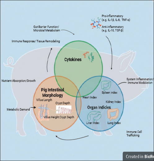

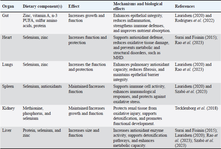

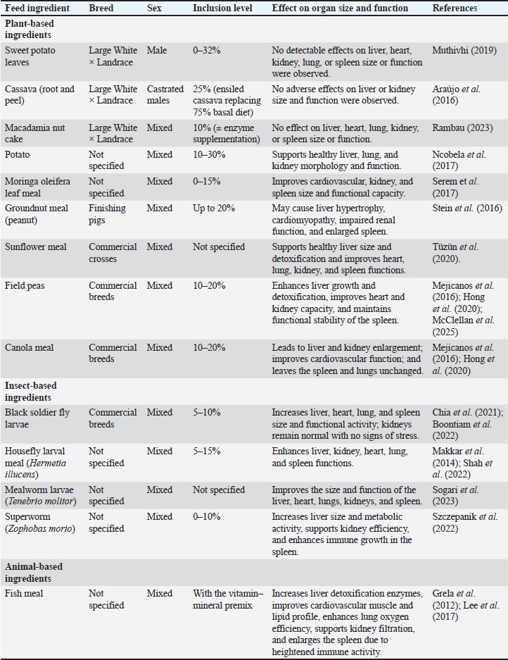
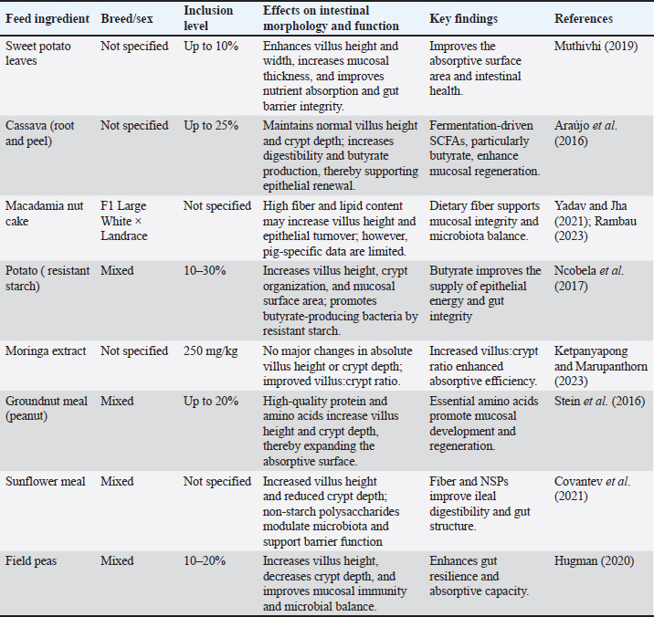
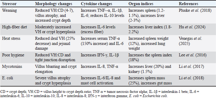

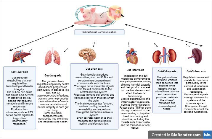
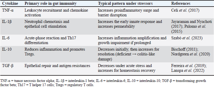
| How to Cite this Article |
| Pubmed Style Khavhadi A, Idowu PA, Modiba MC, Mpofu TJ. Integrating intestinal morphology, cytokine, and organ indices in relation to gut health and physiological adaptation in pigs: Narrative review. Open Vet. J.. 2026; 16(8): 5014-5039. doi:10.5455/OVJ.2026.v16.i8.2 Web Style Khavhadi A, Idowu PA, Modiba MC, Mpofu TJ. Integrating intestinal morphology, cytokine, and organ indices in relation to gut health and physiological adaptation in pigs: Narrative review. https://www.openveterinaryjournal.com/?mno=300144 [Access: August 08, 2026]. doi:10.5455/OVJ.2026.v16.i8.2 AMA (American Medical Association) Style Khavhadi A, Idowu PA, Modiba MC, Mpofu TJ. Integrating intestinal morphology, cytokine, and organ indices in relation to gut health and physiological adaptation in pigs: Narrative review. Open Vet. J.. 2026; 16(8): 5014-5039. doi:10.5455/OVJ.2026.v16.i8.2 Vancouver/ICMJE Style Khavhadi A, Idowu PA, Modiba MC, Mpofu TJ. Integrating intestinal morphology, cytokine, and organ indices in relation to gut health and physiological adaptation in pigs: Narrative review. Open Vet. J.. (2026), [cited August 08, 2026]; 16(8): 5014-5039. doi:10.5455/OVJ.2026.v16.i8.2 Harvard Style Khavhadi, A., Idowu, . P. A., Modiba, . M. C. & Mpofu, . T. J. (2026) Integrating intestinal morphology, cytokine, and organ indices in relation to gut health and physiological adaptation in pigs: Narrative review. Open Vet. J., 16 (8), 5014-5039. doi:10.5455/OVJ.2026.v16.i8.2 Turabian Style Khavhadi, Adivhaho, Peter Ayodeji Idowu, Mamokoma Cathrine Modiba, and Takalani Judas Mpofu. 2026. Integrating intestinal morphology, cytokine, and organ indices in relation to gut health and physiological adaptation in pigs: Narrative review. Open Veterinary Journal, 16 (8), 5014-5039. doi:10.5455/OVJ.2026.v16.i8.2 Chicago Style Khavhadi, Adivhaho, Peter Ayodeji Idowu, Mamokoma Cathrine Modiba, and Takalani Judas Mpofu. "Integrating intestinal morphology, cytokine, and organ indices in relation to gut health and physiological adaptation in pigs: Narrative review." Open Veterinary Journal 16 (2026), 5014-5039. doi:10.5455/OVJ.2026.v16.i8.2 MLA (The Modern Language Association) Style Khavhadi, Adivhaho, Peter Ayodeji Idowu, Mamokoma Cathrine Modiba, and Takalani Judas Mpofu. "Integrating intestinal morphology, cytokine, and organ indices in relation to gut health and physiological adaptation in pigs: Narrative review." Open Veterinary Journal 16.8 (2026), 5014-5039. Print. doi:10.5455/OVJ.2026.v16.i8.2 APA (American Psychological Association) Style Khavhadi, A., Idowu, . P. A., Modiba, . M. C. & Mpofu, . T. J. (2026) Integrating intestinal morphology, cytokine, and organ indices in relation to gut health and physiological adaptation in pigs: Narrative review. Open Veterinary Journal, 16 (8), 5014-5039. doi:10.5455/OVJ.2026.v16.i8.2 |


