





| Research Article | ||
Open Vet. J.. 2026; 16(5): 2792-2802 Open Veterinary Journal, (2026), Vol. 16(5): 2792-2802 Research Article Antimicrobial activity of buffalo milk whey hydrolysates against multidrug-resistant bacteria isolated from dairy productsNoor Adil Abood1, Nawras Kadhum Mahdee Alnakeeb2, Ali Abd Sharad3, Orooba Meteab Faja4*, Ziad M. Alkhozai5, Basima Jasim Mohammed4 and Haifa Jumaa Hassan41College of Pharmacy, AL-Nahrain University, Baghdad, Iraq 2Department of Physiology, Biochemistry, and Pharmacology, College of Veterinary Medicine, University of Al-Qadisiyah, Al-Diwaniyah City, Iraq 3Department of Biotechnology, Science College, Anbar University, Ramadi, Iraq 4Department of Public Health, College of Veterinary Medicine, University of Al-Qadisiyah, Al-Diwaniyah City, Iraq 5College of Science, University of AL-Qadisiyah, Al Diwaniyah, Iraq *Corresponding Author: Orooba Meteab Faja. Department of Public Health, College of Veterinary Medicine, University of Al-Qadisiyah, Al-Diwaniyah City, Iraq. Email: orooba.faja [at] qu.edu.iq Submitted: 28/11/2025 Revised: 27/03/2026 Accepted: 07/04/2026 Published: 31/05/2026 © 2025 Open Veterinary Journal
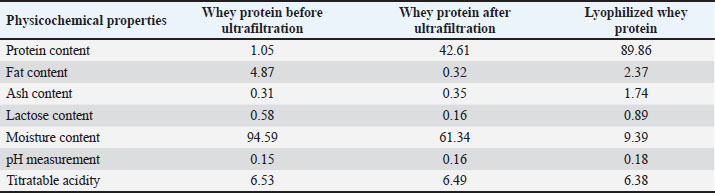
AbstractBackground: Dairy products are nutritionally rich food items that are potentially reservoirs for food pathogenic microorganisms, especially multidrug-resistant (MDR) bacteria, if produced and/or handled under poor hygienic conditions. In addition to the fear of acquiring antimicrobial resistance within the dairy production chain, bioactive peptides from milk whey are attracting attention as natural substitutes for antimicrobial agents and as potential replacements for traditional antibiotics. Aim: The current study attempts to isolate and describe different bacterial species from various frequently consumed products from the dairy industry and determine their antimicrobial resistance patterns. In addition, this study aimed to determine the antimicrobial activity of peptides to be fractionated from enzymatically hydrolyzed buffalo milk whey against selected MDR dairy-derived isolates. Methods: Dairy samples were taken from multiple sites, totaling 290 items, including yogurts, cheeses, and creams, and were then processed in aseptic conditions. Bacterial isolates were cultured and purified using the VITEK®2 Compact System and other biochemical techniques. Using the Kirby-Bauer disk diffusion technique, the antimicrobial susceptibility exhibited 15 antibiotics and a multi-antibiotic resistance (MAR) index was calculated. The 16S rRNA gene was amplified and sequenced for molecular identification and phylogenetic analysis using universal primers. The antimicrobial activities of the whey hydrolysates and peptide fractions from processed, enzymatically hydrolyzed, and fractionated buffalo milk whey were evaluated using minimum inhibitory concentration (MIC) assays. Results: From the dairy products recovered, 12 species were represented by 136 bacterial isolates, with approximately equal levels across cheeses, creams, and yogurts. Significant MAR indices, with most isolates showing >0.2, indicated that many of the isolates came from environments of high contamination risk, showing resistance to tetracycline and erythromycin, ciprofloxacin, and ampicillin. Phylogenetic analysis for all isolates confirmed the closely related genetic relationship to other strains from various parts of the world for the amplified 16S rRNA gene (approximately 1,500 bp). The buffalo milk whey that had been enzymatically hydrolyzed, along with some of the selected chromatographic fractions, exhibited high levels of antibacterial activities against the MDR isolates, although some differences in MIC values were dependent on the specific strain. Conclusion: The use of biochemical and molecular methods proved to be more successful in determining the type of bacteria in dairy products and showed a high frequency of isolates resistant to multiple drugs. The whey peptides derived from buffalo milk exhibited strong antimicrobial properties against the aforementioned resistant bacteria. This highlights the peptides’ ability to be used as a natural antimicrobial to help with food safety and antimicrobial stewardship in dairy production systems. Keywords: Bacteria, Dairy products, Molecular identification, Multidrug resistance. IntroductionDairy products pose microbiological public health risks because they can be easily contaminated with various bacterial species. The production, distribution, and storage of dairy products can support the survival of both gram-positive and gram-negative bacteria, creating significant microbiological hazards. Recent studies have increasingly focused on antimicrobial resistance (AMR) in foodborne bacteria. These studies highlight AMR as a complex and concerning issue due to the spread of multidrug-resistant and potentially fatal strains (McGowan et al., 2006). The lack of sufficient molecular and biochemical systems to monitor bacterial contamination in slaughterhouses underscores the need for improved bacterial identification tools for the dairy industry. Bacterial contamination in slaughterhouses can provide isolates for studies related to dairy production systems. Microbial characterization systems may exhibit variability or reduced reliability when used in milk-based studies. The systems used as instruments may have an acceptable weight. Characterization of bacterial species at the molecular level has become increasingly important in the context of antimicrobial resistance. Evolution is one of nature’s most distinguishing phenomena. Accurate bacterial identification is essential, and automated phenotypic systems, such as VITEK®2, provide rapid preliminary identification. However, definitive identification requires molecular validation, which offers unmatched precision. Numerous studies, such as that of Abdalhamed et al. (2018), have shown that the rapid amplification and electrophoresis of specific 16S rRNA sequences can be used as molecular tools for the differentiation of foodborne bacterial isolates. Foodborne pathogens such as Escherichia coli, Staphylococcus aureus, Salmonella enterica, and Klebsiella spp. are frequently reported and exhibit complex, multidimensional phylogenetic structures. Such complex structures have a great effect on the phylogenetic, ecological, and geographical distribution of such species, as well as their antimicrobial resistance. The AMR profiles of isolates from dairy products highlight the potential of molecular phylogenetic tools to clarify transmission pathways and sources of contamination. Recent work on the hydrolysates of dairy proteins has documented the antimicrobial activities of whey hydrolysates and their peptides against certain clinically relevant pathogens. The broad-spectrum antibacterial activity of whey hydrolysates has been documented and represents a potential strategy to counteract antibiotic resistance (Guilhelmelli et al., 2013; Pan et al., 2021). Peptides derived from whey disrupt bacterial membrane integrity, modulate their metabolism, and chelate certain critical ions, thereby negatively impacting their survival (Kim et al., 2007; Demers-Mathieu et al., 2013). These bioactive peptides demonstrate high potential for food preservation and safety applications. The current evidence highlights the importance of comprehensive molecular characterization of dairy caseins to explain the patterns of contamination, resistance, and connection for the molecular phylogeny of dairy caseins. It also demonstrates the need for monitoring programs to be enhanced for the benefit of public health. This study aimed to use a combination of molecular and biochemical methods to characterize the bacterial species isolated from cheeses, yogurts, and creams and study their antimicrobial resistance patterns. The molecular methods used 16S rRNA gene sequencing and phylogenetic reconstruction, corroborated with gel electrophoresis. Materials and MethodsSampling and collection of samplesOut of all the available dairy products, 290 samples were collected from numerous commercial establishments in Al-Diwaniyah City, including cheese (98), yogurt (95), and creamy products (97), where strict aseptic procedures were followed to avoid external contamination. All samples were collected in sterile containers, and to assist the laboratory in analyzing and processing the samples, all samples were placed in cool containers to keep the samples short legging to the other samples taken. The sample handling procedures were performed under standard microbiological safety precautions. Buffalo milk was obtained from a local dairy farm. Raw milk was received under strict hygienic and dairy farm protocols. Milk samples are transported at 4°C to meet the required laboratory standards and processed within a maximum of 4 hours from collection. The milk was then centrifuged at 4°C for 20 minutes at 4,000 × g to separate the fat and cell to form the skim milk. Separated skim milk was used for whey preparation. Preparation of buffalo milk wheyCasein proteins were precipitated by adjusting the pH of the skimmed milk to 4.6 with the addition of 1 M hydrochloric acid. The mixture was then incubated at room temperature for 30 minutes before centrifugation at 10,000 × g for 20 minutes at 4°C. The whey was then separated, and the supernatant was filtered using Whatman No. 1 filter paper. The separated and filtered whey was stored at 4°C. Protocol for ultrafiltrationThe separated and stored whey was subjected to ultrafiltration using a membrane system with a cut-off of 10 kDa. Ultrafiltration was performed at 4°C under constant pressure until the desired concentration was achieved. Retentate containing the whey proteins was collected while the permeate was discarded. The concentrated whey protein fraction was then processed immediately for lyophilization. Lyophilization conditionsThe ultrafiltered whey protein concentrate was stored at −80°C for 24 hours and then placed in a vacuum lyophilizer (< 0.1 mbar) at −50°C. The resulting dry powder was stored in sealed containers at −20°C for future use. Enzymatic hydrolysis of whey proteinsThe lyophilized whey protein powder was reconstituted in distilled water to obtain a final protein concentration of 5% (w/v). Hydrolysis was performed with pepsin (enzyme-to-substrate ratio 1:100, w/w) and 1 M HCl to adjust the pH to 2.5. The mixture was incubated at 37°C for 3 hours while gently stirring. The mixture was heated to 90°C for 10 minutes to inactivate the enzyme. The hydrolysate was cooled and centrifuged at 10,000 × g for 15 minutes. The supernatant was collected for further analysis. Determination of degree of hydrolysis (DH%)The DH% was assessed using the Trinitrobenzenesulfonic Acid method. After hydrolysis, the free amino groups released were measured by spectroscopy at 340 nm. The following formula was used to determine DH%: DH (%)=(htoth) × 100 where hour represents the number of hydrolyzed peptide bonds and hour<sub>tot</sub> is the total number of peptide bonds in the protein substrate. Chromatographic fractionation (Sephadex G-25)The whey protein hydrolysate was fractionated by gel filtration chromatography using a Sephadex G-25 column (2.5 × 50 cm). The column was equilibrated with distilled water, and elution was performed at a flow rate of 1 ml/minute. Different elution fractions were collected and monitored to determine the absorbance at 214 nm. The fractions were pooled based on the presence of distinct peaks, lyophilized, and stored at −20°C in preparation for antimicrobial assays. Bacterial isolation and phenotypic identificationPotential bacterial contaminants identified in the client-provided samples were isolated by streaking the samples into a number of selective agars, including MacConkey, Eosin Methylene Blue agar, and other selective agars that are customarily used for Enterobacteriaceae and non-enteric pathogens. The samples were kept in an incubator maintained at 37°C for a time duration of 24–48 hours, and successive subculturing was performed to obtain pure isolates. A total of 136 isolates were obtained, and several different genera were formed, including Escherichia, Pseudomonas, Salmonella, Staphylococcus, Proteus, Enterobacter, Klebsiella, Clostridium, Listeria, and Enterococcus. The purified colonies were subjected to classical morphological examinations, including differential motility, Gram’s, and the biochemical tests of catalase and oxidase, and a number of other tests that were ascertained to be further sample identification of the organism by different means of computer automation. Automated biochemical identification (VITEK®2 Compact System)The VITEK®2 Compact System (bioMérieux Inc, France) was used to identify all isolates using miniaturized biochemical reactions together with a sophisticated algorithmic data bank for bacterial identification. A standardized suspension turbidity of 0.5 McFarland was prepared, with GN or GP identification cards chosen upon the Gram reaction. Identification cards were inserted following the insertion protocols provided by the manufacturer, and identification was accepted only if the system recorded confidence parameter values above the threshold level. This method allowed for accurate resolution of bacterial identification to the species level, minimizing user-related variability in the results. Antibiotic susceptibility testing and MAR index calculationAntibiotic susceptibility testing was performed using the Kirby–Bauer disk diffusion method on Mueller–Hinton agar, as directed by the Clinical and Laboratory Standards Institute (CLSI M7–A7). Of the 15 antibiotics tested, ampicillin, tetracycline, erythromycin, ciprofloxacin, gentamicin, chloramphenicol, and others, representing a variety of classes of antibiotics, were selected. After incubation for 18–24 hours at 37°C for 18–24 hour, the diameter of any resulting inhibition was measured, and the results were interpreted according to the CLSI breakpoints. A MAR index was calculated for each isolate as the ratio of the number of antibiotics resisted to the total number tested. Isolates with MAR values >0.2 were considered to originate from high-risk settings, indicating probable exposure to heavy antimicrobial use. We examined the antimicrobial activity of the chromatographic and hydrolysate fractions using the broth microdilution approach. In sterile Mueller–Hinton broth, we prepared two-fold serial dilutions of each sample. The adjusted bacterial suspensions were set to the 0.5 McFarland standard and were diluted to a target 1 × 10⁶ CFU/ml. Then, the microplates were stored and incubated at 37°C for a period of 18–24 hours. The Minimum inhibitory concentration (MIC) is the minimal concentration of the sample that showed no growth. DNA extraction and 16S rRNA gene amplificationDNA was extracted according to the manufacturer’s protocol (Cat. No: K0721). The 16S rRNA gene was amplified using the following primer sequences: 27F (5′-AGAGTTTGATCATGGCTCAG-3′) as the forward primer and 1492R (5′-GGTTACCTTGTTACGACTT-3′) as the reverse primer. Each polymerase chain reaction (PCR) reaction was 25 μl and contained 12.5 μl of 2 × Master Mix (Promega, USA), 1 μl of each primer (10 pmol), 2 μl template DNA, and the remainder was nuclease-free water. The following cycling conditions were used: initial denaturation at 95°C for 5 minutes, followed by 35 cycles of denaturation at 95°C for 30 seconds, annealing at 55°C for 30 seconds, elongation at 72°C for 90 seconds, and a final extension at 72°C for 10 minutes. The expected size of the amplified DNA was approximately 1,500 bp. Gel electrophoresis and visualizationThe PCR products were separated on 1.5% agarose gels prepared in 1 × Tris–Borate–EDTA buffer and stained with ethidium bromide. Thermo Fisher Scientific, USA No. SM0311 1 kb DNA ladder was used as molecular size markers. Bands were visualized under UV light following electrophoresis at 90 V for 60 minutes. All isolates had clear single amplicons of the expected size, which confirmed the successful amplification of 16S rRNA, in accordance with the secondary file’s descriptive molecular analysis. Phylogenetic analysisCleaned PCR products were sequenced using the ABI 3500 Genetic Analyzer (USA), and the chromatograms were subjected to quality control before analysis. The sequences were aligned in MEGA X using the ClustalW method, and the maximum likelihood method was used to construct the phylogenetic trees with 1,000 replications of the bootstrap. The available sequences in the NCBI GenBank database were used as references to determine the position of the sequence in the evolutionary tree. The phylogenetic groups formed demonstrated close proximity of the local isolates and the strains documented elsewhere, confirming the bacterial isolates’ epidemiological relevance. Ethical approvalNot necessary for this manuscript. ResultsPhysicochemical properties of buffalo milk wheyThe ultrafiltration and lyophilization of buffalo milk whey yield measurable compositional changes encapsulating the concentrating effect of the operational steps, as determined by the physicochemical assessment of the whey before and after each of these treatments. Retaining proteins, fat, lactose, minerals, and moisture, whole whey was equilibrated to moderate concentrations. However, during ultrafiltration, the remaining lactose and moisture, in addition to a proportion of the proteins and dry minerals, were reduced, while the protein content was enriched. Lyophilization of the whey concentrate further consolidated the moisture deficits, yielding the most protein-dense concentrate, as evidenced by the moisture and protein ratios remaining in the preparation. While titratable acidity and pH values were essentially indistinguishable across the treatments, the differences were small, and the treatments likely maintained the original whey acidity (Table 1). Table 1. Physicochemical properties of buffalo milk whey before and after ultrafiltration and concentration of lyophilized whey protein.
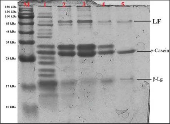
Protein profile of whey hydrolysates as determined by SDS-PAGESDSPAGE banding patterns indicated progressive degradation of whey proteins during pepsin hydrolysis, as evidenced by the distinct banding patterns. The proteins in the whey that had not undergone hydrolysis were initially determined to be intact, as demonstrated by the distinct bands corresponding to the major proteins, including beta-lactoglobulin and lactoferrin. As the hydrolysis time increased from 60 to 240 minutes, the gradual disappearance of high-molecular-weight bands and the emergence of lower-molecular-weight bands were observed, indicating time-dependent enzymatic cleavage. Among all the hydrolysates, the 240-minute hydrolysate exhibited the most extensive fragmentation, showing faint/absent bands of intact proteins and accumulation of low-molecular-weight peptides (Fig. 1).
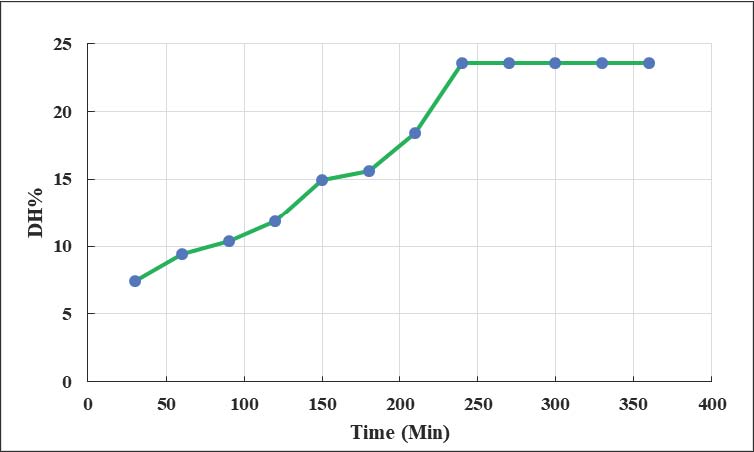
Fig. 1. SDS-PAGE of whey protein hydrolysates from buffalo milk made with pepsin at various times M stands for protein marker; lane 1 shows milk whey that has not been hydrolyzed; lanes 2, 3, 4, and 5 show buffalo whey that has been hydrolyzed for 60, 120, 180, and 240 minutes, respectively. β-Lg, β-Lactoglobulin Monomer; LF, Lactoferrin. Three separate experiments are represented in the data. Degree of hydrolysis of whey proteinsThe DH% increased with longer enzymatic treatment times, showing constant peptide bond cleavage by pepsin activity. A steep ascending curve was observed for the first 120 minutes, indicating high levels of proteolysis early on. A more gradual ascending curve was observed between 180 and 360 minutes, indicating that the levels of reactions approached saturation. A higher DH% was observed in 360 minutes compared with the lower levels of hydrolysis observed. This indicated that higher levels of hydrolysis resulted in more extensive protein rupture compared with lower levels of hydrolysis (Fig. 2).
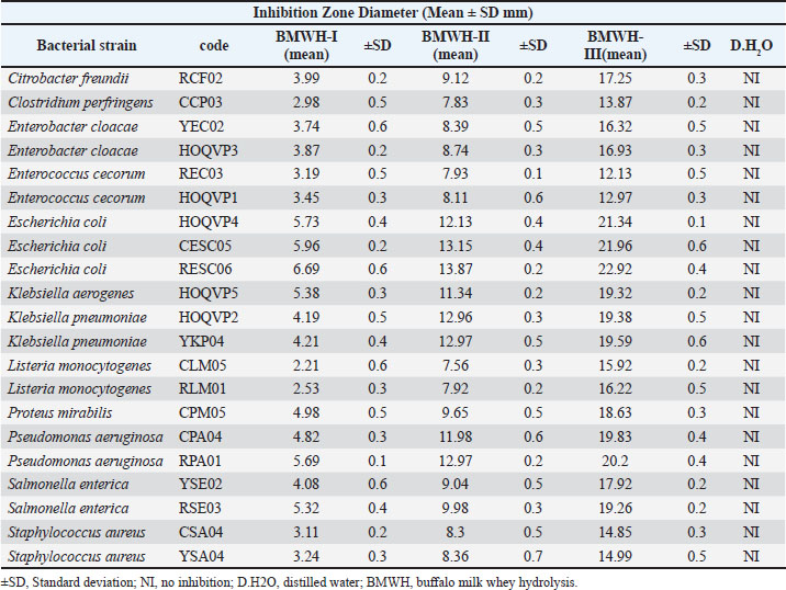
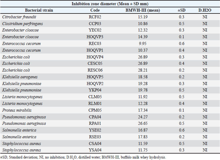
Fig. 2. Effect of buffalo milk whey hydrolysis time (30–360 minutes) on DH (degree of hydrolysis). Antibacterial activity of whey fractions (BMWH-I, II, III)Evaluating the antibacterial activities of BMWH-I, BMWH-II, and BMWH-III against 21 bacterial species showed that BMWH-III had the greatest inhibitory effects, followed by BMWH-II the next, and BMWH-I had the lowest inhibition. BMWH-III exhibited the largest inhibition zone among all the gram-negative and gram-positive bacteria, with the greatest activity against Listeria monocytogenes. Please change the names of S. aureus, S. enterica, and E. coli to italics throughout the entire manuscript. BMWH-I showed the least inhibition, whereas distilled water produced no inhibition, thereby validating the assay. The inhibition ability of BMWH-III was clearly superior, and it appears that the enzymatic hydrolysis step released some active peptide components that had a much greater broad-spectrum activity than the other two (Table 2). Table 2. Antibacterial activity of the whey fractions (BMWH-I, II, and III).
Antibacterial activity of fraction A (BMWH-III Fr. A)Sephadex G-25 chromatography applied to BMWH-III resulted in some subfractions, with Fraction A (Fr. A) having the highest antibacterial activity. Compared with the unfractionated BMWH-III, the inhibition zone diameter of BMWH-III Fr. A was greater, indicating that it was an active peptide component-enriched fraction. This fraction exerted strong inhibitory effects on several microbes, including Proteus mirabilis, Klebsiella pneumoniae, Citrobacter freundii, S. aureus, and L. monocytogenes. The increased activity in Fr. A confirmed the effective capture and partial purification of the bioactive peptide fraction with even stronger antimicrobial properties than the crude hydrolysate (Table 3). Table 3. Antibacterial activity of BMWH-III (Fr. A).
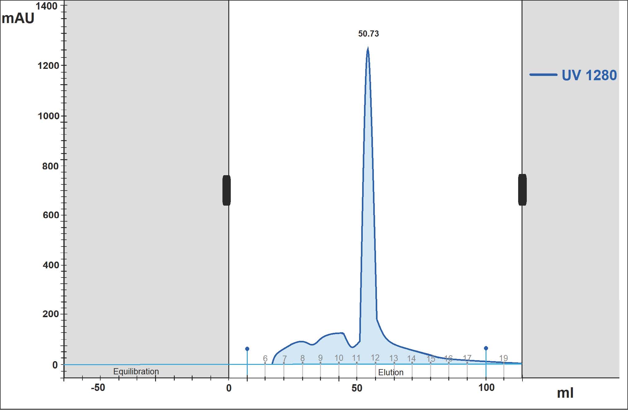
Gel filtration chromatography in a Sephadex G-25 column reported a distinctive elution profile of peptides for the BMWH-III fraction, with the first major peak being a high activity fraction, Fr. A, with the highest antibacterial activity. The rest of the elution consisted of small peaks representing lower fractions of activity (Fig. 3).
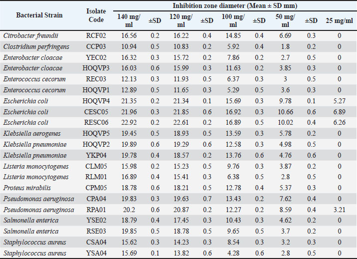
Fig. 3. Elution profiles of buffalo milk whey hydrolysate peptide III (BMWH-III) from a Sephadex G-25 chromatography column using Dextran gel. Minimum inhibitory concentration of BMWH-III Fr. AResults obtained from the MIC testing exhibited that BMWH-III Fr. A confirmed a broad spectrum of bacterial activity for several species, including C. freundii, Klebsiella aerogenes, K. pneumoniae, L. monocytogenes, P. mirabilis, Pseudomonas aeruginosa, and S. enterica with a minimum inhibitory concentration of 25 mg/ml. Only a small set of strains exhibited higher MICs from (50–140 mg/ml), with a species-dependent variation in the antibacterial activity level. All the different microorganisms continued to show low MIC values, confirming that the disrupted, separated, and purified whey peptides did indeed exhibit strong suppression activities and were enriched vectors of bioactive, bioactive peptides of the antimicrobial nature (Table 4). Table 4. MIC values of the antibacterial peptide extracts (BMWH-III, Fr. A).
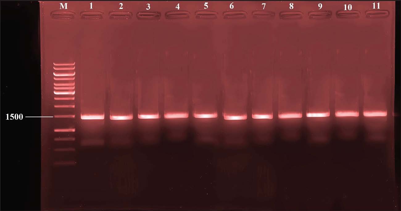
Confirming the presence of a 16S rRNA GeneAs a molecular identification method, the isolates produced unique 16S rRNA amplicon sizes of approximately 1,500 bp. Using primers 27F and 1492R in PCR amplification, strong and distinct bands were produced on 1.5% agarose gels. This indicates that the extracted DNA was intact and usable for further analyses. There were no signs of the generation of non-target byproducts or primer-dimer formations, suggesting that the amplified target was the desired species. The gel electrophoresis patterns confirmed the phenotypical identification of the bacterial species and provided accurate genetic identification of the bacterial species isolated from cheese, yogurt, and cream (Fig. 4).
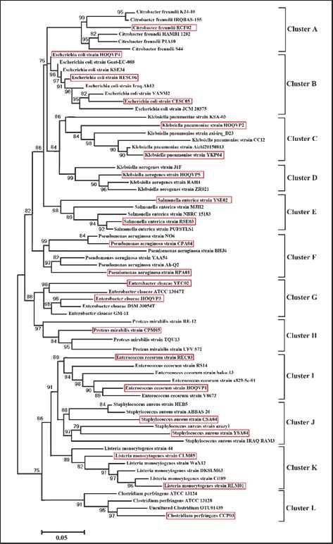
Fig. 4. 16S rRNA PCR products on 1.5% agarose gel. Clear ~1500 bp bands were obtained for all isolates using primers 27F/1492R Lane M: 1 kb ladder; Lanes 1–11: representative isolates. Phylogenetic analysis of dairy-derived isolatesAfter sequencing the amplified 16S rRNA gene and performing phylogenetic reconstruction, the examined isolates clustered closely with reference strains deposited in global genetic databases. The maximum likelihood tree was also able to demonstrate the strains belonging to E. coli, P. aeruginosa, S. enterica, and S. aureus that formed tight monophyletic clades with globally circulating strains. This suggests that these strains underwent evolutionary stasis and are likely to have been transmitted by the same environment or food processed. The phylogenetic data also suggested that these strains were genetically stable, which highlights the widespread antibiotic resistance that is present among the phylogenetically related strains (Fig. 5).
Fig. 5. Phylogenetic tree of 16S rRNA sequences from dairy isolates maximum likelihood tree showing clustering of Al-Diwaniyah City isolates with reference strains. (A) Citrobacter freundii group, (B) Escherichia coli group, (C) Klebsiella pneumoniae group, (D) Klebsiella aerogenes group, (E) Salmonella enterica group, (F) Pseudomonas aeruginosa group, (G) Enterobacter cloacae group, (H) Proteus mirabilis group, (I) Enterococcus cecorum group, (J) Staphylococcus aureus group, (K) Listeria monocytogenes group, and (L) Clostridium perfringens group. DiscussionThis study provides evidence that buffalo milk whey hydrolysates and their isolated peptide fractions are remarkably active antibacterial agents against an extensive variety of pathogenic gram-positive and negative bacteria. Lyophilization and ultrafiltration enhanced the physicochemical properties in a manner consistent with the literature, showing that whey protein concentration steps increase solubility, bioavailability, and functional stability of milk proteins, enhancing their suitability for hydrolysis via enzymatic methods. Ahmed et al. (2021) described comparable observations where the concentration of whey proteins in a milk matrix led to an improvement in the yield of bioactive low molecular weight peptides. Moreover, the increase in whey protein hydrolysis and the concomitant increase in the DH% are in line with the hydrolysate behavior described by Saleh and Mohammed (2022), where a plateau phase due to substrate exhaustion is usually observed after peptide bond cleavage during the rapid phase of primary proteolysis. The DH% and biological activity of the peptides resulting from the enzymatic digestion of whey proteins support the concept that these peptides remain active, indicating that other dairy systems likely provide comparable properties. For the pathogens L. monocytogenes, S. aureus, S. enterica, and P. aeruginosa, greater inhibition was observed from BMWH-III and its purified Fraction A. Also explored were the broad-spectrum antibacterial effects resulting from membrane disruption and interference with metabolic pathways caused by whey-derived peptides were also explored, and these findings were consistent with the findings of Hasan et al. (2020). While some of the isolates from the MIC assay showed weak susceptibility, results from Al-Rubaie and Hamid (2023) showed null detectable MIC values, indicating that variability among isolates was not contradictory. The observed variability may be attributed to differences in peptide composition, factors, and bacterial strain specificity. Nevertheless, the strong inhibitory effects observed at low concentrations indicate that buffalo whey peptides are effective natural antimicrobials. Karim et al. (2021) suggested that bioactive compounds from milk may possess valuable therapeutic and food-preserving attributes. The antimicrobial results and strain identities were confirmed by molecular analysis and 16S rRNA amplification. Regarding molecular analysis and the importance of 16S rRNA gene amplification in determining the bacterial diversity of food samples, the work of Qasim and Jaber (2022) is thorough and notable. The rapid and efficient amplification of ~1500 bp fragments and the tight clustering of local isolates with the global reference strains corroborated the 16S amplification results and the phylogenetic hypothesis made by analyzing the reference to the 16S rRNA gene, which appears misplaced in the context of bacterial phylogeny, as bacteria do not possess mitochondria. Clarification of the genetic markers used for phylogenetic analysis is needed. The molecular analysis further suggested that the isolates were epidemiologically significant and that the whey peptides were likely effective against strains present in both local and global food chains. The enriched buffalo whey peptides are likely to be beneficial as natural antimicrobial agents that could be adopted for application in food safety, functional nutrition, and bio-preservation. ConclusionConcentrated, enzyme-hydrolyzed buffalo milk whey contains bioactive fractions with strong antimicrobial actions against many foodborne pathogens. Ultrafiltration and lyophilization enhanced the functional properties of whey proteins BMWH-III, particularly fraction A, had the highest antimicrobial activity after pepsin hydrolysis and chromatographic purification. The identification of bacterial strains was validated using molecular techniques and phylogenetic analysis. This study concludes that buffalo whey-derived peptides are potential natural antimicrobials that can be integrated into food safety, food preservation, and health supplement applications. AcknowledgmentThe authors would like to express their gratitude to the College of Veterinary Medicine for providing laboratory facilities and technical support throughout the study. Conflict of interestThe authors declare that there are no conflicts of interest existed in this work. FundingThis research was self-funded by the authors with no external financial support. Authors’ contributionsAll authors have contributed to this study. Data availabilityData are available upon request from the corresponding author. ReferencesAbdalhamed, A.M., Zeedan, G.S. and Zeina, H.A. 2018. Isolation and identification of bacteria causing mastitis in small ruminants and their susceptibility to antibiotics, with special reference to molecular diagnosis of Staphylococcus aureus. J. Anim. Health. Prod. 6(2), 57–65. Abdel-Hamid, M., Goda, H.A., De Gobba, C., Jenssen, H. and Osman, A. 2016. Antibacterial activity of papain hydrolysed camel whey and its fractions. Int. Dairy. J. 61, 91–98. Abdel-Hamid, M., Otte, J., De Gobba, C., Osman, A. and Hamad, E. 2017. Angiotensin I-converting enzyme inhibitory activity and antioxidant capacity of bioactive peptides derived from enzymatic hydrolysis of buffalo milk proteins. Int. Dairy. J. 66, 91–98. Adamson, N.J. and Reynolds, E.C. 1996. Characterization of casein phosphopeptides prepared using alcalase: determination of enzyme specificity. Enzyme Microb. Technol. 19(3), 202–207. Ahmed, R., Farhan, S. and Younis, M. 2021. Bioactive whey peptides and their functional applications in food systems. Int. J. Dairy Sci. 16, 45–53. Al-Rubaie, S.M. and Hamid, A.T. 2023. Antibacterial potential of enzymatically hydrolyzed bovine whey proteins. Food Biotechnol. 37, 122–134. Demers-Mathieu, V., Gauthier, S.F., Britten, M., Fliss, I., Robitaille, G. and Jean, J. 2013. Antibacterial activity of peptides extracted from tryptic hydrolysate of whey protein by nanofiltration. Int. Dairy. J. 28(2), 94–101. Guilhelmelli, F., Vilela, N., Albuquerque, P., Derengowski, L.D.S., Silva-Pereira, I. and Kyaw, C.M. 2013. Antibiotic development challenges: mechanisms of action of antimicrobial peptides and bacterial resistance. Front. Microbiol. 4, 353. Hasan, H.A., Ibrahim, N.A. and Saleem, R.J. 2020. Functional properties and antimicrobial activity of whey protein hydrolysates. J. Food Prot. 83, 214–221. Karim, A.J., Rasheed, S.M. and Mahmood, F.S. 2021. Therapeutic relevance of dairy-derived peptides: a review of antimicrobial and immunomodulatory activities. Nutr. Food. Res. 5, 88–97. Kilonzo-Nthenge, A. et al. 2020. Prevalence and antimicrobial resistance of enterococci isolated from retail foods. J. Food Prot. 83(8), 1335–1343 McGowan, L.L., Jackson, C.R., Barrett, J.B., Hiott, L.M. and Fedorka-Cray, P.J. 2006. Prevalence and antimicrobial resistance of enterococci isolated from retail fruits, vegetables, and meats. Journal of Food Protection 69(12), 2976–2982; doi: 10.4315/0362-028x-69.12.2976 Kim, S.B., Seo, I.S., Khan, M.A., Ki, K.S., Lee, W.S., Lee, H.J., Shin, H.S. and Kim, H.S. 2007. Enzymatic hydrolysis of heated whey: iron-binding ability of peptides and antigenic protein fractions. J. Dairy Sci. 90(9), 4033–4042. Pan, Y., Shi, Y., Chen, J., Li, X. and Wang, L. 2021. Antibacterial and antibiofilm activity of camel whey hydrolysates against foodborne pathogens. J. Dairy. Sci. 104(4), 3854–3865. Qasim, L.M. and Jaber, R.K. 2022. Phylogenetic profiling of foodborne pathogens using 16S rRNA gene sequencing. Microb. Diagn. 9, 201–210. Saleh, M.H. and Mohammed, R.K. 2022. Enzymatic hydrolysis kinetics of milk whey proteins and formation of low-molecular-weight peptides. J. Dairy Technol. 75, 98–109 Wakabayashi, H., Takase, M. and Tomita, M. 2003. Lactoferricin derived from milk protein lactoferrin. Curr. Pharm. Des. 9(16), 1277–1287. McGowan, L.L., Jackson, C.R., Barrett, J.B., Hiott, L.M. and Fedorka-Cray, P.J. 2006. Prevalence and antimicrobial resistance of enterococci isolated from retail fruits, vegetables, and meats. Journal of Food Protection 69(12), 2976–2982; doi: 10.4315/0362-028x-69.12.2976 | ||
| How to Cite this Article |
| Pubmed Style Abood NA, Alnakeeb NKM, Sharad AA, Faja OM, Alkhozai ZM, Mohammed BJ, Hassan HJ. Antimicrobial activity of buffalo milk whey hydrolysates against multidrug-resistant bacteria isolated from dairy products. Open Vet. J.. 2026; 16(5): 2792-2802. doi:10.5455/OVJ.2026.v16.i5.21 Web Style Abood NA, Alnakeeb NKM, Sharad AA, Faja OM, Alkhozai ZM, Mohammed BJ, Hassan HJ. Antimicrobial activity of buffalo milk whey hydrolysates against multidrug-resistant bacteria isolated from dairy products. https://www.openveterinaryjournal.com/?mno=299976 [Access: June 26, 2026]. doi:10.5455/OVJ.2026.v16.i5.21 AMA (American Medical Association) Style Abood NA, Alnakeeb NKM, Sharad AA, Faja OM, Alkhozai ZM, Mohammed BJ, Hassan HJ. Antimicrobial activity of buffalo milk whey hydrolysates against multidrug-resistant bacteria isolated from dairy products. Open Vet. J.. 2026; 16(5): 2792-2802. doi:10.5455/OVJ.2026.v16.i5.21 Vancouver/ICMJE Style Abood NA, Alnakeeb NKM, Sharad AA, Faja OM, Alkhozai ZM, Mohammed BJ, Hassan HJ. Antimicrobial activity of buffalo milk whey hydrolysates against multidrug-resistant bacteria isolated from dairy products. Open Vet. J.. (2026), [cited June 26, 2026]; 16(5): 2792-2802. doi:10.5455/OVJ.2026.v16.i5.21 Harvard Style Abood, N. A., Alnakeeb, . N. K. M., Sharad, . A. A., Faja, . O. M., Alkhozai, . Z. M., Mohammed, . B. J. & Hassan, . H. J. (2026) Antimicrobial activity of buffalo milk whey hydrolysates against multidrug-resistant bacteria isolated from dairy products. Open Vet. J., 16 (5), 2792-2802. doi:10.5455/OVJ.2026.v16.i5.21 Turabian Style Abood, Noor Adil, Nawras Kadhum Mahdee Alnakeeb, Ali Abd Sharad, Orooba Meteab Faja, Ziad M. Alkhozai, Basima Jasim Mohammed, and Haifa Jumaa Hassan. 2026. Antimicrobial activity of buffalo milk whey hydrolysates against multidrug-resistant bacteria isolated from dairy products. Open Veterinary Journal, 16 (5), 2792-2802. doi:10.5455/OVJ.2026.v16.i5.21 Chicago Style Abood, Noor Adil, Nawras Kadhum Mahdee Alnakeeb, Ali Abd Sharad, Orooba Meteab Faja, Ziad M. Alkhozai, Basima Jasim Mohammed, and Haifa Jumaa Hassan. "Antimicrobial activity of buffalo milk whey hydrolysates against multidrug-resistant bacteria isolated from dairy products." Open Veterinary Journal 16 (2026), 2792-2802. doi:10.5455/OVJ.2026.v16.i5.21 MLA (The Modern Language Association) Style Abood, Noor Adil, Nawras Kadhum Mahdee Alnakeeb, Ali Abd Sharad, Orooba Meteab Faja, Ziad M. Alkhozai, Basima Jasim Mohammed, and Haifa Jumaa Hassan. "Antimicrobial activity of buffalo milk whey hydrolysates against multidrug-resistant bacteria isolated from dairy products." Open Veterinary Journal 16.5 (2026), 2792-2802. Print. doi:10.5455/OVJ.2026.v16.i5.21 APA (American Psychological Association) Style Abood, N. A., Alnakeeb, . N. K. M., Sharad, . A. A., Faja, . O. M., Alkhozai, . Z. M., Mohammed, . B. J. & Hassan, . H. J. (2026) Antimicrobial activity of buffalo milk whey hydrolysates against multidrug-resistant bacteria isolated from dairy products. Open Veterinary Journal, 16 (5), 2792-2802. doi:10.5455/OVJ.2026.v16.i5.21 |


