





| Short Communication | ||
Open Vet. J.. 2026; 16(8): 5501-5508 !!
Open Veterinary Journal, (2026), Vol. 16(8): 5501–5508 Short communication Evaluation of microscopy-based interlaboratory comparison programs for Trypanosomosis and Paratuberculosis in veterinary laboratories of United Arab EmiratesShameem Habeeba$*, Ghada Abdel Wahab$, Hamida Al Chemissi, Wissam Sorhan,Mohd Al Breiki and Asma Abdi ShahBiosecurity Affairs Division, Development and Innovation Sector, Abu Dhabi Agriculture and Food Safety Authority (ADAFSA), Abu Dhabi, UAE. $Both authors contributed equally to this study *Corresponding Author: Shameem Habeeba. Biosecurity Affairs Division, Development and Innovation Sector, Abu Dhabi Agriculture and Food Safety Authority (ADAFSA), Abu Dhabi, UAE. Email: shameem.beevi [at] adafsa.gov.ae Submitted: 24/11/2025 Revised: 25/06/2026 Accepted: 09/07/2026 Published: 08/08/2026 © 2025 Open Veterinary Journal
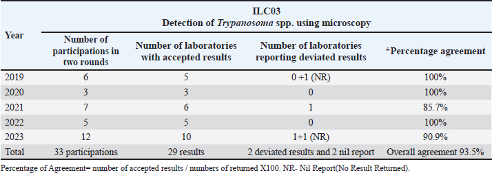
AbstractBackground: Quality control programs are designed to provide a regular, objective, and independent assessment of a laboratory’s ability to provide an acceptable standard of service. For the diagnosis of trypanosomosis and paratuberculosis (Johne’s disease), microscopy is a simple and inexpensive technique that requires a standard or in-house diagnostic method and competent staff. Aim: This study aimed to evaluate laboratory performance on blood and fecal smear examination among public and private veterinary diagnostic laboratories in the United Arab Emirates (UAE) along with a questionnaire survey analysis from participant laboratories. Methods: Interlaboratory comparison programs were organized by the Biosecurity Affairs Division of the Abu Dhabi Agriculture and Food Safety Authority (ADAFSA). Each program was run by sending out fixed blood smears for detecting trypanosomosis and fixed fecal smears for detecting positive and negative types of paratuberculosis. A retrospective assessment of the data obtained for a period of five years (2019–2023) was conducted using Cohen’s kappa and percentage agreement of results. A questionnaire survey was also conducted among the participant laboratories to understand the needs and impact of ILC on quality control programs. Results: Data assessment demonstrated good performance in the identification of target pathogens, namely Trypanosoma spp. in blood smears and Mycobacterium avium subsp. paratuberculosis (MAP) acid fast bacilli in fecal smears. In addition, the staining method and quality of stains used by the different participating laboratories were also good. The overall percentage of agreement with Cohen’s kappa of 95% CI indicated substantial reliability in both ILC programs. Questionnaire analysis revealed a satisfactory report from the participating laboratories for the two ILC programs. Conclusion: The present study on the retrospective data assessment showed highly satisfactory performance results from the majority of participating laboratories and also has a good impact on improving the quality assurance and competence of participating laboratories in terms of trypanosomosis and paratuberculosis diagnosis. Among the participants, only three deviated results were found in this five-year period, and further investigation and improvement in performance were advised. Our study provides the first multiyear evaluation of veterinary microscopy-based ILCs in the UAE, addressing a regional gap in microscopy proficiency testing. Keywords: Interlaboratory, Microscopy, Performance evaluation, Quality control. IntroductionDiagnostic laboratories play a central role in providing information about the health of patients for appropriate disease prevention, diagnosis, and treatment. Quality control (QC) plays a pivotal role in determining the reliability and confidence of laboratory reports (Farr, 2004; Mishra et al., 2023). Laboratory diagnosis in veterinary medicine is vital for establishing clinical diagnosis and providing high-quality evidence-based care in the treatment and prevention of animal diseases. Blood smear examination is an essential technique for the diagnosis and management of various blood-borne parasitic diseases, including trypanosomosis. Microscopic examination of fecal smears using acid fast stain is a quick alternative method to detect MAP for confirmation of clinical cases. The lack of basic biologists in the examination of samples using conventional methods has made microscopy a challenging procedure, which is otherwise a gold standard technique. Accurate identification of disease-causing organisms and differentiation of normal and abnormal cells is important for determining the appropriate treatment. Acceptable and reliable results are the key determinants in the performance of any diagnostic laboratory. A recent review underscored that a well-organized parasitology quality assurance program, encompassing both internal and external assessments, is fundamental to achieving this diagnostic accuracy (Parija, 2025). ILC is the schematic assessment of laboratory quality through an external agency using material of known value but undisclosed results (Chamroon, 2008). ISO/IEC 17043:2023, 3.4 defines ILC as the “design, performance, and evaluation of measurements or tests on the same or similar items by two or more laboratories under predetermined conditions.” Participation in inter-laboratory comparisons provides laboratories with the opportunity to independently evaluate their analytical performance, both in absolute terms and compare to other techniques. Section 7.7 of ISO/IEC 17025:2017 requires that laboratories plan and undertake quality assurance procedures for monitoring the validity of tests and calibrations and shall include participation in inter-laboratory comparison or proficiency testing schemes wherever available and appropriate. This assessment measures the accuracy of the results to obtain reliability. Comparison of results provides insight into one’s own performance that is not available to individual laboratories. This provides a tangible basis for quality improvement initiatives. In the past 5 years, the Biosecurity Affairs Division of ADAFSA (earlier Veterinary Laboratories Division-VLD) has organized ILC schemes in various diagnostic tests for public and private laboratories in the UAE. These programs are designed to provide regular, objective, and independent assessment of a laboratory’s ability to provide an acceptable standard of service by comparison with peers in the field to develop a laboratory quality system. Since a formal interlaboratory program is not available for the microscopic detection of Trypanosoma in blood smear and acid-fast bacilli, namely MAP in fecal smear, this was developed to challenge test performance (ILAC-P9:06/2014). A comparative study in 2023 confirmed the value of such programs for trypanosomosis, finding that many laboratories committed reading errors when not subject to regular external quality assessments (Akinbobola et al., 2023). The present study aimed to evaluate the laboratory performance on blood smear and fecal smear examination among public and private laboratories throughout the UAE from 2019 to 2023 by data assessment of the respective ILC programs organized by the ADAFSA lab. A questionnaire survey of the participating laboratories was conducted to evaluate the ILC samples and to determine the need and impact of ILC programs on the performance of these laboratories. Materials and MethodsWe conducted a retrospective review of the results from the laboratories (nine public and four private laboratories) that participated in two ILC programs, namely, 1) detection of Trypanosoma spp. using microscopy and 2) detection of acid-fast bacilli using microscopy for a period of five years from 2019 to 2023. Interlaboratory comparison programs for respective blood smear and fecal smear examination were organized by the Parasitology and Bacteriology Units of Biosecurity Affairs Division, ADAFSA. The scheme started in 2019 by sending out blood smears and fecal smears in triplicates with positive and negative types. Those laboratories that showed willingness to participate were included in the ILC programs. Participants were from nine public and four private laboratories throughout the UAE. The workflow for the preparation of the samples for the two ILC programs is as follows. Preparation of the ILC samplesILC-03: detection of Trypanosoma spp. by microscopyBlood samples were collected from the jugular vein in ethylenediaminetetraacetic acid tubes and brought to the laboratory within 24 hours of collection in cool packs. The specimens for ILC sample preparation were obtained from routine blood samples of camel origin received in the ADAFSA laboratory and found to be positive for trypanosomosis. Reference slides were produced using standardized parasitological methods and independently verified by two senior microscopists. Only consensus-validated slides were released to the participants. Slide preparation followed controlled quality-assurance procedures, including standardized fixation and staining, smear uniformity and organism visibility checks, and environmental monitoring during preparation and storage to ensure consistency across rounds. Thin blood smears were prepared from whole blood samples within 24 hours of collection as per standard laboratory methods, air-dried, and fixed in absolute methanol for two minutes. Control slides (positive and negative) for each batch were stained with Giemsa stain for 30 minutes, and the smear quality was ascertained by careful screening for blood parasites in a minimum of 300 fields using a 100-x oil immersion objective under a light microscope (WOAH, 2021). Samples with a parasite density score of +++(≥10/HPF-high-power field) were selected as positive ILC samples (Matovu et al., 2019). Negative ILC samples were prepared from routine samples that were confirmed negative by microscopy and molecular methods. The fixed but unstained smears of quality tested batches form the ILC samples. There were two cycles per year. Each cycle had three blood smears for investigation including positives and negatives. ILC-06: microscopic detection of acid fast bacilliCamel fecal samples collected from confirmed positive and negative cases using q-PCR were used as a reference sample for preparing negative and positive slides for the ILC program. The reference samples were aliquoted, labelled, and kept at −80°C. Reference samples (positive and negative) were validated for fit of purpose before the launch of each ILC program. Triplicate slides from each sample were stained using acid-fast bacilli stain (cold stain) and examined microscopically by two competent staff at our laboratory for quality assurance. After passing the quality check, a thin smear was prepared from each used sample, air-dried, and fixed by passing the slides under a benzene flame for three quick passes. Paratuberculosis can be diagnosed if clumps (three or more organisms) of small (0.5–1.5 µm), strongly acid-fast bacilli are found (WOAH, 2021). There were two cycles of ILC-06 per year, each cycle of three slides. Standardized methods have been established for preparing such smears for proficiency testing, ensuring consistency in the concentration of acid fast bacilli and the authentic appearance of the smear (Smithwick and Stratigos, 1978). Scheme design and result interpretationThe VLD-BSAD organizer distributed ILC samples to its participating laboratories via regular mail. An instruction document was accompanied with the specimens for analysis. Laboratories carried out specimen testing as with other routine samples. The due date and a mailing address were included in the instruction for the participants to return their results. To evaluate the performance of each laboratory on blood smear and fecal smear examination, the organizer prepared ILC samples twice a year. Six samples were prepared and divided into two cycles. Both the ILC programs were qualitative with positive/negative results. Hence, the performance assessment of the participating laboratory was based on the agreement of the results with the ILC provider. The classification of laboratory performance followed a binary scheme in which results matching the reference diagnosis were deemed accepted, while any discrepancy—including false-positive, false-negative, or incorrect identification, was recorded as deviated. For a robust validation and to assess the inter-rater reliability, we calculated Cohen’s kappa, and precision was evaluated using 95% confidence intervals (CI). Kappa was selected because it quantifies agreement beyond chance and is widely used in proficiency testing and diagnostic accuracy evaluations. Kappa values were interpreted according to Landis and Koch, (1977) criteria, where values 0.61–0.80 indicate substantial agreement and 0.81–1.00 indicate almost perfect agreement, respectively. The participating laboratories received performance reports with recommendations from the organizer. A laboratory that failed to submit any test result was recorded as a “no result returned/nil report” (NR). Non-returned results (NR) were treated as missing data and excluded from agreement and kappa calculations to avoid biasing performance estimates. The performance data of the participating laboratories for both ILC programs were retrospectively collected from the laboratory data management system and analyzed to record the percentage of agreement of the results. Questionnaire surveyA structured questionnaire was used to collect data from the participating laboratories. Questions were based on understanding the purpose of the ILC program, satisfaction on communication of the program, quality of ILC samples, evaluation methods, frequency of the ILC program, the need for other ILC programs, and the impact of ILC on the quality control programs of the participating laboratories. Ethical approvalThe study used retrospective, fully anonymized laboratory quality-assurance data and did not involve human subjects or live animal experimentation. Table 1. Performance of the participating laboratories for ILC03 during the five-year period (2019–2023).
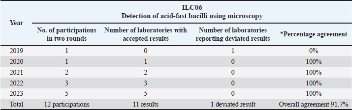
Table 2. Performance of participating laboratories for ILC06 during the five-year period (2019–2023).
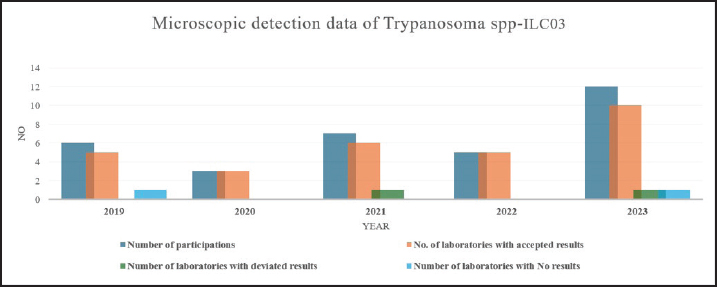
Fig. 1. Participatory results for ILC03 during the five-year period (2019–2023).
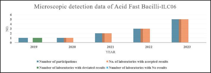
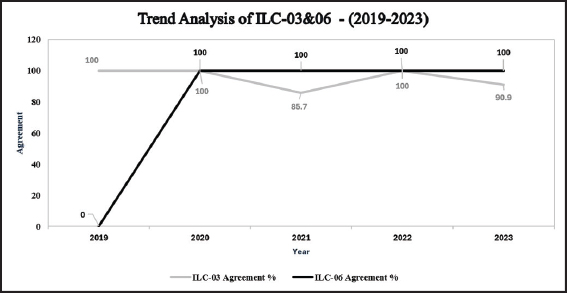
Fig. 2. Participatory results for ILC06 during the five-year period (2019–2023). Results and DiscussionThe test results of the public and private laboratories from the different emirates of United Arab Emirates that took part in ILC programs are shown in Tables 1 and 2 and Figures 1 and 2. The performance data of the participating laboratories for both ILC programs are summarized in Table 3. Among the 13 participants, four laboratories (30.8%) voluntarily participated in the schemes ILC03 and ILC06 regularly in all the 5 years. The overall percentage of agreement with Cohen’s kappa of 95% CI indicated substantial reliability in both ILC programs (Fig. 3). Table 3. Summary performance of participating laboratories for ILC03 and ILC-06.
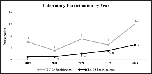
Fig. 3. Trend analysis and percentage agreement of ILC 03 and ILC 06 (2019–2023). Identification of the target pathogen and the staining method were the major parameters for assessing the laboratory performance of blood and fecal smear examination. The numbers of reports with accepted results are shown in Table 1 and were calculated as the percentages in proportion to the total reports received. Cohen’s kappa analysis for ILC-03 program showed almost perfect agreement between the laboratory results and the reference standard (kappa=0.942; observed agreement=97.3%; approximate 95% CI: 0.895–0.988). Similarly, for ILC-06, Cohen’s kappa analysis showed almost perfect agreement (kappa=0.873; observed agreement=93.8%; 95% CI: 0.788–0.958). A major methodological limitation of this study is the small number of participating laboratories in the ILC rounds. These sample size constraints also affect the interpretation of Cohen’s kappa. Although the overall kappa values for both programs were high (ILC03: 0.942; ILC06: 0.873), kappa is unstable when the number of observations is small. This aligns with prior methodological work showing that when sample sizes fall below 30 observations, kappa confidence intervals become unstable and less meaningful (Sim and Wright, 2005). Increasing participation in future ILC rounds would improve the stability of agreement metrics, enhance the interpretability of kappa statistics, and support more reliable longitudinal comparisons. Studies on proficiency testing for microscopy-based diagnosis have consistently reported variable performance across laboratories, highlighting the importance of regular external quality control assessment (Mamuye et al., 2018). The high agreement rates observed in our study for Trypanosoma detection (97.3%) and MAP detection (93.8%) demonstrate that well-designed interlaboratory comparison programs with rigorous pre-distribution quality assurance can achieve excellent participant performance. This high agreement in both programs is consistent with expectations for laboratories operating under structured quality systems aligned with ISO 15189 and ISO/IEC 17043, which emphasize standardized procedures, internal verification, and traceability in proficiency testing processes. Reference slides were prepared using controlled methods, independently verified by senior microscopists, and distributed under quality-assured conditions. Such measures are essential to ensure that performance outcomes reflect laboratory diagnostic capability rather than inconsistencies in slide preparation (Milne et al., 2013; Zambrano et al., 2020). Mesganaw et al. (2025) found that the overall TB smear microscopy performance level of laboratory professionals at peripheral diagnostic laboratories in Ethiopia was satisfactory, indicating a good level of competence even though notable technical errors related to smear reading and reporting were observed. Our high agreement rates align with recent calls for systematic quality assurance in parasitology (Parija, 2025). Akinbobola et al. (2023) reported reading errors in bovine trypanosomosis microscopy in Nigeria, reinforcing the need for regular ILC participation to mitigate diagnostic pitfalls. For acid fast bacilli, the WHO (2022) practical manual recommends proficiency testing, blinded rechecking, and on site evaluation as key EQA components—our ILC 06 program successfully incorporated the first two elements and achieved 93.8% agreement. Our findings show that both public and private laboratories demonstrated satisfactory performance on identification and staining methods for both ILC programs, whereas only three laboratories (23.1%) reported deviated results in the five-year retrospective data evaluation. Our study provides the first multiyear evaluation of veterinary microscopy-based ILCs in the UAE, addressing a regional gap in microscopy proficiency testing. The participants showed a high ability to identify and differentiate the pathogen following appropriate staining techniques. Across the five-year period, two cases of no result returned/nil report (NR) were recorded in the ILC-03 program in 2019 and 2023. Both NR cases were excluded from the denominator when calculating the percentage agreement. Of the 13 participants, three laboratories (23.1%) reported deviated results: one in 2019 for ILC-06 and two in 2021 and 2023 for ILC-03. The participants were notified by the organizer to implement corrective actions. Although no formal root cause investigations were undertaken for these three laboratories, several common microscopy related factors may have contributed to the findings. For ILC-03, error of false positive for Trypanosoma spp., can be due to misinterpretation of stain precipitate, platelet aggregates, or other artifacts simulating parasites, which is a well-recognized pitfall in blood smear examination. For ILC-06, the potential cause of false-negative acid-fast bacilli could involve inadequate decolorization during the acid-fast staining process, insufficient screening of the smear, or challengeable interpretation due to the low number of MAP organisms in the examined field. Without an on-site assessment or structural feedback, these remain speculative; however, both laboratories successfully participated in subsequent cycles with acceptable results, suggesting that awareness of the deviation alone may have prompted informal corrective actions, such as re-reading or staff discussion. In this study, laboratories participated on a voluntary basis. Laboratories with higher confidence in their analytical precision may have been more likely to participate, potentially resulting in an underestimation of the true interlaboratory variance found in routine practice. This is a significant limitation that voluntary participants are often more confident in their quality standards. Despite its voluntary nature, the study provides a critical baseline of motivated laboratories that can serve as a benchmark for future mandatory ILC programs. Participating in such external quality control programs in a periodic manner is important to understand the competence of each laboratory. Common errors in microscopy techniques that can result in deviated results including competence and knowledge of microscopist, quality of stain, staining technique, incorrect reading of slides, and most importantly, the ability to differentiate between the features of the pathogen and the artifact, which is one of the common causes of false positives. Therefore, capacity building and supervision should be continuously provided to laboratory personnel to improve their knowledge, skills, and quality of blood/fecal smear examination. Participating in internal and external quality control programs should be a routine practice in a laboratory. Interestingly and importantly, microscopy remains a simple and inexpensive technique for the diagnosis of several important diseases. Hence, special emphasis needs to be given to the training of laboratory personnel and should be repetitive and self-sustaining.
Fig. 4. ILC 03 and ILC 06: detection of Trypanosoma spp. and acid fast bacilli using microscopy (tThe number of participant laboratories 2019). In 2019, six out of 13 laboratories participated (46.2%) in both ILC programs, whereas in 2023, this number increased to 10 out of 13 laboratories in 2023 (76.9%). Our study showed that regular participation for both ILC03 and ILC06 was found in only 4 laboratories throughout the study period. There was a significant increase (66.7%) in the number of participating laboratories in 2023 (Fig. 4) compared to that in 2019–2022, which may indicate the trust in our ILC program in addition to the increased awareness towards the importance of quality programs in assuring high-quality results relying on microscopy analysis. A significant driver for participation in ILC programs for testing and calibration labs may be the mandatory requirements in clause 7.7.2 of ISO/IEC 17025, where laboratories must monitor their performance by comparing results with those of other laboratories. Several accreditation bodies view ILC/PT (proficiency test) data as the primary objective evidence of a laboratory’s technical competence. Increased participation is noted in specialized areas, such as veterinary parasitology, where standardized reference materials might be scarce, making peer-comparison the only reliable way to ensure accuracy. Practicing day-to-day internal quality control and regular external quality assessment will improve performance based on the knowledge and expertise obtained through job experience rather than sound knowledge. Mamuye et al. (2018) reported that laboratory application of their knowledge was obtained mostly through on-job experience. The questionnaire survey helped the organizer to assess the effectiveness of the ILC program. However, only nine (69.2%) out of the participating laboratories responded to the questionnaire. Despite this limitation, all nine labs reported good quality of the ILC samples and were satisfied with the way of communication of the ILC program, the number of ILC slides, the frequency of ILC distribution, and the result evaluation procedure. Participant laboratories had a profound impact by improving the quality of testing methods and the competence of training personnel. Participant labs expressed their willingness to join new ILC programs and suggested their specific ILC requirements, such as enzyme-linked immunosorbent assay (ELISA) tests for the detection of food and mouth disease (FMD), avian influenza, and Rift Valley fever, as well as molecular tests for FMD, bluetongue, and peste des petits ruminants (PPR). These requests highlight a clear demand for external quality assessment beyond microscopy, particularly for economically important viral diseases in the UAE. This will help the organizer plan new programs that meet the industrial needs. Well-developed and planned quality control programs will ensure that the reports generated within the laboratory are accurate, reliable, and reproducible (Mcfarlane et al., 2015). Moreover, a regular survey of medical laboratories that question the accuracy and precision of laboratory analysis should be conducted to sustain improvements in laboratory quality (Teka and Kibatu, 2012). It is necessary to strengthen the laboratory’s quality control system through internal quality practice, proper and regular training and education of laboratory personnel, and attending educational seminars on total quality management. These measures collectively reduce errors at every step of the testing process. ConclusionParticipation in external quality control programs leads to understanding the competency of each laboratory. The retrospective evaluation of the five-year dataset indicated that most participating laboratories consistently delivered highly satisfactory performance results. Questionnaire survey analysis revealed that ILC programs had a strong positive impact on improving the quality assurance and competence of participant laboratories in the diagnosis of trypanosomosis and paratuberculosis. Increasing participation in future ILC rounds would enhance the robustness of agreement metrics and provide more meaningful comparisons. Quality assurance in a laboratory is multifactorial, including the education level, work experience, responsibility of the laboratory personnel, participation in various accreditation programs, and total quality management systems. The study underscores the importance of robust quality control processes for the effective functioning of diagnostic laboratories and highlights that any identified errors must be promptly corrected to enhance overall laboratory performance. Moreover, the ADAFSA-designed ILC program helped fill the longstanding gap in proficiency testing for microscopic techniques used to detect both pathogens, which is considered a critical component for strengthening laboratory quality control systems. Our five-year experience demonstrates that well-designed ILC programs are effective tools for sustaining diagnostic accuracy in veterinary microscopy. Regular participation, even on a voluntary basis, helps laboratories identify common pitfalls, improve staining and reading practices, and build confidence in their results. We recommend that future ILC initiatives in the region expand to include molecular and serological tests, as requested by survey participants, and transition toward mandatory enrolment to further elevate the standard of veterinary diagnostics across the UAE. AcknowledgmentsWe thank Ms. Asma Abdi Mohamed Shah, Director, Biosecurity Affairs Division, ADAFSA, for the facilities and approvals. Conflict of interest The authors declare no conflict of interest. FundingThis study received no specific grant. Authors’ contributionsSH and GAW: Conceptualization, Data Analysis, Writing the article, Reviewing, Editing and Submission. HAC: Data Retraction. WS: Data Analysis. MAB and AAS: Review, supervision, and approval. Data availabilityAll data supporting the findings of this study are available within the manuscript. ReferencesAkinbobola, A., Adeyemi, O. and Ogundele, F. 2023. Comparative performance evaluation of blood film microscopy for bovine trypanosomosis among diagnostic laboratories in Nigeria. Vet. Parasitology: Regional Studies and Rep. 42, 100887; doi: 10.1016/j.vprsr.2023.100887 Farr, J.M. 2004. Best Jobs for the 21st Century. Indianapolis, IN: JIST Works. 460 p. ILAC-P9:06 2014 ILAC Policy for Participation in Proficiency Testing Activities and ISO 17011:2017 Conformity assessment—Requirements for accreditation bodies accrediting conformity assessment bodies. Position Statement - Inter-laboratory Comparison Surveillance in Accreditation. Version Date: 2017-12-15 Institute for Quality Management in Healthcare ISO/IEC 17025:2017 General requirements for the competence of testing and calibration laboratories, Reference number ISO/IEC 17025:2017(E). ISO 15189 2022. Medical laboratories—Requirements for quality and competence. International Organization for Standardization. ISO/IEC 17043 2023 Conformity assessment—General requirements for the competence of proficiency testing providers. Landis, J., Koch, G. 1977a. The measurement of observer agreement for categorical data 159–174. Mamuye, G.D., Merga, D.Y., Terfa, K.K., Fatuma, H. and Aster, H. 2018. Assessment of knowledge, attitude and practices of medical laboratory professionals on the use of internal quality control for laboratory tests among selected health centers in Addis Ababa Ethiopia. Prim. Health Care 8(02). Matovu, E., Edielu, A., Ojom, J., Nanteza, A., Kato, C.D., Biéler, S. and Ndung’u, J.M. 2019. Field evaluation of LED fluorescence microscopy for demonstration of Trypanosoma brucei rhodesiense in patient blood. Microsc. Res. 7(1), 1–9; doi:10.4236/mr.2019.71001 Mcfarlane, A., Aslan, B., Raby, A., Moffat, K.A., Selby, R. and Padmore, R. 2015. Internal quality control practices in coagulation laboratories: recommendations based on a patterns-of-practice survey. Int. J. Lab. Hematol. 37, 729–738. Mesganaw, B., Belayneh, M., Hibstu, Z. and Fenta, A. 2025. Proficiency and associated factors of laboratory professionals in sputum smear microscopy at selected peripheral public and private diagnostic laboratories in Ethiopia: cross-sectional study. BMC Infect. Dis. 25(1), 510; doi:10.1186/s12879-025-10906-6 Milne, L.M., Kyi, M.S., Chiodini, P.L., Warhurst, D.C., Lalloo, D.G., Bailey, J.W., al, E. and et al. 2013. Quality assurance for parasitology diagnostics. Clin. Microbiol. Rev. 26(3), 559–589. Mishra, B., Das, B.K.L., Khan, S.A., Gelal, B., Niraula, A., Chaudhari, R.K. and Lamsal, M. 2023. Knowledge of internal quality control for laboratory tests among laboratory personnel working in department of biochemistry in a tertiary care center: a descriptive cross-sectional study. JNepal. Med. Asso. 61, 167–170; doi:10.31729/jnma.8040 Parija, S.C. 2025. From the desk of editor-in-chief: ensuring quality assurance in parasitology laboratories. Trop. Parasitology 15(2), 69–70. Sim, J. and Wright, C. C. 2005. Reviews the statistical behaviour of kappa and emphasizes that small sample sizes produce wide, unstable confidence intervals. Smithwick, R.W. and Stratigos, C.B. 1978. Preparation of acid fast microscopy smears for proficiency testing and quality control. J. Clin. Microbiol. 8(1), 110–111; doi:10.1016/j.jcm.1978.11.015 Teka, A. and Kibatu, G. 2012. Quality of liver and kidney function tests among public medical laboratories in western region of amhara national regional state of Ethiopia. Ethiop. J. Health. Sci. 22, 19–26. WOAH Manual of diagnostic tests and vaccines for terrestrial animals, thirteenth edition 2024. PARATUBERCULOSIS (JOHNE’S DISEASE) Chapter 3.1.17.—Paratuberculosis (Johne’s disease) WOAH Terrestrial manual of diagnostic tests and vaccines for terrestrial animals, thirteenth edition 2024. Chapter 3.1.22—Surra in all species (Trypanosoma evansi infection) World Health Organization. 2022. Practical manual on tuberculosis laboratory strengthening, 2022 update. Geneva: World Health Organization. Zambrano, L.D., Ortega, O., Bornay-Llinares, F.J., González, L.M., De Lucio, A., Fuentes, I., Carmena, D., Gárate, T., Valladares, B. and López Vélez, R. 2020. Quality assurance in parasitology laboratories: challenges and strategies. Am. J. Trop. Med. Hyg. 103(3), 1005–1012. | ||

| How to Cite this Article |
| Pubmed Style Habeeba S, Wahab GA, Chemissi HA, Sorhan W, Breiki MA, Shah AA. Evaluation of microscopy-based interlaboratory comparison programs for Trypanosomosis and Paratuberculosis in veterinary laboratories of United Arab Emirates. Open Vet. J.. 2026; 16(8): 5501-5508. doi:10.5455/OVJ.2026.v16.i8.42 Web Style Habeeba S, Wahab GA, Chemissi HA, Sorhan W, Breiki MA, Shah AA. Evaluation of microscopy-based interlaboratory comparison programs for Trypanosomosis and Paratuberculosis in veterinary laboratories of United Arab Emirates. https://www.openveterinaryjournal.com/?mno=298914 [Access: August 08, 2026]. doi:10.5455/OVJ.2026.v16.i8.42 AMA (American Medical Association) Style Habeeba S, Wahab GA, Chemissi HA, Sorhan W, Breiki MA, Shah AA. Evaluation of microscopy-based interlaboratory comparison programs for Trypanosomosis and Paratuberculosis in veterinary laboratories of United Arab Emirates. Open Vet. J.. 2026; 16(8): 5501-5508. doi:10.5455/OVJ.2026.v16.i8.42 Vancouver/ICMJE Style Habeeba S, Wahab GA, Chemissi HA, Sorhan W, Breiki MA, Shah AA. Evaluation of microscopy-based interlaboratory comparison programs for Trypanosomosis and Paratuberculosis in veterinary laboratories of United Arab Emirates. Open Vet. J.. (2026), [cited August 08, 2026]; 16(8): 5501-5508. doi:10.5455/OVJ.2026.v16.i8.42 Harvard Style Habeeba, S., Wahab, . G. A., Chemissi, . H. A., Sorhan, . W., Breiki, . M. A. & Shah, . A. A. (2026) Evaluation of microscopy-based interlaboratory comparison programs for Trypanosomosis and Paratuberculosis in veterinary laboratories of United Arab Emirates. Open Vet. J., 16 (8), 5501-5508. doi:10.5455/OVJ.2026.v16.i8.42 Turabian Style Habeeba, Shameem, Ghada Abdel Wahab, Hamida Al Chemissi, Wissam Sorhan, Mohd Al Breiki, and Asma Abdi Shah. 2026. Evaluation of microscopy-based interlaboratory comparison programs for Trypanosomosis and Paratuberculosis in veterinary laboratories of United Arab Emirates. Open Veterinary Journal, 16 (8), 5501-5508. doi:10.5455/OVJ.2026.v16.i8.42 Chicago Style Habeeba, Shameem, Ghada Abdel Wahab, Hamida Al Chemissi, Wissam Sorhan, Mohd Al Breiki, and Asma Abdi Shah. "Evaluation of microscopy-based interlaboratory comparison programs for Trypanosomosis and Paratuberculosis in veterinary laboratories of United Arab Emirates." Open Veterinary Journal 16 (2026), 5501-5508. doi:10.5455/OVJ.2026.v16.i8.42 MLA (The Modern Language Association) Style Habeeba, Shameem, Ghada Abdel Wahab, Hamida Al Chemissi, Wissam Sorhan, Mohd Al Breiki, and Asma Abdi Shah. "Evaluation of microscopy-based interlaboratory comparison programs for Trypanosomosis and Paratuberculosis in veterinary laboratories of United Arab Emirates." Open Veterinary Journal 16.8 (2026), 5501-5508. Print. doi:10.5455/OVJ.2026.v16.i8.42 APA (American Psychological Association) Style Habeeba, S., Wahab, . G. A., Chemissi, . H. A., Sorhan, . W., Breiki, . M. A. & Shah, . A. A. (2026) Evaluation of microscopy-based interlaboratory comparison programs for Trypanosomosis and Paratuberculosis in veterinary laboratories of United Arab Emirates. Open Veterinary Journal, 16 (8), 5501-5508. doi:10.5455/OVJ.2026.v16.i8.42 |


