





| Research Article | ||
Open Vet. J.. 2026; 16(5): 3130-3143 Open Veterinary Journal, (2026), Vol. 16(5): 3130-3143 Research Article Effect of Boswellia serrata and Trigonella foenum-graecum seed extracts on wound healing of partial pancreatectomy-induced diabetic ratSaddam Khalid Hummadi*Department of Veterinary Surgery, College of Veterinary Medicine, Tikrit University, Tikrit, Iraq *Corresponding Author: Saddam Khalid Hummadi. Department of Surgery and Obstetrics, Faculty of Veterinary Medicine, Tikrit University, Tikrit, Iraq. Email: surgeon [at] tu.edu.iq Submitted: 22/11/2025 Revised: 03/03/2026 Accepted: 16/03/2026 Published: 31/05/2026 © 2025 Open Veterinary Journal
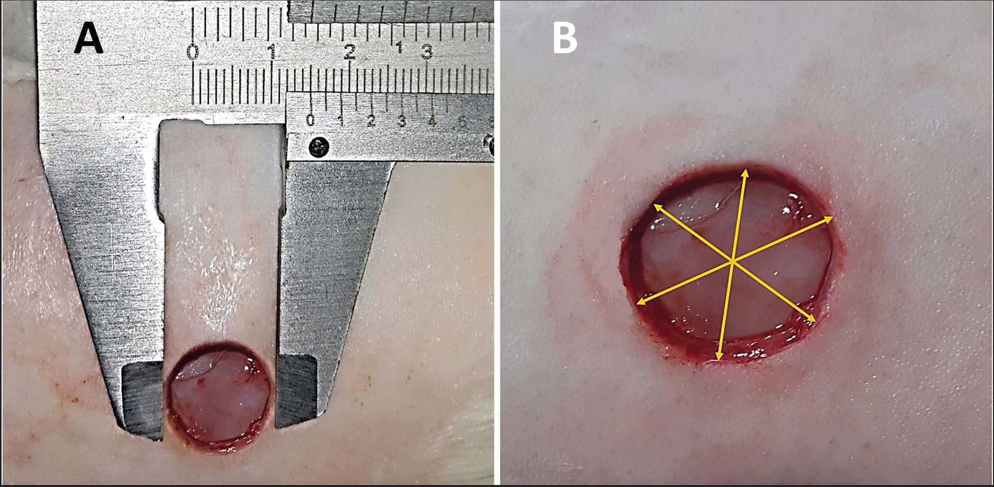
ABSTRACTBackground: The treatment of wounds associated with diabetes is among the common issues encountered in veterinary healthcare. Aim: This research endeavor sought to prepare and evaluate the impact of the composition of Boswellia serrata extract (BSE) and Trigonella foenum-graecum seed extract (TFGE) on the healing of induced diabetic wounds in rats. Methods: Thirty-two clinically healthy adult male rats, aged 6–8 months and weighing 280 ± 20 g, were used in this study. After partial pancreatectomy, 2 equal groups (n=16) were randomly selected among the 32 experimental animals and exposed to a full-thickness excisional wound. Group Ι, represented control, was treated with the topical application of penicillin-streptomycin antibiotic, and group II was treated with ointment prepared from 20% BSE and 10% TFGE. Each group was treated daily for 21 days. Results: Macroscopic analysis exhibited significant superiority (p < 0.05) of wounds in the treated group compared to the control group at the 7th, 14th, and 21st day. The histopathological findings of the treated group (GII) revealed accelerated revascularization, modulation of inflammatory response, enhancement of collagen fiber formation and maturation, regeneration of epidermal tissue, and hair follicles with improved reepithelialization. Gene expression values showed significantly (p < 0.05) upregulation of Vascular Endothelial Growth Factor and Transforming Growth Factor beta 1 and downregulation of tumor necrosis factor-α on days 3, 7, 14, and 21 post-wound treatment, in contrast to the wounds of the control group (GI). Conclusion: Topical application of the Boswellia serrata and T. foenum-graecum seed extracts substantially enhanced excisional wound healing in diabetic animals. Keywords: Boswellia Serrata; Diabetic rat; Gene expression; T. foenum-graecum seed extract; Wound healing. IntroductionThe wound healing involves a process of a highly coordinated cascade of cellular and immunological responses, which occurs immediately after injury and continues to seal the wound (Masoko et al., 2010). Middle-aged and older pet animals are more likely to have diabetes mellitus, and 7%–34% of diabetic dogs also have dermatological disorders (Peikes et al., 2001). The most common complications combined with diabetic wounds are poor or delayed healing and wound infections, which represent a prevalent issue for individuals exhibiting inadequate regulation of blood glucose levels (Amin and Doupis, 2016). The inability to regulate elevated blood glucose levels precipitates neurovascular impairment that initiates within the microcirculation and ultimately affects regional blood circulation, thereby resulting in a diminished delivery of oxygen and nutrients to the body tissue and nerves, which is necessary for wound healing (Bournival et.al., 2012). Diabetes associated infections can range from simple cellulitis to osteomyelitis (Uckaya et al., 2015). There are several therapeutic options for wound care, including anti-inflammatory medications and antibiotics, but most of these medications have a number of undesirable side effects. Numerous studies have demonstrated that applying herbal medication externally has a variety of advantages, including leukocyte chemotaxis, activation of local macrophages, enhancement of local immunity, regulation of tissue metabolism, promotion of microcirculation, and antibacterial properties (Yang, et al., 2015). Several natural remedies have shown promise in treating wounds as a substitute for the synthetic medications (Xu et al., 2023). Boswellia serrata contains a wide range of phytochemicals, polysaccharides, and digestive enzymes. Furthermore, it contains essential oils (Ayub et al., 2023). The localized administration of oleo-gum-resins derived from B. serrata onto the surface of excisional wounds in a singular dosage influences the distinct stages of the wound healing process, including fibroplasia, collagen biosynthesis, and the contraction of the wound (Bansal, et al., 2013). Trigonella foenum-graecum seed extract (TFGE) exhibits antimicrobial properties against prevalent bacteria present in diabetic wounds, including Staphylococcus aureus (Sharma, et al., 2016), along with accelerating the proliferative stage of diabetic wounds (Muralidharan, et al., 2016). The polysaccharide found in the seed extract of T. foenum-graecum has strong antioxidant activities and enhanced wound closure and promoted epidermal regeneration, with complete re-epithelialization (Ktari et al., 2017). The anti-inflammatory, proproliferative, and antioxidant properties of Boswellia serrata extract (BSE) and TFGE encourage the researcher to hypothesize that the combination of these extracts would improve wound healing more than either extract alone. Hence, this study aimed to determine the influence of topical application of B. serrata and T. foenum-graecum seed extracts on wound repair in diabetic animals. Materials and MethodsExperimental animalsThirty-two adult male rats, weighing 280 ± 20 g and aged 6–8 months, were used in the study. The animals underwent a preconditioning phase within the animal facility for a duration of 14 days preceding the start of the study. Following this period, they were housed individually in 40 × 20 × 20 centimeter crates at standard room temperature (22°C ± 3°C) with unrestricted access to food and water throughout the experiment. Preparation of B. serrata extractBoswellia serrata was extracted according to Kumar et al. (2023), with the following modification: 250 g of B. serrata was manually ground into powder and suspended in 500 ml of aqueous ethanol (95%) for 24 hours with stirring at room temperature. After filtration, the residue was resuspended in 500 ml of aqueous ethanol (95%) for an additional 24 hours to maximize the phytoconstituent recovery, followed by re-filtrating. Then, the amalgamated liquid-phase filtrate was subjected to concentration using a rotary vacuum evaporator (Buchi, Switzerland) at 40°C under low pressure to completely remove ethanol. The percentage yield of the dried extract (11.4 g) in relation to the initial material weight (250 g) was 4.56%. In accordance with a previously published protocol (Chevrier et al., 2005), the extract was chemically standardized according to the overall amount of boswellic acid (38.6%). The final dried extract was stored in an airtight amber glass container at 4°C until used. Preparation of T. foenum-graecum seed extractPurified T. foenum-graecum seeds were pulverized into a fine powder. Pulverized material (50 g) was immersed in 500 ml of absolute methanol (10% w/v) at room temperature (22°C ± 3°C) for 24 hours and then filtered. The filtrate was centrifuged at 8,000 rpm for 15 minutes, after which the clear supernatant was carefully pipetted off. The methanol is subsequently removed by placing the supernatant into a rotary evaporator, where the temperature is calibrated to 45°C, and the system is run under reduced pressure to facilitate evaporation (Hasona et al., 2016). Preparation of B. Serrata and T. foenum-graecum seeds extract ointmentCrude BSE and TFE powders were incorporated into the ointment matrix at final concentrations of 20% and 10% w/w, respectively. We measured 20 g of BSE and 10 g of TFE and then added the powders to the required ointment base (white petrolatum) to reach a total mass of 100 g. The mixture was processed in a homogenizer until a homogeneous blend was achieved, yielding a smooth product free of visible particles and with a consistent distribution of active ingredients across the batch (Allen and Ansel, 2014). Partial pancreatectomySurgery of 90% partial pancreatectomy was performed according to the protocol of Laybutt et al. (2003), with the following modifications: food and water were withheld for 8 hours prior to surgery. The rats were anesthetized intramuscularly using a combination of 70 mg/kg ketamine hydrochloride (NexGen®) and 10 mg xylazine hydrochloride (Xylapan® Switzerland) (Pennasilico et al., 2025). Immediately following the induction of anesthesia, the abdominal region from the xiphoid to the pubis was shaved free of hair and disinfected with 10% polyvinylpyrrolidone-iodine to prepare the skin for aseptic surgical operation. Subsequently, the animals were immobilized in a dorsal recumbent position. The animals were subjected to a 10-mm midline incision to expose and identify the pancreatic tissue. The upper part of the pancreatic head remained intact, anatomically extending from 2 mm of the common bile duct to the first part of the duodenum (Kara, 2005), and then the other portion of the pancreas (approximately 90% of it) was abraded gently with circular sweeping motions across the target tissue by a sterile cotton applicator (SURTEX®) to protect the major blood vessels of other organs. The animals were fed ad libitum after surgery, and their blood glucose levels were assessed 10 days later, with measurements repeated every 10 days until the conclusion of the study. Following surgery, the animals received subcutaneous injections of 0.1 mg/kg buprenorphine (Hestehave et al., 2017) every 12 hours for 48 hours. Induction of the excision woundTen days after the partial pancreatectomy, the animals were anesthetized, and the back skin of the animals was prepared surgically as mentioned above in the operation of the partial pancreatectomy. A full thickness of the excision wound (circular area about 15 ± 0.5 mm in diameter) (Fig. 1A) was created along the marking area using a sterile metallic punch cylinder (Hummadi and Al-Falahi, 2024).
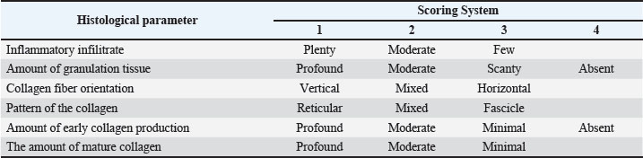
Fig. 1. A. Induced excisional full-thickness cutaneous wound, measuring 15 mm in diameter, created after a partial pancreatectomy. B. Measurement of three distinct wound diameters using a Vernier caliper to assess wound closure. Experimental designAfter inducing partial pancreatectomy, two equal groups (n=16) were randomly selected from among the 32 experimental animals and exposed to a full-thickness excisional wound. The control group was treated with the topical application of penicillin-streptomycin antibiotic, and group II was treated with ointment prepared from 20% B. serrata and 10% T. foenum-graecum seed extract. Each group was treated daily for 21 days. During the wound healing period and at the intervals of 3, 7, 14, and 21 days post-treatment (n=4 per group for each time point), wounds in all groups were traced macroscopically and histopathologically, in addition to quantitative real-time polymerase chain reaction (qRT-PCR) for measuring gene expression for estimation of Vascular Endothelial Growth Factor A (VEGFa), Transforming Growth Factor beta 1 (TGF-β1) and tumor necrosis factor-α (TNF-α). Macroscopic evaluationVernier calipers are used to measure three distinct diameters, one of which crosses the other at the center of the with an equivalent distance between either (Fig. 1B). The mean wound diameter was used to calculate the wound area, which was subsequently incorporated into the following formula: Wound closure (%)=[(W0 - Wt) / W0] × 100 The initial wound area was measured as W0, while Wt represents the wound area measured on the assessment days (3rd, 7th, 14th, and 21st days) (Hummadi and Al-Falahi, 2024). A statistical analysis of the daily measurement data was conducted. Histopathological evaluationThe rats were euthanized intraperitoneally by a combination of 300 mg/kg ketamine hydrochloride and 30 mg xylazine hydrochloride (Underwood and Anthony, 2020) at intervals of 3, 7, 14, and 21 days post-wound treatment; each interval consisted of four rats for each group. Biopsies of 2 mm³ were collected from the periphery toward the center of the wound. Subsequently, the specimens were rinsed with physiological saline and subsequently fixed in a solution of 10% neutral buffered formalin, processed for sectioning at a thickness of 5 microns, and stained using hematoxylin-eosin (H&E) staining methodology (Carriel et al., 2017), then scored semi-quantitatively according to Sultana et al., (2009) as shown in Table 1. Table 1. The Semi-quantitatively scores of histological sections according to Sultana et al., (2009).
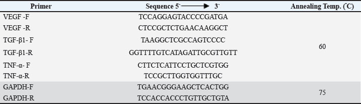
Analysis and calculation of gene expression levelsBiopsies were collected from wound tissue and normal uninjured skin tissue of each animal in the study at intervals of 3, 7, 14, and 21 days following wound care. Each interval consisted of four rats for each group, as described above in biopsies for microscopic evaluation. The tissue biopsies were kept at −20°C after being submerged in a 1.5 ml Eppendorf tube that had been precooled and contained 1 ml of TRIzol reagent (Thermo Scientific, USA). Then, the total RNA was extracted from the tissue biopsies by following the manufacturer’s instructions for the Promega RNA extraction Kit (Promega, USA kit), and one-step qRT-PCR was performed using Bio Molecular System (Australia) to estimate the gene expression of VEGFa, TGF-β1, and TNF-α. PrimersThe primers used in the present study included forward and reverse glyceraldehyde-3-phosphate dehydrogenase (GAPDH) gene primer, which were required as a housekeeping gene, forward and reverse Vascular Endothelial Growth Factor (VEGF) gene primer, TGF-β1, as well as Forward and Reverse TNF-α gene primer (Chen et al., 2019), as genes of interest (Table 2). Table 2. Details of the primers.
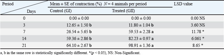
Data analysis using qRT–PCR (Livak Method)A qRT-PCR assay was performed to amplify the synthetic cDNA of the VEGF, TGF-β1, and TNF-α genes (Macrogen) in rats. The rat GAPDH gene (Macrogen) served as an endogenous control to calibrate the target genes. According to Jensen (2012), the relative changes in gene expression from real-time quantitative polymerase chain reaction were analyzed using the 2−ΔΔCT method. Statistical analysisThe Statistical Analysis System (SAS) was used to conduct the statistical analysis of the data. Two-way analysis of variance and least significant difference (LSD) were performed to assess significant differences among means in this study. P < 0.05 is the significance level that was considered indicative of a meaningful difference. The histomorphological scoring results were accepted at p ≤ 0.001 (SAS, 2018). Ethical approvalThe care and use committee of the College of Veterinary Medicine, University of Tikrit, authorized every procedure employed in this investigation. Approval was granted under the number "Tu. Vet.106" on 5/1/2025. ResultsClinical observationsMeasurement of blood glucose levels at distinct time intervals (every 10 days) after partial pancreatectomy showed clear elevation ranging from 267 to 320 mg/dl in all experimental animals. Macroscopic evaluationThe percentage values of wound closure are recorded in Table 3. The obtained values related to the treated group (B. serrata and T. foenum-graecum seed extract) at periods of 7th day (59.53 ± 2.28), 14th day (82.23 ± 0.97), and 21st day (98.91 ± 1.36). These data indicated significant superiority (p < 0.05) for the treated group compared with the control group, which had values of 26.54 ± 5.65, 59.36 ± 2.86, and 64.10 ± 2.67 at the 7th, 14th, and 21st day, respectively. Table 3. Average wound contraction values (% of Mean ± SE) for each group over the study period.
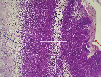
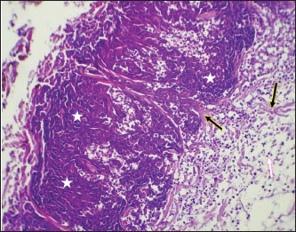
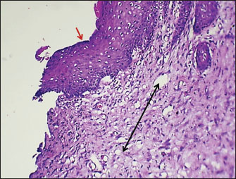
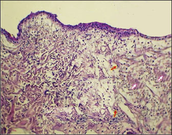
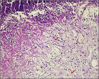
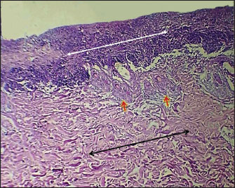
Histopathological evaluationIn the control group, the histopathological section on day 3 expresses massive necrosis of both the epidermal and dermal layers with fibrinopurulent exudate that covered the upper dermal tissue. In addition, a wide area of necrotizing suppurative inflammation of dermal tissue that rested on newly formed granulation tissue, including acellular hyalinized collagenous stroma (Fig. 2). On day 7, the skin section exhibited necrotic tissue alongside immature granulation tissue formation within the dermal layer, with newly synthesized collagen fibers extending into the upper dermis and permeating the necrotic tissue, surrounded by polymorphonuclear leukocyte infiltration (Fig. 3). The main characteristic finding at 14 days was mild regeneration of epidermal epithelia and proliferation of immature granulation tissue with numerous fibroblasts and newly formed capillaries accompanied by mild infiltration of mononuclear cells (Fig. 4). At 21 days post-wounding, the epidermal layer with a thin loose dermal stroma accompanied by moderate breakdown of dermal collagen fibers associated with moderate proliferation of fibroblast (Fig. 5).
Fig. 2. The skin of the control group at 3 days post-wounding shows a wide area of dermal suppurative inflammation (white arrow) that is covered by fibrinopurulent exudate (red arrow) and resting on newly formed granulation tissue (blue arrow) (H&E stain, × 10).
Fig. 3. Skin of the control group at 7 days post-wounding shows moderate extension of collagenous fibers into the dermal tissue (black arrow) through necrotic tissue (stars), with multifocal infiltration of inflammatory cells (white arrow) (H&E stain, × 10).
Fig. 4. Skin of the control group at 14 days post-wounding shows incomplete regeneration of epidermal epithelium (red arrow) and proliferation of immature granulation tissue (black arrow) in the upper dermis (H&E stain, × 20).
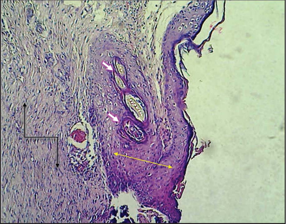
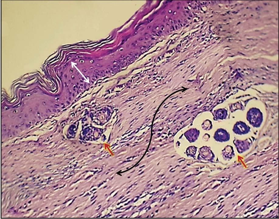
Fig. 5. Skin of the control group at 21 days post-wounding shows a thin epidermal layer and thin loose dermal stroma accompanied by moderate breakdown of dermal collagen fibers (white arrow) associated with moderate proliferation of fibroblasts (red arrow) (H&E stain, × 10). In the treated group at 3 days period, the histopathological results revealed a large amount of immature vascular granulation tissue with newly formed collagen fibers in the dermal tissue and under the necrotic tissue (Fig. 6). The main characteristic finding at 7 days post-wounding was obvious dermal replacement by rich collagen granulation tissue with regeneration of epidermal tissue and the number of regenerated hair follicles accompanied by mild MNC infiltration (Fig. 7). The wound manifestation on the 14th day post-treatment showed complete reepithelialization of the epidermis, accompanied by superficial keratin production, and dermal characteristics of thick, irregular collagen bundles with minimal granulation tissue infiltrated by a few inflammatory cells (Fig. 8). In the treated group at 21 days, beneath the full-thickness epidermal layer, there is a remarkable increase in dermal thickness, characterized by abundant collagen fibers and evidence of mature fibrous connective tissue devoid of inflammatory response. Additionally, the follicular system (Fig. 9).
Fig. 6. The skin of the treated group at 3 days post-wounding shows a large amount of immature vascular granulation tissue (black arrow), with newly formed collagen fibers (red arrows) in the dermal tissue and beneath the suppurative necrotic tissue (H&E stain, × 10).
Fig. 7. Skin of the treated group at 7 days post-wounding shows obvious dermal replacement by rich collagen granulation tissue (black arrow), with regeneration of epidermal tissue (white arrow) and several regenerated hair follicles (red arrow) accompanied by mild MNC infiltration (H&E stain, × 10).
Fig. 8. Skin of the treated group at 14 days post-wounding shows scanty granulation tissue (black arrow) infiltrated with few inflammatory cells, horizontal collagen fiber orientation, and fascicle pattern beneath the full thickness of the epidermal layer (yellow arrow) with hair follicle development (white arrow) (H&E stain, × 10).
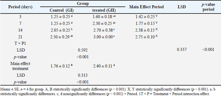
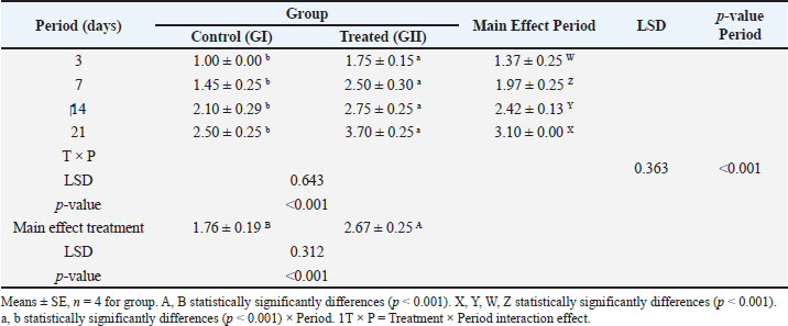
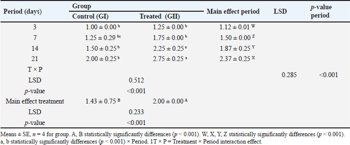
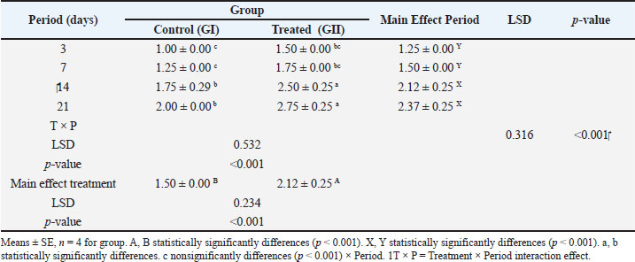
Fig. 9. The skin of the treated group at 21 days post-wounding shows a remarkable increase in dermal thickness with abundant collagen fibers (black arrow) and slight epidermal hyperplasia (white arrow), along with a will-developed follicular system (red arrow) (H&E stain, × 20). In the present study, the scoring of inflammatory infiltration at days 3 and 7 post-wounding showed a nonsignificant decrease between the control group (GI) and treated group (GII), whereas at days 14 and 21, the inflammatory infiltration was significantly (p < 0.001) decreased in the treated group (GII) (2.70 ± 0.38 and 3.00 ± 0.00) compared with the control group (GI), which graded 2.05 ± 0.25 and 2.50 ± 0.29 respectively (Table 4). In terms of granulation tissue amount scores, the results indicated a significant decrease (p < 0.001) in the treated group (GII) at all study periods (days 3, 7, 14, and 21) compared with the wounds in the control group (GI) (Table 5). The scoring of collagen fiber orientation was clearly elevated in the wounds of the treated group (GII) at days 14 and 21 (2.25 ± 0.25 and 2.75 ± 0.25, respectively) compared with the wounds of the control group (GI), which graded 1.50 ± 0.25 and 2.00 ± 0.25, respectively (Table 6). The assessment of collagen patterns on the 3rd and 7th day post-wounding revealed no differences between the two groups, but on the 14th and 21st days, there was a significant increase (p < 0.001) in the treated group (GII) with scores of 2.50 ± 0.25 and 2.75 ± 0.25, respectively, compared to the control group (GI), which had scores of 1.75 ± 0.29 and 2.00 ± 0.00, respectively, at the same periods (Table 7). Table 4. Scores of inflammatory infiltration related to histopathological sections between groups during the study periods.
Table 5. The results of scoring the amount of granulation tissue related to histopathological sections between groups during the study periods.
Table 6. Scores of collagen fiber orientation related to histopathological sections between groups during the study period.
Table 7. Scores of collagen pattern related to histopathological sections between groups during study periods.
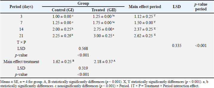
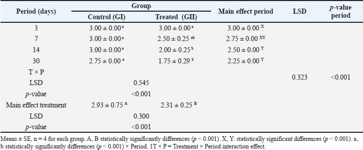
Furthermore, the amount of early collagen scored in the wounds of the treated group (GII) was markedly decreased, especially at the 14th and 21st days (2.75 ± 0.00 and 3.00 ± 0.25, respectively), as compared to the control group (G1), which graded 2.00 ± 0.25 and 2.25 ± 0.29, respectively, at the same period. Additionally, the main effect of treatment exhibited a statistically significant disparity (p < 0.001) between the treated and control groups with scores of 2.18 ± 0.37 and 1.62 ± 0.25, respectively (Table 8). The amount of mature collagen also exhibited a significant increase in the treated group (GII) at the 14th and 21st days, which graded 2.00 ± 0.25 and 1.75 ± 0.29, respectively, in comparison with the control group (G1) during the same intervals, which recorded scores of 3.00 ± 0.00 and 2.75 ± 0.00, respectively (Table 9). Table 8. Scores of the amount of early collagen related to histopathological sections between groups during the study periods.
Table 9. Scores of the amount of mature collagen related to histopathological sections between groups during the study periods.
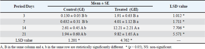
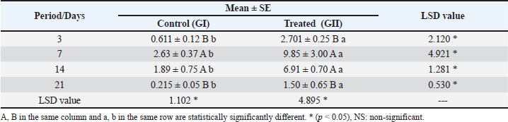
RT-PCR analysisThe mean values of VEGF gene levels are listed in Table 10. The levels of VEGF gene expression in the wound of the treaded group (GII) were found to be significantly (p < 0.05) upregulated on days 3, 7, 14, and 21 post-treatment, with levels of 1.91 ± 0.63, 4.01 ± 1.12, 12.21 ± 2.21, and 9.82 ± 1.65, respectively, relative to the wounds of the control group (GI), with levels of 0.130 ± 0.05, 0.632 ± 0.31, 2.61 ± 0.45 and 1.94 ± 0.60, respectively. The (TGF-β1) expression exhibited a statistically significant increase (p < 0.05) upregulation in the wounds of the treaded group (GII) on days 3, 7, 14, and 21 post-treatment (2.701 ± 0.25, 9.85 ± 3.00, 6.91 ± 0.70, and 1.50 ± 0.65, respectively) compared with those in the wounds of the control group (GI) (0.611 ± 0.12, 2.63 ± 0.37, 1.89 ± 0.75, and 0.215 ± 0.05, respectively) at the same periods (Table 11). Table 10. The mean values of VEGF in the wound site of groups at different time points.
Table 11. The mean values of TGF-β1 in wound site of groups at different time points.
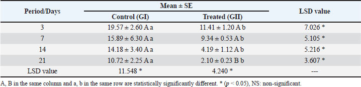
The current investigation demonstrated a significant (p < 0.05) upregulation of TNF-α) (Table 12) in the wounds of the control group (GI), with levels of 19.57 ± 2.60, 15.89 ± 6.30, 14.18 ± 3.40, and 10.72 ± 2.25 on days 3, 7, 14, and 21 post-treatment, respectively, in contrast to the treated group (GII), which exhibited levels of 11.41 ± 1.20, 9.34 ± 0.53, 4.19 ± 1.12, and 2.10 ± 0.23, respectively. Table 12. The mean values of TNF-α in wound site of the groups at different time points.
DiscussionIn the current study, the blood glucose levels at distinct time intervals (every 10 days) after partial pancreatectomy were clearly elevated in all animals, ranging from 267 to 320 Mg/dl. Animals were designated as diabetic if their blood glucose levels reached 250 mg/dl (Kim et al., 2014; Fekrazad et al., 2018). Macroscopically, the wound closure percentage values at the period of 3rd day revealed no discernible variations between the two groups. This finding aligns with a recent study by Marouf et al. (2025) that investigated the impact of various concentrations of BSE on excisional wounds and revealed no discernible variation between treated groups at day four post-treatment. This is often attributed to the beginning of the proliferative phase, which usually starts 2–3 days after wounding (Bartold and Ivanovski, 2025). This proposed interpretation is in line with that of a previous study (Cañedo-Dorantes and Cañedo-Ayala, 2019), which suggested that the onset of the proliferation phase marked the beginning of the wound closure. The obtained values related to the treated group at periods of 7th day, 14th day, and 21st day. This data indicated significant superiority (p < 0.05) for the treated group in relation to group I. This result was in accordance with Marouf et al. (2025), who investigated the impact of various concentrations of BSE on excisional wounds and attributed that to the activity of phytochemical contents of B. serrata extract, particularly 3-O-acetyl-11-keto-β-boswellic acid, which has antioxidant, antimicrobial, and anti-inflammatory effects (Maraghehpour et al., 2016 and Beghelli et al., 2017). In addition to inhibiting pro-inflammatory products (Gilbert et al., 2020). On the other hand, Khathayer and Ray (2020) indicated that fenugreek seeds reduce the expression of matrix metalloproteinase-2 (MMP2), which will lead to reduced collagen fiber degradation and enhanced healing of diabetic wounds (Alvarez et al., 2019). According to Sharma et al. (2016), fenugreek has antibacterial activity against Staphylococcus aureus, which frequently invades diabetic wounds. Furthermore, Ktari et al. (2017) determined that fenugreek seed extract enhances cell proliferation in diabetic wounds and accelerates re-epithelialization. Therefore, the aforementioned mechanisms may be responsible for the high wound closure rate observed in the present study. The histopathological findings of wounds in the treated group (GII) of the current study were interpreted as an acceleration of wound bed revascularization, modulation of inflammatory response, enhancement of collagen fiber formation and maturation, regeneration of epidermal tissue and hair follicles with improved re-epithelialization. This outcome aligns with Pengzong et al. (2019), who investigated the effectiveness of B. serrata extract on the healing of diabetic wounds and reported the reduction of inflammatory cell infiltration with increased revascularization of wound tissue with increased collagen synthesis, in addition to the suppression of oxidative-inflammatory markers. Alam et al. (2012) connected the anti-inflammatory activity of B. serrata extract to the different phytochemicals associated, including α, β, and γ boswellic acids. Accordingly, a recent study by Han et al. (2025) revealed that boswellic acids modulate the inflammatory response and inhibit fibroblast ferroptosis by targeting STAT3, thereby improving wound healing in diabetic rats. Furthermore, Namjou and Rouhi-Broujeni (2021), who investigated the healing of diabetic wounds after treatment with 2.5% B. serrata cream, indicated enhancement of wound size reduction with re-epithelialization. The reconstruction of damaged tissue indicated the deposition of high-quality collagen (Dong et al., 2022). On the other hand, Sumitra et al. (2000) found that applying a suspension of T. foenum-graecum seeds topically reduced the size of wounds and promoted re-epithelization. The researchers attributed these effects to the aldehyde content, which is responsible for the deposition and maturation of new collagen. The increase in the amount of collagen in wounds promotes tissue regeneration and enhances wound healing (Liu et al., 2023). In addition to its anti-inflammatory and antioxidant properties as a result of the existence of saponins, alkaloids, flavonoids, polysaccharides, and phenolic compounds (Aylanc et al., 2020; Tewari et al., 2020). In the current study, the mean values of VEGF gene expression in the wound of the treaded group (GII) were found to be significantly (p < 0.05) upregulated on days 3, 7, 14, and 21 post-treatment. In contrast to the wounds of the control group (GI). The expression of TGF-β1 demonstrated a considerable increase (p < 0.05) in the wounds of the treated group (GII) on days 3, 7, 14, and 21 post-treatment compared with those in the wounds of the control group (GI) at the same periods. The current investigation demonstrated a significant (p < 0.05) downregulation of TNF-α in the wounds of the treated group (GII) on days 3, 7, 14, and 21 post-treatment, in contrast to the wounds of the control group (GI). The in vivo investigation conducted by Pengzong et al. (2019) on the effects of B. serrata extract on diabetic wounds also supported the gene expression of the present study, which reflected promoting the TGF-β and VEGF together with inhibition of TNF-α and Bcl-2-associated X protein apoptosis (a crucial indicator of cellular apoptosis under demanding circumstances like oxidative stress and hyperglycemia). Another investigation by Li et al. (2018) revealed that the upregulation of TNF-α delayed healing, linked to an aggravated inflammatory reaction and suppression of TGF-β and thus inhibited type I collagen synthesis. Clinically, Brem and Tomic-Canic (2007) showed that hyperglycemia inhibits TGF-β expression, this analysis validated the histopathological findings of the current study. Cabral-Pacheco et al. (2020) indicated that excessive signaling of TNF-α suppresses the synthesis of tissue inhibitors of metalloproteinases and increases the synthesis of matrix metalloproteinases, thereby retarding wound healing. Additionally, the experimental study by Khathayer and Ray (2020) showed that the fenugreek seeds reduce the expression of MMP2. Furthermore, Bao et al. (2009) demonstrated that VEGF enhances the proliferation of fibroblasts and collagen production, leading to improved wound healing. Moreover, the extract of T. foenum graecum has anti-inflammatory effects and significantly decreased TNF-α (Suresh et al., 2012). Salimabad et al. (2023) investigated the effects of T. foenum graecum extract on the healing of diabetic wounds in rats. They observed modulation of inflammatory cells and enhanced neovascularization in the wound bed, which was attributed to the expression of VEGF. Otherwise, Diab et al. (2023) indicated that T. foenum graecum extract enhanced the expression of TGF-β. The simultaneous suppression of TNF-α and upregulation of VEGF and TGF-β reflects a complementary mechanistic interaction between the phytochemicals in BSE and TFGE. Boswellic acids, the main bioactive components of BSE, are known to inhibit nuclear factor kappa B pathway, a key transcriptional regulator of pro-inflammatory cytokines like TNF-α (Maouche et al., 2024). TNF-α expression also reduced inflammation-mediated inhibition of tissue repair pathways (Dichtl et al., 2022). Concurrently, the TFGE contains several bioactive substances, including flavonoids, steroidal saponins, and 4-hydroxyisoleucine, that activate antioxidant and anti-inflammatory signaling (Coban et al., 2025), as well as immunomodulatory and neovascularization enhancement (Salimabad et al., 2023). Collectively, these mechanisms point to a complementary approach in which anti-inflammatory suppression and proregenerative signaling simultaneously support angiogenesis and tissue remodeling. In accordance with the abovementioned findings and the results of gene analysis in the treated group of the present study, which displayed upregulation of VEGF, TGF-β, and downregulation of TNF-α, the diabetic wound of the group that received treatment revealed enhanced healing. ConclusionIn conclusion, the topical application of the B. serrata and T. foenum-graecum seed extracts revealed a superior effect in enhancing the healing of excisional wounds in diabetic animals by promoting VEGF and TGF-β expression and inhibiting TNF-α. This orchestration may result in revascularization augmentation, inflammatory response modulation, collagen fiber formation and maturation enhancement, epidermal tissue regeneration, and hair follicle regeneration with improved reepithelialization. AcknowledgmentsThe author expresses gratitude to the animal house personnel at the College of Veterinary Medicine, Tikrit University. Conflict of interestNo specific grant was obtained for this study. FundingNone. Authors' ContributionsThis manuscript is solely authored by Hammadi. Data availabilityAll data are available in the revised manuscript. ReferencesAlam, M., Khan, H., Samiullah, L. and Siddique, K.M. 2012. A review on phytochemical and pharmacological studies of Kundur (Boswellia serrata Roxb ex Colebr.)-A Unani drug. J. Appl. Pharm. Sci. 2012, 148–156. Allen, L. and Ansel, H.C. 2014. Ansel’s pharmaceutical dosage forms and drug delivery systems. Tenth edition. Philadelphia: Wolters Kluwer Health; p: 794, 2014. Alvarez, O.M., Markowitz, L., Onumah, N. and Wendelken, M. 2019. Swift Downregulation of Gelatinases (MMP-2, MMP-9) in Neuropathic Diabetic Foot Ulcers Treated with Total Contact Cast. Wounds 31(5), 39–41. Amin, N. and Doupis, J. 2016. Diabetic foot disease: from the evaluation of the 'foot at risk' to the novel diabetic ulcer treatment modalities. World. J. Diabetes. 7(7), 153–164. Aylanc, V., Eskin, B., Zengin, G., Dursun, M. and Cakmak, Y.S. 2020. In vitro studies on different extracts of fenugreek (Trigonella spruneriana BOISS.): phytochemical profile, antioxidant activity, and enzyme inhibition potential. J. Food Biochem. 44(11), e13463. Ayub, M.A., Hanif, M.A., Blanchfield, J., Zubair, M., Abid, M.A. and Saleh, M.T. 2023. Chemical composition and antimicrobial activity of Boswellia serrata oleo-gum-resin essential oil extracted by superheated steam. Nat. Prod. Res. 37(14), 2451–2456. Bao, P., Kodra, A., Tomic-Canic, M., Golinko, M.S., Ehrlich, H.P. and Brem, H. 2009. The role of vascular endothelial growth factor in wound healing. J. Surg. Res. 153(2), 347–358. Bartold, M. and Ivanovski, S. 2025. Biological processes and factors involved in soft and hard tissue healing. Periodontology. 2000. 97(1), 16–42. Beghelli, D., Isani, G., Roncada, P., Andreani, G., Bistoni, O., Bertocchi, M., Lupidi, G. and Alunno, A. 2017. Antioxidant and ex vivo immune system regulatory properties of Boswellia serrata extracts. Oxid. Med. Cell. Longev. 1(1), 7468064. Bournival, J., Francoeur, M.A., Renaud, J. and Martinoli, M.G. 2012. Quercetin and sesamin protect neuronal PC12 cells from high-glucose-induced oxidation, nitrosative stress, and apoptosis. Rejuvenation Res. 15(3), 322–333. Brem, H. and Tomic-Canic, M. 2007. Cellular and molecular basis of wound healing in diabetes. J. Clin. Invest. 117(5), 1219–1222. Cabral-Pacheco, G.A., Garza-Veloz, I., Castruita-de La Rosa, C., Ramirez-Acuña, J.M., Perez-Romero, B.A., Guerrero-Rodriguez, J.F., Martinez-Avila, N. and Martinez-Fierro, M.L. 2020. The Roles of Matrix Metalloproteinases and Their Inhibitors in Human Diseases. Int. J. Mol. Sci. 21(24), 9739. Cañedo-Dorantes, L. and Cañedo-Ayala, M. 2019. Skin acute wound healing: a comprehensive review. Int. J. Inflam. 1(1), 3706315. Carriel, V., Campos, A., Alaminos, M., Raimondo, S. and Geuna, S. 2017. Staining methods for normal and regenerative myelin in the nervous system. Methods Mol. Biol. 1560, 207–218. Chen, L., Zheng, Q., Chen, X., Wang, J. and Wang, L. 2019. Low-frequency ultrasound enhances vascular endothelial growth factor expression, thereby promoting the wound healing in diabetic rats. Exp. Ther. Med. 18(5), 4040–4048. Chevrier, M.R., Ryan, A.E., Lee, D.Y., -W.., Zhongze, M., Wu-Yan, Z. and Via. 2005. Boswellia carterii Extract Inhibits TH1 Cytokines and Promotes TH2 Cytokines in vitro. Clin. Vaccine Immunol. 12(5), 575–580. Coban, F., Ozer, H., Yilmaz, B. and Lan, Y. 2025. Characterization of bioactive compounds in fenugreek genotypes in varying environments: diosgenin, trigonelline, and 4-hydroxyisoleucine. Front. Plant. Sci. 16, 1562931. Diab, A.M., Al-Khefa, B.T., Khalafallah, M.M., Salah, A.S., Farrag, F.A. and Dawood, M.A.O. 2023. Dietary methanolic extract of fenugreek enhanced the growth, haematobiochemical, immune responses, and resistance against Aeromonas hydrophila in Nile Tilapia, Oreochromis niloticus. Aquac. Res. 1(1), 3055476. Dichtl, S., Sanin, D.E., Koss, C.K., Willenborg, S., Petzold, A., Tanzer, M.C., Dahl, A., Kabat, A.M., Lindenthal, L., Zeitler, L., Satzinger, S., Strasser, A., Mann, M., Roers, A., Eming, S.A., El Kasmi, K.C., Pearce, E.J. and Murray, P.J. 2022. Gene-selective transcription promotes the inhibition of tissue reparative macrophages by TNF. Life. Sci. Alliance. 5(4), e202101315. Dong, Q., Zu, D., Kong, L., Chen, S., Yao, J., Lin, J., Lu, L., Wu, B. and Fang, B. 2022. Construction of antibacterial nano-silver embedded bioactive hydrogel to repair infectious skin defects. Biomater. Res. 26(1), 36. Fekrazad, R., Sarrafzadeh, A., Kalhori, K.A., Khan, I., Arany, P.R. and Giubellino, A. 2018. Improved wound remodeling correlates with modulated TGF‐beta expression in skin diabetic wounds following combined red and infrared photobiomodulation treatments. Photochem. Photobiol. 94(4), 775–779. Gilbert, N.C., Gerstmeier, J., Schexnaydre, E.E., Börner, F., Garscha, U., Neau, D.B., Werz, O. and Newcomer, M.E. 2020. Structural and mechanistic insights into 5-lipoxygenase inhibition by natural products. Nat. Chem. Biol. 16(7), 783–790. Han, Z., Wu, W., Bai, Z., Xiu, Y. and Zhou, D. 2025. Beta-boswellic acid facilitates diabetic wound healing by targeting STAT3 and inhibiting ferroptosis in fibroblasts. Front. Pharmacol. 16, 1578625. Hasona, N.A., Ahmed, M.Q., Alghassab, T.A., Alghassab, M.A. and Alghabban, A.A. 2016. Antihyperlipidemic effect of pomegranate peel and Iranian fenugreek extracts on cholesterol-rich diet induced hypercholesterolemia in Guinea pigs. Merit. Res. J. Med. Sci. 4(4), 196–203. Hestehave, S., Munro, G., Christensen, R., Brønnum Pedersen, T., Arvastson, L., Hougaard, P. and Abelson, K.S.P. 2017. Is there a reasonable excuse for not providing post-operative analgesia when using animal models of peripheral neuropathic pain for research purposes?. PLos One. 12(11), e0188113. Hummadi, S. and Al-Falahi, N. 2024. Macroscopic effect of small intestine submucosa hydrogel-silver nanoparticles composite on healing of infected wounds. Adv. Anim. Vet. Sci. 12(2), 216–225; doi:10.17582/journal.aavs/2024/12.2.216.225 Jensen, E.C. 2012. Real‐time reverse transcription polymerase chain reaction to measure mRNA: use, limitations, and presentation of results. Anatomical Rec. Adv. Integr. Anat. Evol. Biol. 295(1), 1–3. Kara, M.E. 2005. The anatomical study on the rat pancreas and its ducts with emphasis on the surgical approach. Ann. Anat. 187(2), 105–112. Khathayer, F. and Ray, S.K. 2020. Diosgenin as a novel alternative therapy for inhibition of growth, invasion, and angiogenesis abilities of different glioblastoma cell lines. Neurochem. Res. 45(10), 2336–2351. Kim, T.H., Cho, H.Y. and Lee, S.M. 2014. High-voltage pulsed current stimulation enhances wound healing in diabetic rats by restoring the expression of collagen, α-smooth muscle actin, and TGF-β1. Tohoku. J. Exp. Med. 234(1), 1–6. Ktari, N., Trabelsi, I., Bardaa, S., Triki, M., Bkhairia, I., Ben Slama-ben Salem, R., Nasri, M. and Ben Salah, R. 2017. Antioxidant and hemolytic activities, and effects in rat cutaneous wound healing of a novel polysaccharide from fenugreek (Trigonella foenum-graecum) seeds. Int. J. Biol. Macromol. 95, 625–634. Kumar, L., Verma, R., Sharma, G., Nishad, A. and Sharma, S. 2023. Evaluation of Anti-Inflammatory Activity of Gum Resin Extract of Boswellia Serrata. Int. J. Pharm. Sci. Med. 8(4), 110–131. Laybutt, D.R., Glandt, M., Xu, G., Ahn, Y.B., Trivedi, N., Bonner-Weir, S. and Weir, G.C. 2003. Critical reduction in beta-cell mass results in two distinct outcomes over time. Adaptation with impaired glucose tolerance or decompensated diabetes. J. Biol. Chem. 278(5), 2997–3005. Li, W., Kandhare, A.D., Mukherjee, A.A. and Bodhankar, S.L. 2018. Hesperidin, a plant flavonoid, accelerated cutaneous wound healing in streptozotocin-induced diabetic rats: role of TGF-ß/Smads and Ang-1/Tie-2 signaling pathways. EXCLI. J. 17, 399. Liu, N., Zhu, S., Deng, Y., Xie, M., Zhao, M., Sun, T., Yu, C., Zhong, Y., Guo, R., Cheng, K., Chang, D. and Zhu, P. 2023. Construction of multifunctional hydrogel with metal-polyphenol capsules for infected full-thickness skin wound healing. Bioact. Mater. 24, 69–80. Maouche, A., Boumediene, K. and Baugé, C. 2024. Bioactive compounds in osteoarthritis: molecular mechanisms and therapeutic roles. Int. J. Mol. Sci. 25(21), 11656. Maraghehpour, B., Khayamzadeh, M., Najafi, S. and Kharazifard, M. 2016. Traditionally used herbalmedicines with antibacterial effect on Aggegatibacter actinomycetemcomitans: Boswellia serrata and Nigella sativa. J. Indian. Soc. Periodontol. 20(6), 603–607. Marouf, B.H., Ismaeel, D.O. and Abdulqader, G.M.R. 2025. Effect of Different Concentrations of Topically Applied Boswellia Serrata Extract on Excisional Wound Healing in Rats. Iraqi J. Pharm. Sci. 34(1), 72–84. Masoko, P., Picard, J. and Eloff, J.N. 2010. The use of a rat model to evaluate the in vivo toxicity and wound healing activity of selected Combretum and Terminalia (Combretaceae) species extracts. Onderstepoort J. Vet. Res. 77(1), 1–7. Muralidharan, P., Thenmozhi, M. and Prakash, R. 2016. Cell proliferative action of hydroalcoholic extract of Trigonella foenum graecum in rats. Int. J. Pharm. Sci. Res. 7(2), 708. Namjou, A. and Rouhi-Broujeni, H. 2021. Antihyperglycemic, antihyperlipidemic and wound healing of Boswellia serrata on experimentally induced diabetic rats. Abanico. Vet. 10(1), 1–17. Peikes, H., Morris, D.O. and Hess, R.S. 2001. Dermatologic disorders in dogs with diabetes mellitus: 45 cases (1986–2000). J. Am. Vet. Med. Assoc. 219(2), 203–208. Pengzong, Z., Yuanmin, L., Xiaoming, X., Shang, D., Wei, X., Zhigang, L., Dongzhou, D., Wenjing, Y., Jianbiao, Y., Yang, X. and Xia, L. 2019. Wound healing potential of the standardized extract of Boswellia serrata on experimental diabetic foot ulcer via inhibition of inflammatory, angiogenetic and apoptotic markers. Planta Med. 85(08), 657–669. Pennasilico, L., Serino, F., Galosi, M., Piccionello, A.P., Angorini, A., Dini, F. and Bella, C.D. 2025. Anesthetic effects of a mixture of xylazine, ketamine, and buprenorphine in laboratory rats subjected to short surgical procedures. Open Vet. J. 15(3), 1370. Salimabad, F., Fathi, A.N. and Babaei, S. 2023. Effect of hydroalcoholic extract of Trigonella foenum-graecum leaves on wound healing in type 1 diabetic rats. J. Wound. Care. 32(11), S24–S35. SAS. 2018. Statistical Analysis System, User's Guide. Statistical. Version 9.1th ed. Cary. N.C:SAS. Inst. Inc. Sharma, V., Singh, P. and Rani, A. 2016. Antimicrobial activity of Trigonella foenum-graecum L. (Fenugreek). Eur. Exp. Biol. 7(1), 1–4. Sultana, J., Molla, M.R., Kamal, M., Shahidullah, M., Begum, F. and Bashar, M.A. 2009. Histological differences in wound healing in maxillofacial region in patients with or without risk factors. Bangladesh. J. Pathol. 24(1), 3–8. Sumitra, M., Manikandan, P., Suguna, L. and Cehittar, G. 2000. Study of dermal wound healing activity of Trigonella foenum graceum seeds in rats. J. Clin. Biomed. Nutr. 28(2), 59–67. Suresh, P., Kavitha, C., Babu, S.M., Reddy, V.P. and Latha, A.K. 2012. Effect of ethanol extract of Trigonella foenum graecum (Fenugreek) seeds on Freund's adjuvant-induced arthritis in albino rats. Inflammation 35(4), 1314–1321. Tewari, D., Jóźwik, A., Łysek-Gładysińska, M., Grzybek, W., Adamus-Białek, W., Bicki, J., Strzałkowska, N., Kamińska, A., Horbańczuk, O.K. and Atanasov, A.G. 2020. Fenugreek (Trigonella foenum-graecum L.) seeds dietary supplementation regulates liver antioxidant defense systems in aging mice. Nutrients 12(9), 2552. Uckaya, I., Aragon-Sanchezc, J., Lewa, D. and Lipskya, B.A. 2015. Diabetic foot infections: what have we learned in the last 30 years?. Int. J. Infect. Dis. 40, 81–91. Underwood, W. and Anthony, R., 2020. AVMA guidelines for the euthanasia of animals: 2020 edition. 2013(30), pp.2020–1. Xu, F., Wu, J., Hu, Y., Chu, C., Liu, W., Li, X. and Xiao, W. 2023. Mechanisms of action underlying the effect of Tongsaimai on wound healing based on experimental and network pharmacology. J. Ethnopharmacol. 311, 116451. Yang, Y., Wang, F., Yin, D., Fang, Z. and Huang, L. 2015. Astragulus polysaccharide-loaded fibrous mats promote the restoration of microcirculation in/around skin wounds to accelerate wound healing in a diabetic rat model. Colloids Surf. B. Biointerfaces 136, 111–118. | ||
| How to Cite this Article |
| Pubmed Style Saddam Khalid Hummadi. Effect of Boswellia serrata and Trigonella foenum-graecum seed extracts on wound healing of partial pancreatectomy-induced diabetic rat. Open Vet. J.. 2026; 16(5): 3130-3143. doi:10.5455/OVJ.2026.v16.i5.54 Web Style Saddam Khalid Hummadi. Effect of Boswellia serrata and Trigonella foenum-graecum seed extracts on wound healing of partial pancreatectomy-induced diabetic rat. https://www.openveterinaryjournal.com/?mno=298649 [Access: June 26, 2026]. doi:10.5455/OVJ.2026.v16.i5.54 AMA (American Medical Association) Style Saddam Khalid Hummadi. Effect of Boswellia serrata and Trigonella foenum-graecum seed extracts on wound healing of partial pancreatectomy-induced diabetic rat. Open Vet. J.. 2026; 16(5): 3130-3143. doi:10.5455/OVJ.2026.v16.i5.54 Vancouver/ICMJE Style Saddam Khalid Hummadi. Effect of Boswellia serrata and Trigonella foenum-graecum seed extracts on wound healing of partial pancreatectomy-induced diabetic rat. Open Vet. J.. (2026), [cited June 26, 2026]; 16(5): 3130-3143. doi:10.5455/OVJ.2026.v16.i5.54 Harvard Style Saddam Khalid Hummadi (2026) Effect of Boswellia serrata and Trigonella foenum-graecum seed extracts on wound healing of partial pancreatectomy-induced diabetic rat. Open Vet. J., 16 (5), 3130-3143. doi:10.5455/OVJ.2026.v16.i5.54 Turabian Style Saddam Khalid Hummadi. 2026. Effect of Boswellia serrata and Trigonella foenum-graecum seed extracts on wound healing of partial pancreatectomy-induced diabetic rat. Open Veterinary Journal, 16 (5), 3130-3143. doi:10.5455/OVJ.2026.v16.i5.54 Chicago Style Saddam Khalid Hummadi. "Effect of Boswellia serrata and Trigonella foenum-graecum seed extracts on wound healing of partial pancreatectomy-induced diabetic rat." Open Veterinary Journal 16 (2026), 3130-3143. doi:10.5455/OVJ.2026.v16.i5.54 MLA (The Modern Language Association) Style Saddam Khalid Hummadi. "Effect of Boswellia serrata and Trigonella foenum-graecum seed extracts on wound healing of partial pancreatectomy-induced diabetic rat." Open Veterinary Journal 16.5 (2026), 3130-3143. Print. doi:10.5455/OVJ.2026.v16.i5.54 APA (American Psychological Association) Style Saddam Khalid Hummadi (2026) Effect of Boswellia serrata and Trigonella foenum-graecum seed extracts on wound healing of partial pancreatectomy-induced diabetic rat. Open Veterinary Journal, 16 (5), 3130-3143. doi:10.5455/OVJ.2026.v16.i5.54 |


