





| Research Article | ||
Open Vet. J.. 2026; 16(5): 2911-2921 Open Veterinary Journal, (2026), Vol. 16(5): -2921 Research Article Molecular detection and phylogenetic characterization of Leptospira interrogans in sheltered dogs from Iraq’s Kurdistan RegionHardi Fattah Marif*, Basim Abdulwahid Ali, Shadman Othman Husin, Sana Jamal Mohammed, Mahran Hssen Ali, and Sivan Othman AliDepartment of Clinic and Internal Medicine, College of Veterinary Medicine, University of Sulaimani, Sulaimaniyah, Iraq *Corresponding Author: Hardi Fattah Marif. Department of Clinic and Internal Medicine, College of Veterinary Medicine, University of Sulaimani, Sulaimaniyah, Iraq. Email: hardi.marif [at] univsul.edu.iq Submitted: 09/11/2025 Revised: 11/03/2026 Accepted: 18/03/2026 Published: 31/05/2026 © 2025 Open Veterinary Journal
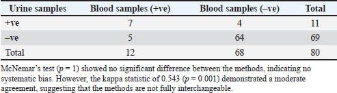
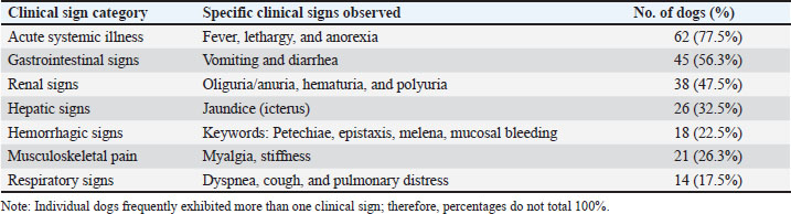
ABSTRACTBackground: Leptospirosis is a major worldwide health concern caused by pathogenic spirochetes from the genus Leptospira. Dogs are a significant reservoir host, particularly in shelter environments, where overcrowding and poor sanitation increase the risk of transmission. Aim: This study aimed to find Leptospira in protected dogs for the first time, define the leptospira molecularly, and perform a phylogenetic analysis to ascertain the similarities and differences in the sequenced gene among strains from nearby nations. Methods: Eighty blood and urine samples were collected from sheltered dogs under general anesthesia between May and August 2025. The samples were then subjected to DNA extraction and conventional polymerase chain reaction that targeted the conserved secY gene using phylogenetic analysis and sequencing. Results: Leptospirosis DNA was found in 23 samples (28.75%), including 11 urine samples (13.75%) and 12 blood samples (15%). Male dogs aged 2–3 years had the highest incidence of infection (23.1%), whereas female dogs had the highest rate (23.5%). Conclusion: These results highlight the critical need for comprehensive surveillance programs, enhanced biosecurity procedures, and vaccination plans in shelter settings throughout the Kurdistan Region. Keywords: Canine leptospirosis, Kurdistan Region, Molecular detection, SecY gene, Zoonosis. IntroductionLeptospirosis is a zoonotic disease caused by spirochetes of the Leptospira genus (Hagedoorn et al., 2023; Sykes et al., 2023; Suzuki et al., 2024). It is one of the common bacterial infections in human and animal populations worldwide. Genomic studies have established that the genus Leptospira is a heterogeneous collection of species, which can be grouped into three categories: pathogenic, intermediate, and saprophytic. Over 350 distinct serovars have been described in the pathogenic species (Goarant et al., 2022). Currently, 38 pathogenic species of Leptospira have been recognized, and ongoing studies are ongoing to identify new species (Harran et al., 2022). Pathogenic Leptospira are located within the renal tubules of host reservoirs, and they are most commonly shed by rodents through urine into the environment (Piredda et al., 2021). Pathogenic Leptospira can survive in moist soil and on the water surface for extended periods, possibly as long as several months (Smith et al., 2022; Ricardo et al., 2024; Sayanthi and Susanna, 2024). Humans can become infected through the urine of diseased animals or through exposure to contaminated soil and water (Da Costa et al., 2021; Mosquera et al., 2024). When exposed to contaminated environments, pathogenic Leptospira can enter humans and other animals through dermal and mucosal surfaces (Da Costa et al., 2021). Leptospirosis is a serious zoonotic disease that is found worldwide, causing approximately one million cases and close to 60,000 deaths each year (Da Costa et al., 2021; Cagliero et al., 2022; Mosquera et al., 2024). It mainly infects at-risk populations and livestock, causing a gross underestimation of the disease’s public health effects and a significant economic loss (Cagliero et al., 2022). Recent findings from Middle Eastern nations indicate variable prevalence and seroprevalence rates of leptospirosis among various hosts. In Iraq, dark-field microscopy and direct microscopic examination revealed a staggering prevalence of 94.3%. In Turkey, prevalence rates ranged from very low, such as 0.05% among humans, to as high as 74.4% in some populations based on the method of diagnosis used. In Iran, both low and very high levels of seroprevalence have been reported, such as 1.1% seroprevalence among humans and up to 71% among dogs, while bovine infections resolved up to 78.4%. These results suggest that although the prevalence of Leptospira infection varies among hosts and methods of diagnosis, it is clear that it extends throughout the region, and some regions also have very high levels of infection, leading to the need for further epidemiological and molecular research in surrounding nations (Harran et al., 2022). Dogs play a major role in leptospirosis transmission, especially in urban and suburban areas where humans and animals frequently interact (Piredda et al., 2021; Smith et al., 2022; Guzmán et al., 2023). When dogs come into contact with contaminated water and soil, they can become infected and subsequently shed Leptospira bacteria in their urine, contaminating the environment and putting other animals and humans at risk (Esteves et al., 2023). This cycle is a key factor in the persistence and spread of leptospirosis (Da Costa et al., 2021; Guzmán et al., 2023; Vyn et al., 2024). Stray dog populations, as well as those in shelters, are considered to be more susceptible to acquiring leptospirosis because of their higher exposure to pathogenic Leptospira in their environment. These populations had increased seroprevalence on a worldwide basis, thus posing a significant public health issue (Miotto et al., 2018). In recent times, there has been increasing evidence of a notable rise in dogs in shelters, especially after legislative changes that support sterilization and adoption over euthanasia as more humane measures for controlling stray dog populations. Thus, shelter overcrowding has emerged as a common issue, leading to compromised cleanliness and increased rodent infestation (Esteves et al., 2023; Ricardo et al., 2024). Many shelters have structural inadequacies and are confronted with resource constraints, often not adhering to developed guidelines for ensuring cleanliness during the intake and adoption procedures (Ricardo et al., 2024). The shelters in Sulaymaniyah face challenges similar to those in the rest of the country, where a large population of feral dogs is found in areas with limited resources and unhygienic sanitation. Such conditions promote the spread and multiplication of disease-causing pathogens. Despite the epidemiological importance of dog populations in shelters, molecular studies on leptospirosis have not yet been conducted in the Kurdistan Region of Iraq to determine the prevalence of leptospirosis in such canine populations. Lack of such a study means a serious gap in the knowledge of the ecological dynamics of the disease in the region, together with the zoonosis risks. The objective of this research is to detect Leptospira in dogs in a Kurdistan shelter for the first time, in addition to performing characterization and phylogenetic studies. The similarities and differences between the sequenced gene and those from neighboring countries will be further described in this study. The results of this study will be crucial for future prevention and control measures in shelter settings, local public health surveillance initiatives, and the advancement of scientific knowledge regarding leptospirosis epidemiology in the Middle East. Materials and MethodsCollection sampleBlood and urine samples were obtained from 80 suspected male and female sheltered dogs (80 blood and 80 urine samples) from the shelter of Sulaimani province in Razyana village (Fig. 1A and B) between 05/2025 and 8/2025 from the same animal according to several clinical signs (Table 5). The study area was located between 34 and 35° North and 45 and 46° East (Fig. 1). The district is characterized by four distinct seasons with rainfall ranging from 400 to 700 mm in autumn, winter, and early spring. This city is crucial as it is located on the border of Iran and is a large city. The shelter is moderately dense and near many villages and population, meaning it is not far from people. We chose this shelter because it is the only one in Sulaimaniyah City. The animals were classified into three age categories (1–2, 2–3, and >3 years old. Samples were collected from dogs under general anesthesia using a sterile syringe, blood samples were directly taken from the radial vein, and urine samples were taken from the urinary bladder. Samples were transported to the laboratory in a cooled container, and each sheltered dog’s age and sex were documented (Table 1). Table 1. Distribution of positive and negative test results.
Table 5. Clinical signs used to classify dogs with suspected leptospirosis (n=80).
Fig. 1. A: Geographical location of (Sulaimani location) in Sulaymaniyah province on the map, where blood and urine samples were collected from sheltered dogs for polymerase chain reaction (PCR) test (30). B: Razyana village is the exact place of shelter. DNA extractionGenomic DNA samples were extracted from whole blood and urine using the Genomic DNA Kit (Add-bio, Korea) according to the manufacturer’s instructions. This kit correctly separates whole genomic DNA using a silica membrane column method. The entire purified DNA was eluted with 50–100 μl) of an elution buffer and was kept at −20°C until use. Polymerase chain reactionThe secY gene was amplified using conventional polymerase chain reaction (PCR) with species-conserved primers: G1 (forward, CTG AAT CGC TGT ATA AAA GT) and G2 (reverse, GGA AAA CAA ATG GTC GGA AG) (De Abreu Fonseca et al., 2006; González et al., 2012). PCR was performed using Add Start Taq Master Mix (Add Bio, Korea). For amplification 285 bp primer sec Y gene, the reaction mix consisted of 10 µl of master mix, 5 µl of DNA template, 1 µl (10 pmol) each of forward and reverse primers, and 3 µl of ultra-pure water for a final volume of 20 µl. Amplification was performed using a Hervuan thermal cycler (USA). The cycling conditions were initial denaturation at 94°C for 30 seconds, followed by 40 cycles of denaturation at 94°C for 30 seconds, annealing at 60°C for 30 seconds, and extension at 72°C for 30 seconds. This was followed by a final extension at 72°C for another 5 minutes. PCR success was confirmed via agarose gel electrophoresis. Gel electrophoresisPCR amplicons for the secY gene were separated using 1% agarose gel in TAE 1 × buffer, containing safety dye. 10 µl of each PCR product and a standard 100 bp DNA ladder were loaded. Electrophoresis lasted for 30 minutes at 150 V, and DNA bands were visualized using a UV transilluminator. Sequencing and alignmentPositive PCR products were purified and sequenced bidirectionally. For comparative analysis, unprocessed sequences were edited and trimmed and then aligned using ClustalW in MEGA X software (Saitou and Nei, 1987). Phylogenetic analysisPhylogenetic relationships were reconstructed using the Neighbor-Joining with the Kimura two-parameter model (Kimura, 1980; Felsenstein, 1985), implemented in MEGA X. The robustness of the tree topology was assessed with 1,000 bootstrap replications. Statistical analysisAll data were analyzed using IBM SPSS statistics (Version ‘27.0’, Windows). Descriptive statistics are presented as frequencies and percentages. Chi-square tests were used to assess associations between categorical variables (e.g., sex and blood sample result). When the expected cell numbers were few, Fisher’s exact test was applied (e.g., categories of age and blood sample results). The degree of agreement between the diagnostic methods was examined using McNemar’s test and Cohen’s kappa statistic with corresponding 95% confidence intervals. Statistical significance was set at p < 0.05. Ethical approvalAll procedures involving animal subjects were conducted in accordance with local ethical committee laws and regulations regarding the care and use of animals in research. The study adhered to the ethical standards for veterinary research at the College of Veterinary Medicine, University of Sulaimani (Ethic, commit. A-125). The ethical approval date was 15/06/2025. ResultsOf the 80 urine and blood samples, Leptospira species DNA was detected in 23 samples. Twelve positive blood samples and 11 positive urine samples were observed in all samples (Table 1). Positive samples produced an amplicon of approximately 545 bp, consistent with the targeted secY fragment (Fig. 2).
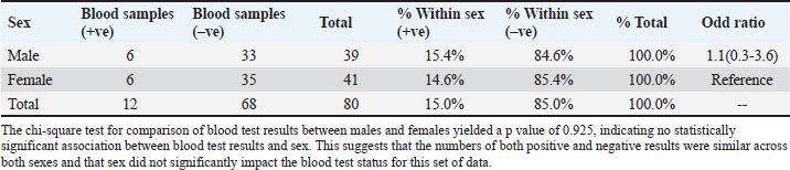
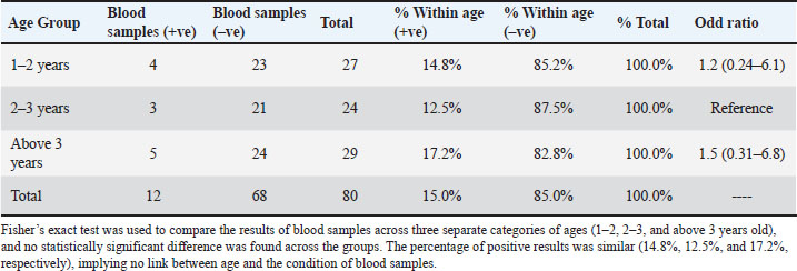
Fig. 2. Gel electrophoresis of PCR products amplified using secY gene primers targeting Leptospira spp. A 285 bp band indicates a positive result. Lanes 3 and 5 show clear amplification of the expected 285-bp band, confirming the presence of Leptospira DNA. The Lane M DNA ladder (100 bp marker) was used for MW estimation. Lanes 1 and 2: negative samole, Lane 4: negative control. The prevalence of Leptospira spp. in relation to age and sex (Table 4) showed that the suspected dogs were divided into three age groups (1–2, 2–3, and >3 years old), with 39 males and 41 female dogs. A higher infection rate (17.2%) was observed in suspected dogs aged >3 years, whereas a lower infection rate (12.5%) was observed in those aged 2–3 years old (Tables 2–4). Table 2. Blood sample results between males and females and the percent positivity of leptospirosis in suspected sheltered dogs related to sex with the odd ratio.
Table 3. Blood sample outcomes across the three age groups with the odd ratio.
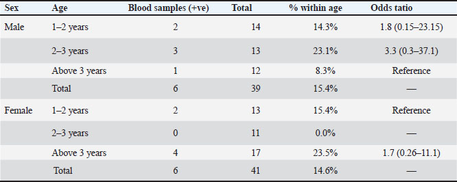
Table 4. Percent positivity of leptospirosis in suspected sheltered dogs according to sex and age with the odd ratio.
The results of the study demonstrated that the infection with Leptospira spp. differed based on the sex and age of the animals; therefore, leptospirosis was slightly higher in male dogs, with those aged 2–3 years having the highest infection rate (23.1%), OR: 3.3 [0.3–37.1], and male dogs above 3 years old having the lowest infection rate (8.3%) (Table 4). Female dogs above 3 years old have the highest percentage of infection, which is 23.5% [OR: 1.7 (0.26–11.1)], while female dogs between 2 and 3 years old have the lowest percentage of infection, which is 0.0% (Tables 4 and 2). Sequence confirmationBLAST analysis of the model amplicons revealed 99% similarity with the sequences of Leptospira interrogans in GenBank. The sequences obtained for Leptospira isolates in this study have also been uploaded to GenBank and can be accessed under the following accession numbers: PX250983.1 and PX250984.1. Leptospira DNA detection in urine and blood indicates active or recent infection. Blood positivity indicates early-stage bacteremia, whereas urine positivity indicates renal colonization and bacterial shedding. The results indicate the potential of Leptospira for zoonotic transmission from dogs to humans. Phylogenetic tree analysisPhylogenetic reconstruction based on the partial secY gene revealed that both field isolates (BH/2025 and BH2/2025) clustered closely within the L. interrogans clade. These two strains, grouped in the same branch (99%), are identical, indicating a high degree of genetic relatedness. The clustering pattern suggests that the isolates derived from whole blood and urine samples of dogs represent the same or very closely related L. interrogans (Fig. 3).
Fig. 3. Neighbor-joining phylogenetic tree of the secY gene sequences from L. interrogans. Field isolates from dog blood and urine (red circles) clustered within the L. interrogans lineage. Bootstrap values (1,000 replicates) are shown for the major nodes. The reference sequences were obtained from GenBank and annotated with their accession numbers. The field isolates (BH/2025 and BH2/2025) were positioned near other L. interrogans reference strains, including serovar Canicola (CP197415.1, PV251996.1) and isolates (TH-60-PQ376782 and LW-2-PQ376775), with the highest identities being 99%, which are recognized globally as pathogenic serovars associated with canine infections. Owing to the limitations of our study, we only sequenced a partial sequence of secY. Therefore, we cannot conclusively confirm that the strain belongs to the Canicola serovar. The close relationship with these strains indicates that the circulating genotypes in the study area may be closely related to canine-adapted serovars such as Canicola. The close association of these field strains, supported by bootstrap values, demonstrates that systemic infection detected in the bloodstream corresponds to the same strain subsequently shed in urine. This confirms the link between acute bacteremia and renal colonization in dogs and highlights their role as pathogenic Leptospira carriers and environmental shedders. The alignment demonstrates that the PX250983.1-BH/2025 and PX250984.1-BH/2/2025 field isolates share a high degree of sequence identity with the reference strains. No major substitutions or deletions were observed in the analyzed segment (positions 1–95), indicating the secY gene region was conserved (Fig. 4).
Fig. 4. Multiple sequence alignment of the secY gene from Leptospira isolates. Field strains (PX250983.1-BH/2025) and (PX250984.1-BH/2/2025) were compared with the reference sequences. Alignment was performed using ClustalW in MEGA X. Dots indicate residues identical to the consensus sequence. Both field strains showed high sequence similarity and conserved amino acid motifs across the region analyzed. Strong sequence conservation highlights secY gene’s reliability as a molecular marker for species identification and phylogenetic studies in Leptospira. Moreover, the close similarity between the field isolates and reference strains supports their placement within pathogenic Leptospira lineages. DiscussionThis research is the first molecular identification of Leptospira in dogs from the Sulaymaniyah province, Kurdistan, Iraq. Out of the 80 dogs tested (urine and blood), 23 samples (28.75%) were found positive for Leptospira species DNA using PCR with secY gene targeting, including 12 positive blood samples (15%) and 11 positive urine samples (13.75%). Phylogenetic analysis showed that the isolates from the field were closely clustered with L. interrogans serovar Canicola, with a 99.65% similarity with reference strains. Leptospira DNA detection in urine and blood samples is proof of active or recent infection in shelter dog groups. Blood positivity is evidence of early bacteremia, and urine positivity is evidence of renal colonization and active shedding of pathogenic Leptospira spp. in the environment (Mosquera et al., 2024). This dual-detection profile confirms that infected dogs pose an enormous risk of zoonotic transmission to shelter workers, vets, and the public at large. The prevalence established in our research, 28.75% in general, is consistent with data from earlier research from surrounding nations and Iraq. In Iraq, an earlier study tested 218 dogs and discovered that 37 (16.97%) were seropositive upon testing with indirect Enzyme-Linked Immunosorbent Assay on stray dogs from the governorates of Baghdad, Al-Qadisiyah, and Dhi-Qar. Of the 37 seropositive dogs, only 5 (13.51%) were confirmed positive by PCR, representing only 2.3% of all tested dogs. The high prevalence we recorded might be due to the use of PCR detection directly as compared to serological screening, because PCR can detect active infections regardless of antibody status. Regional research from other Middle Eastern countries shows appreciable variability. For example, in Turkey, surveys identified a substantially high seroprevalence of 43.96% in free-roaming dogs in Ankara using the microscopic agglutination test, with Bratislava being predominant instead of Canicola. In addition, another study in Iran estimated an even higher prevalence of 71% among stray dogs from the North Khorasan province, with L. interrogans Grippotyphosa being the most dominant serovar. Variation in prevalence from study to study may also have been explained by an assortment of factors. Detection rates are strongly influenced by the diagnostic assays applied; serological tests disclose past infection, and PCR identifies active infection. The diagnostic strategy applied sequentially in the present study from Iraq may have resulted in an underestimate of actual prevalence due to the suboptimal sensitivity of serological screening (Alfattli and Al-Mohamed, 2017). Geographic and climatic factors are also prominent contributors. The distinct rainy seasons of the Kurdistan Region (700–400 mm from autumn to early spring) create favorable conditions for the survival of Leptospira in the environment. Living conditions, such as overcrowding, poor hygiene, and infestation with rats, which were typically mentioned during our study, possibly boost transmission more effectively than freely moving populations of strays studied elsewhere. The predominance of serovar Canicola in our study differs from the Turkish results, wherein Bratislava was dominant (Aslantaş et al., 2005), and the Iranian results, wherein Grippotyphosa was dominant (Arzamani et al., 2022). This serovar distribution may reflect different reservoir host populations and environmental conditions specific to each region. (Khamesipour et al., 2014). Brucella sp. and Leptospira sp. detection in dogs using conventional PCR. The study was performed in Iran, and the result was 19.15% in dogs by using PCR. Moreover, the results of other studies were higher (22%; Zakeri et al., 2010). Leptospira wolffii, a potential new pathogenic Leptospira species detected in humans, sheep, and dogs. This fluctuation in positive results of PCR from years to years and this variation due to environmental factors, time of sampling depending on studies, the peak of leptospirosis in late of summer to fall or owner consciousness and for these reasons more tests may be proposed for a broader range of potential cases. The prevalence and epidemiology of leptospirosis have been investigated through many studies worldwide. Many studies worldwide investigated the epidemiology and prevalence of leptospirosis in dogs. Alterations in seroprevalence and infection rates of leptospirosis are intensely determined by environmental factors, including high rainfall, flooding, natural disasters, population growth, urbanization, and poor sanitation and hygiene (Khalili et al., 2020). Evaluation of the diagnostic value of a PCR assay compared with a serologic micro-agglutination test for canine leptospirosis (Martin et al., 2022). However, the results of our study are in agreement with those of Miotto et al. (2018). The sensitivity of PCR was 100%, and the specificity was 88.3%. Testing a Leptospiral-specific PCR assay with urine samples from dogs with clinical suspicion of leptospirosis. It is important to emphasize that the application of PCR in a clinical setting, notably when using urine specimens, may provide false-positive results, and the association with DNA sequencing, as performed in the present study, is highly recommended to provide more reliable results for proper comparison with other diagnostic techniques. Most dogs diagnosed by PCR and DNA sequencing were identified using urine samples, and even though the detection of Leptospiral DNA in urine specimens taken from dogs with multiple signs of leptospirosis is highly suggestive of acute Leptospiral infection, it may not distinguish dogs with acute infection from those with chronic renal carriage of leptospires associated with other underlying diseases causing clinical manifestations similar to leptospirosis. Instead, the detection of Leptospiral DNA from blood and serum samples must be considered a more reliable strategy for diagnosing acute leptospirosis in dogs using molecular methods. Our results also showed no distinction in laboratory findings and clinical outcomes between confirmed and non-confirmed cases of leptospirosis, highlighting the non-specificity of clinical and laboratory evaluations for the diagnosis of the disease. Our findings indicated that the infection rates in dogs over the age of 3 years were significantly higher (17.2%) than in younger animals, which corresponds with patterns identified in the Iraqi study, where dogs aged 2 years or younger demonstrated greater seropositivity (24.16%) than their older counterparts (Alfattli and Al-Mohamed, 2017). Similarly, the Turkish study found age-related differences, exhibiting statistical significance in different age groups (Aslantaş et al., 2005). This trend most likely portrays the aggregated exposure risk over time, coupled with the consequent development of immunity in some animals. The modest prevalence found in males (15.4% vs. 14.6% in females) also confirms the findings from the Iraqi study (19.4% vs. 15.89%) (Alfattli and Al-Mohamed, 2017). This finding is likely due to behavioral variations, as males tend to move around more and come across pathogen-contaminated places (Ladd et al., 2023). Characterization of the L. interrogans serovar Canicola from dog shelters is particularly noteworthy considering the amount of adaptation within canine hosts, which itself is a critical zoonosis threat. In comparison to the Turkish study, wherein numerous serovars were identified, with Bratislava being the most dominant (Aslantaş et al., 2005), the marked dominance of Canicola within our dataset suggests a possibly increased risk for person-to-person transmission to people who come into contact with these dogs. The shelter environment provides unique epidemiological challenges not wholly addressed in studies aimed at free-ranging stray aggregations. Closely confined animal environments, communal water, and frequent human contact create intense transmission opportunities. Leptospirosis is a zoonosis that can spread through direct or indirect contact with the urine or blood of an infected animal, as well as through contaminated water, soil, and mud. In addition to dogs, rodents, farm livestock, pigs, horses, and wild animals can be transmission sources (Wang and Dunn, 2025). A variety of preventative activities are recommended to reduce the hazard of leptospirosis to humans. Vaccination of domestic livestock and companion animals with available bacterin vaccines effectively reduces urinary shedding, and consequently, the opportunity for transmission diminishes. In addition, it is important to adhere to strict hygiene practices and to use protective procedures in animal contact or when working under damp and contaminated conditions. In conclusion, efficient rodent control is an integral part of prevention, given the prominent role of rodents as Leptospira reservoirs (Sykes et al., 2022). ConclusionThis study offers the first molecular confirmation of L. interrogans serovar Canicola among shelter dogs in Sulaymaniyah province, with a prevalence of 28.75%, suggesting a significant zoonotic risk. While highlighting particular difficulties inherent to shelter settings, our results conform to regional patterns of canine leptospirosis. The prevalence and confirmation of the Canicola serovar in blood and urine suggest the active infection and environmental shedding of the organism, underscoring the need for prompt implementation of preventative and control measures in shelter management. The establishment of targeted surveillance and intervention efforts to safeguard the health of people and animals in the Kurdistan Region depends on these initial data. AcknowledgmentsWe would like to thank the Veterinary Teaching Hospital at the College of Veterinary Medicine, University of Sulaymaniyah, Sulaymaniyah, for their support in conducting and processing all blood and urine samples. Conflict of interestThe authors have no conflicts of interest related to this work. FundingNo funding was received for this study. Authors’ contributionsConceptualization: Hardi Fattah Marif. Data Curation: Hardi Fattah Marif, Basim Abdulwahid Ali, Sana Jamal Mohammed, and Shadman Othman Husin. Funding acquisition: Hardi Fattah Marif, Basim Abdulwahid Ali, Shadman Othman Husin, Sana Jamal Mohammed, Mahran Hssen Ali, Sivan Othman Ali. Methodology: Hardi Fattah Marif, Basim Abdulwahid Ali, Shadman Othman Husin. Project administration and supervision: Hardi Fattah Marif and Basim Abdulwahid Ali. Validation: Hardi Fattah Marif, Basim Abdulwahid Ali, Shadman Othman Husin, and Sana Jamal Mohammed. Visualization: Mahran Hssen Ali and Sivan Othman Ali. Original draft: Hardi Fattah Marif, Basim Abdulwahid Ali, Shadman Othman Husin, Sana Jamal Mohammed. Hardi Fattah Marif, Shadman Othman Husin, Sana Jamal Mohammed: Writing, review, and editing. Data availabilityThe generated sequence data have been deposited in GenBank (accession numbers: PX250983.1 and PX250984.1). All other data supporting the findings are available upon reasonable request from the corresponding author. ReferencesAlfattli, H.H.H. and Al-Mohamed, S.A.A. 2017. First serological and molecular detection of Leptospira interrogans serovar canicola bacteria in dogs in some Iraqi governorates. J. Coll. Educ. 1(28), 621–636; doi:10.31185/eduj.Vol1.Iss28.28 Arzamani, K., Abdollahpour, G., Azimian, A., Van Belkum, A. and Ghasemzadeh-Moghaddam, H. 2022. High-level seroprevalence against Leptospira interrogans serovars among wild foxes, jackals and stray dogs in the North Khorasan Province, Iran. Vet. Med. Sci. 8(5), 1915–1921; doi:10.1002/vms3.890 Aslantaş, O., Özdemir, V., Kiliç, S. and Babür, C. 2005. Seroepidemiology of leptospirosis, toxoplasmosis, and leishmaniosis among dogs in Ankara, Turkey. Vet. Parasitology 129(3-4), 187–191; doi:10.1016/j.vetpar.2004.11.037 Cagliero, J., Vernel-Pauillac, F., Murray, G., Adler, B., Matsui, M. and Werts, C. 2022. Pathogenic leptospires limit dendritic cell activation through avoidance of TLR4 and TRIF signaling. Front. Immunol. 13, 911778; doi:10.3389/fimmu.2022.911778 Centers for Disease Control and Prevention. Atlanta, Georgia: Centers for Disease Control and Prevention, 2025. Available via https://www.cdc.gov/leptospirosis/about/index.html De Abreu Fonseca, C., Teixeira De Freitas, V.L., Caló Romero, E., Spinosa, C., Arroyo Sanches, M.C., Da Silva, M.V. and Shikanai-Yasuda, M.A. 2006. Polymerase chain reaction in comparison with serological tests for early diagnosis of human leptospirosis. Trop. Med. Int. Health. 11(11), 1699–1707; doi:10.1111/j.1365-3156.2006.01727.x Esteves, S.B., Santos, C.M., Silva, B.C.S., Salgado, F.F., Guilloux, A.G.A., Cortez, A., Lucco, R.C. and Miotto, B.A. 2023. Time for change? A systematic review with meta-analysis of leptospires infecting dogs to assess vaccine compatibility in Brazil. Prev. Vet. Med. 213, 105869; doi:10.1016/j.prevetmed.2023.105869 Felsenstein, J. 1985. Confidence limits on phylogenies: an approach using the bootstrap. Evolution 39, 783–791; doi:10.1111/j.1558-5646.1985.tb00420.x Goarant, C., Adler, B. and De La Peña Moctezuma, A. 2022. Leptospira.In Pathogenesis of Bacterial Infections in Animals. Adler Hoboken, NJ: Wiley, pp: 502–27; 10.1002/9781119754862.ch23 González, F.H.D., Oliveira, S.T., Messick, J.B., Biondo, A.W., Dos Santos, A.P., Stedile, R., Dalmolin, M.L., De Sá Guimaraes, A.M., Mohamed, A.S. and Riediger, I.N. 2012. Exposure to Leptospira spp. in sick dogs, shelter dogs and stray dogs. Vet. Clin. Pathol. 41, 772–776. Guzmán, D.A., Diaz, E., Sáenz, C., Álvarez, H., Cueva, R., Zapata-Ríos, G., Prado-Vivar, B., Falconí M., Pearson, T. and Barragán, V. 2023. Domestic dogs in indigenous Amazonian communities: key players in Leptospira cycling and transmission? BioRxiv. 09.19.558554; doi: 10.1101/2023.09.19.558554 Hagedoorn, N.N., Maze, M.J., Carugati, M., Cash-Goldwasser, S., Allan, K.J., Chen, K., Cossic, B., Demeter, E., Gallagher, S., German, R., Galloway, R.L., Habuš, J., Rubach, M.P., Shiokawa, K., Sulikhan, N. and Crump, J.A. 2023. Global distribution of Leptospira serovar isolations and detections from animal host species: a systematic review and online database. medRxiv. 10 (03), 23296503; doi: 10.1101/2023.10.03.23296503 Harran, E., Hilan, C., Djelouadji, Z. and Ayral, F. 2022. Epidemiology of leptospirosis: the first literature review of the neglected disease in the Middle East. Trop. Med. Infect. Dis. 7(10), 260; doi:10.3390/tropicalmed7100260 Khalili, M., Sakhaee, E., Bagheri Amiri, F., Asadabadi Safat, A., Afshar, D. and Esmaeili, S. 2020. Serological evidence of leptospirosis in Iran; a systematic review and meta-analysis. Microb. Pathog. 138, 103833; doi:10.1016/j.micpath.2019.103833 Khamesipour, F., Doosti, A., Emadi, M.F. and Awosile, B. 2014. Detection of Brucella sp. and Leptospira sp. in dogs using conventional polymerase chain reaction. J. Vet. Res. 58(4), 527–531. Kimura, M. 1980. A simple method for estimating evolutionary rates of base substitutions through comparative studies of nucleotide sequences. J. Mol. Evol. 16(12), 111–120; doi:10.1007/bf01731581 Ladd, R., Meek, P., Eames, J.C. and Leung, L.K.P. 2023. Activity range and patterns of free-roaming village dogs in a rural Cambodian village. Wildlife Res. 51(1), WR23024; doi:10.1071/WR23024 Martin, E.A., Heseltine, J.C. and Creevy, K.E. 2022. The evaluation of the diagnostic value of a PCR assay when compared to a serologic micro-agglutination test for canine leptospirosis. Front. Vet. Scie. 9, 815103. Maphill. 2013. Team: Savanna style panoramic map of Sulaymaniyah. Maphill. Available via http:// www.maphill.com/iraq/sulaymaniyah/panoramicmaps/savanna-style-map/(Accessed 13 October2025) Miotto, B.A., Guilloux, A.G.A., Tozzi, B.F., Moreno, L.Z., Da Hora, A.S., Dias, R.A., Heinemann, M.B., Moreno, A.M., Filho, A.F.D.S., Lilenbaum, W. and Hagiwara, M.K. 2018. Prospective study of canine leptospirosis in shelter and stray dog populations: identification of chronic carriers and different Leptospira species infecting dogs. PLos One 13(7), e0200384; doi:10.1371/journal.pone.0200384 Miotto, B.A., Tozzi, B.F., Penteado, M.D.S., Guilloux, A.G.A., Moreno, L.Z., Heinemann, M.B. and Hagiwara, M.K. 2018. Diagnosis of acute canine leptospirosis using multiple laboratory tests and characterization of the isolated strains. J. Clin. Biol. Physiol. 14(1), 222. Mosquera, P., Mejia, L., Ortiz, G., Pazmino, G., Pearson, T., Barragan, V. and Trueba, G. 2024. Mixed Leptospira infections in domestic animals from a rural community with high leptospirosis endemicity. PLoS One 19(10), e0312556; doi:10.1371/journal.pone.0312556 Piredda, I., Ponti, M.N., Piras, A., Palmas, B., Pintore, P., Pedditzi, A. and Chisu, V. 2021. New insights on Leptospira infections in a canine population from North Sardinia, Italy: a sero-epidemiological study. Biology 10(6), 507; doi:10.3390/biology10060507 Ricardo, T., Azócar-Aedo, L.I., Previtali, M.A. and Monti, G. 2024. Seroprevalence of pathogenic Leptospira serogroups in asymptomatic domestic dogs and cats: systematic review and meta-analysis. Front. Vet. Sci. 11, 1301959; doi:10.3389/fvets.2024.1301959 Saitou, N. and Nei, M. 1987. The neighbor-joining method: a new method for reconstructing phylogenetic trees. Mol. Biol. Evol. 4(4), 406–425; doi:10.1093/oxfordjournals.molbev.a040454 Sant’anna Da Costa, R., N. Di Azevedo, M.I., Dos Santos Baptista Borges, A.L., Carvalho-Costa, F.A., Martins, G. and Lilenbaum, W. 2021. Persistent high leptospiral shedding by asymptomatic dogs in endemic areas triggers a serious public health concern. Animals 11(4), 937; doi:10.3390/ani11040937 Sayanthi, Y. and Susanna, D. 2024. Pathogenic Leptospira contamination in the environment: a systematic review. Infect. Ecol. Epidemiol. 14(1), 2324820; doi:10.1080/20008686.2024.2324820 Smith, A.M., Stull, J.W. and Moore, G.E. 2022. Potential drivers for the re-emergence of canine leptospirosis in the United States and Canada. Trop. Med. Infect. Dis. 7(11), 377; doi:10.3390/tropicalmed7110377 Suzuki, R., Suzuki, S., Sato, O.S., Alfonso, R., Kitalong, C., Nwe, Y.Y., Takabe, K., Morita, M., Akeda, Y. and Koizumi, N. 2024. Bacteriological and serological investigation of leptospirosis in dogs and pigs in Palau. Am. J. Trop. Med. Hyg. 111(2), 246–250; doi:10.4269/ajtmh.24-0129 Sykes, J.E., Francey, T., Schuller, S., Stoddard, R.A., Cowgill, L.D. and Moore, G.E. 2023. Updated ACVIM consensus statement on leptospirosis in dogs. J. Vet. Intern. Med. 37(6), 1966–1982; doi:10.1111/jvim.16903 Sykes, J.E., Haake, D.A., Gamage, C.D., Mills, W.Z. and Nally, J.E. 2022. A global one health perspective on leptospirosis in humans and animals. J. Am. Vet. Med. Assoc. 260(13), 1589–1596; doi:10.2460/javma.22.06.0258 Vyn, C.M., Libera, K.C., Jardine, C.M. and Grant, L.E. 2024. Canine leptospirosis: a One Health approach for improved surveillance, prevention, and interdisciplinary collaboration. Can. Vet. J. 65, 609–612. Wang, S. and Dunn, N. 2025. Leptospirosis. In: StatPearls, Treasure Island, FL: StatPearls Publishing. Available via https://www.ncbi.nlm.nih.gov/books/NBK441858/. Zakeri, S., Khorami, N., Ganji, Z.F., Sepahian, N., Malmasi, A.A., Gouya, M.M. and Djadid, N.D. 2010. Leptospira wolffii, a potential new pathogenic Leptospira species detected in human, sheep and dog. Inf. Gen. Evol. 10, 273–277 | ||




| How to Cite this Article |
| Pubmed Style Marif HF, Ali BA, Husin SO, Mohammed SJ, Ali MH, Ali SO. Molecular detection and phylogenetic characterization of Leptospira interrogans in sheltered dogs from Iraq’s Kurdistan Region. Open Vet. J.. 2026; 16(5): 2911-2921. doi:10.5455/OVJ.2026.v16.i5.33 Web Style Marif HF, Ali BA, Husin SO, Mohammed SJ, Ali MH, Ali SO. Molecular detection and phylogenetic characterization of Leptospira interrogans in sheltered dogs from Iraq’s Kurdistan Region. https://www.openveterinaryjournal.com/?mno=295449 [Access: June 26, 2026]. doi:10.5455/OVJ.2026.v16.i5.33 AMA (American Medical Association) Style Marif HF, Ali BA, Husin SO, Mohammed SJ, Ali MH, Ali SO. Molecular detection and phylogenetic characterization of Leptospira interrogans in sheltered dogs from Iraq’s Kurdistan Region. Open Vet. J.. 2026; 16(5): 2911-2921. doi:10.5455/OVJ.2026.v16.i5.33 Vancouver/ICMJE Style Marif HF, Ali BA, Husin SO, Mohammed SJ, Ali MH, Ali SO. Molecular detection and phylogenetic characterization of Leptospira interrogans in sheltered dogs from Iraq’s Kurdistan Region. Open Vet. J.. (2026), [cited June 26, 2026]; 16(5): 2911-2921. doi:10.5455/OVJ.2026.v16.i5.33 Harvard Style Marif, H. F., Ali, . B. A., Husin, . S. O., Mohammed, . S. J., Ali, . M. H. & Ali, . S. O. (2026) Molecular detection and phylogenetic characterization of Leptospira interrogans in sheltered dogs from Iraq’s Kurdistan Region. Open Vet. J., 16 (5), 2911-2921. doi:10.5455/OVJ.2026.v16.i5.33 Turabian Style Marif, Hardi Fattah, Basim Abdulwahid Ali, Shadman Othman Husin, Sana Jamal Mohammed, Mahran Hssen Ali, and Sivan Othman Ali. 2026. Molecular detection and phylogenetic characterization of Leptospira interrogans in sheltered dogs from Iraq’s Kurdistan Region. Open Veterinary Journal, 16 (5), 2911-2921. doi:10.5455/OVJ.2026.v16.i5.33 Chicago Style Marif, Hardi Fattah, Basim Abdulwahid Ali, Shadman Othman Husin, Sana Jamal Mohammed, Mahran Hssen Ali, and Sivan Othman Ali. "Molecular detection and phylogenetic characterization of Leptospira interrogans in sheltered dogs from Iraq’s Kurdistan Region." Open Veterinary Journal 16 (2026), 2911-2921. doi:10.5455/OVJ.2026.v16.i5.33 MLA (The Modern Language Association) Style Marif, Hardi Fattah, Basim Abdulwahid Ali, Shadman Othman Husin, Sana Jamal Mohammed, Mahran Hssen Ali, and Sivan Othman Ali. "Molecular detection and phylogenetic characterization of Leptospira interrogans in sheltered dogs from Iraq’s Kurdistan Region." Open Veterinary Journal 16.5 (2026), 2911-2921. Print. doi:10.5455/OVJ.2026.v16.i5.33 APA (American Psychological Association) Style Marif, H. F., Ali, . B. A., Husin, . S. O., Mohammed, . S. J., Ali, . M. H. & Ali, . S. O. (2026) Molecular detection and phylogenetic characterization of Leptospira interrogans in sheltered dogs from Iraq’s Kurdistan Region. Open Veterinary Journal, 16 (5), 2911-2921. doi:10.5455/OVJ.2026.v16.i5.33 |


