





| Research Article | ||
Open Vet. J.. 2026; 16(5): 2922-2928 Does the spleen regenerate in rabbits after partial splenectomy?Asmaa Bouknine1*, Mira Chiraoui2, Abdelhak Bouknine3, Hemida Houari2 and Sidi Ali Selmani11Institute of Veterinary Science, University of Blida, Blida, Algeria 2Institute of Veterinary Sciences, Ibn Khaldoun University of Tiaret, Tiaret, Algeria 3Laboratory of Biodiversity, Health and Resource Valorization, Faculty of Natural and Life Sciences, Ibn Khaldoun University of Tiaret, Tiaret, Algeria © 2025 Open Veterinary Journal
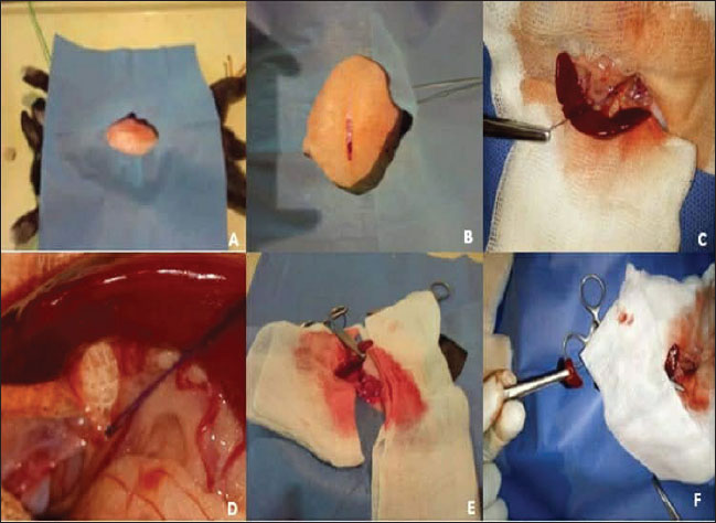
ABSTRACTBackground: The spleen plays a vital role in the regulation of immunity and blood complete splenectomy can increase vulnerability to infection, whereas partial splenectomy preserves function. However, the ability of residual tissue to regenerate remains uncertain. Aim: Hematological and histological evaluation of spleen regeneration following partial splenectomy in Algerian rabbits. Methods: Fifteen adult male rabbits were divided into three groups, with five animals in each group. Clinical evaluation, hematological analysis, and histological examination were performed to assess the extent of splenic regeneration following partial splenectomy. Results: All animals recovered uneventfully. On day 15, mild anemia and inflammatory changes were observed. By day 30, the splenic tissue showed reorganization with active lymphoid follicles, angiogenesis, and restored hematologic values. Conclusion: Partial splenectomy in rabbits allows for structural and functional regeneration within 1 month, indicating that it a safe and effective approach to preserve splenic activity. Keywords: Histology, Partial splenectomy, Rabbit, Regeneration, Spleen. IntroductionThe spleen is a lymphoid organ that plays an important role in the defense of an organism, participating in filtration processes, phagocytosis, and immunoglobulin production. Therefore, their main functions are: hematopoietic, immunological in the production of lymphocytes, plasmacytes, and macrophages. Phagocytosis is also denoted as hemocatheresis and is part of the "pool'' outlying of blood storage (Lewis et al., 2019). Total spleen resection is an option in some clinical situations, including traumatic injury, thrombocytopenia, and severe portal hypertension. Despite the apparent simplicity of the procedure, 2–10 years in the aftermath, a number of patients develop complications collectively referred to as post-splenectomy syndrome, most typically manifested by recurrent infections of varying severity (Malagó et al., 2008; Buzelé et al., 2019). The study of spleen regeneration after splenectomy is often limited to patients and small laboratory animals, whereas splenectomy has only been studied in the context of survival in larger species. Thus, previous studies reported a postoperative survival rate of 52% in dogs and 87.5% in splenectomized horses infected with Theileria haneyi, while earlier work in cattle described an almost complete postoperative recovery (Andrey et al., 2005). Partial splenectomy is often preferred to total splenectomy as a method to prevent serious postoperative sepsis. However, most previous studies on spleen function following partial splenectomy failed to provide accurate data on the minimum increase in residual spleen weight necessary for protection against postoperative sepsis (Knowles et al., 2022). Performing partial splenectomy with a safe, simple, and definite technique in patients with trauma with hemodynamic instability and accompanying intra-abdominal injury could play an important role in the preservation of immune function and reducing morbidity (Chiba et al., 1984). This study aimed to evaluate the clinical and histological evolution of the residual spleen after partial splenectomy in local Algerian rabbits to determine whether the spleen has the capacity to regenerate following partial resection. Materials and MethodsAnimalsFifteen apparently healthy adult male local Algerian rabbits were used in this experimental study. Animals were randomly assigned to three groups (n=15 in total, 5 rabbits per group): Group I (Control): A midline laparotomy without splenic resection was performed. The incision was routinely closed after the abdominal exploration. Group II (15 days): A partial splenectomy was performed, and relaparotomy was performed on postoperative day 15 to evaluate macroscopic and histological changes in the residual spleen. Group III (30 days): The same partial splenectomy procedure was performed as in Group II, but re-exploration and tissue sampling were conducted on day 30 to assess long-term healing and regeneration. All animals were apparently healthy based on physical and clinical examination before the experiment, maintained under identical environmental conditions, and provided with a standard laboratory diet and free access to water. Anesthesia and the surgical procedureFood was withheld for 12 hours before surgery. Anesthesia was induced intramuscularly using a combination of acepromazine (0.8 mg/kg), buprenorphine (0.05 mg/kg), and ketamine (40 mg/kg). After shaving and disinfecting the abdomen with povidone-iodine solution, a midline incision was made to expose the spleen. The caudal pole of the spleen was ligated and partially resected in Groups II and III following the standard small-animal surgical technique described by Slatter (2003). Hemostasis was achieved using fine 3–0 silk ligatures. The abdominal wall was closed in two layers: the muscular layer with 3–0 Polyglactin 910 (Vicryl) and 30 Nylon interrupted sutures (Fig. 1).
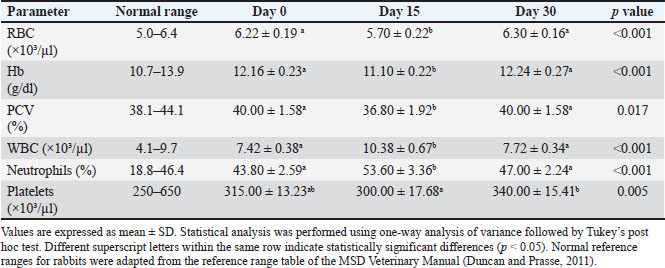
Fig. 1. Partial splenectomy in rabbits. (A) Aseptic preparation of the surgical site. (B) Skin and linea alba incision. (C) Spleen isolation (D) Ligation of the splenic vessels. (E) Application of hemostatic forceps. (F) Splenic parenchyma sectioning. Postoperative careAll animals were observed daily for activity, wound healing, and body temperature. Buprenorphine (0.05 mg/kg) was used for three consecutive days to maintain postoperative analgesia. The rabbits were kept under hygienic conditions at 22°C and monitored for either 15 or 30 days, according to the experimental group. Blood sampling and hematological analysisBlood samples (0.5 ml) were collected from the middle ear vein at each time point: day 0 (baseline), day 15, and day 30. Samples were placed in Ethylenediaminetetraacetic acid tubes and analyzed for hematological parameters, including red blood cell (RBC) count, white blood cell (WBC) count, hemoglobin concentration (Hb), packed cell volume (PCV), differential leukocyte count, and platelet count. Histological examination of the specimenHistological evaluation was performed on postoperative days 15 and 30 only, as no histological samples were collected from the control group (laparotomy without biopsy). The normal rabbit spleen architecture was used as a reference. Splenic samples were fixed in 10% neutral-buffered formalin, dehydrated through graded alcohols, cleared in xylene, and embedded in paraffin. Sections of 5 µm thickness were stained with hematoxylin and eosin and examined under a light microscope for structural and cellular evaluation. Histologic regeneration score and image-based measuresA semiquantitative histologic regeneration score (0–8) was used to assess the extent of tissue remodeling, which was adapted from previously established scoring systems for mucosal regeneration (Ishii et al., 2012). The evaluation included four parameters: hemorrhage congestion, inflammatory infiltration, lymphoid follicle regeneration, and neo-angiogenesis, each graded from 0 (absent) to 2 (prominent). The total score reflected the degree of regenerative activity within the tissue. Statistical analysis resultsStatistical analysis was performed using IBM SPSS Statistics. Haematological data were analyzed using analysis of variance, and Tukey’s post-hoc test was applied for multiple comparisons when significant differences were detected. The hematological results are expressed as mean ± standard deviation (SD). Histological regeneration and inflammation scores are presented as median [interquartile range (IQR)], and were compared between groups using the Mann–Whitney U test. A value of p < 0.05 was considered statistically significant. Ethical approvalAll procedures in this study complied with Algerian animal protection law (Law No. 95-322/1995) and followed the guidelines of the Algerian Association of Experimental Animal Sciences (AASEA). The experimental protocol was conducted under the supervision and authorization of senior academic staff in accordance with institutional practices. ResultsClinical resultsThe anesthetic procedure was secure during the surgical procedure, and the patient tolerated the anesthetic protocol well and recovered uneventfully. A mild decrease in appetite and activity was observed on the first postoperative day, followed by rapid normalization. Proper use of analgesics before and during surgery provided better pain relief during and after surgery. Body temperature and wound healing remained within normal ranges surgery was successfully performed in both groups, and all rabbits recovered without serious complications. No intraoperative complications occurred in both groups, and all rabbits survived the surgery. At the second laparotomy, the residual spleen appeared healthy with visible vascularization and moderate enlargement. Hematological findingsHematological parameters were recorded before surgery at baseline (day 0). The overall analysis revealed significant time-dependent changes in several hematological parameters (one-way ANOVA, p < 0.05). Erythrocytic parameters showed a transient decline at day 15 after surgery. The RBC count decreased significantly at day 15 (5.70 ± 0.22) compared with the baseline values (6.22 ± 0.19; p=0.003) and subsequently recovered by day 30 (6.30 ± 0.16), with no significant difference between days 0 and 30 (p=0.793). A similar pattern was observed for hemoglobin concentration, which was reduced at day 15 (11.10 ± 0.22) compared with day 0 (12.16 ± 0.23; p < 0.001) and day 30 (12.24 ± 0.27; p < 0.001), while baseline and day 30 values did not differ significantly (p=0.862). Packed cell volume also showed a significant reduction at day 15 (36.80 ± 1.92) compared with day 0 (40.00 ± 1.58) and day 30 (40.00 ± 1.58) (p=0.029), with similar values between baseline and day 30 (p=1.000). In contrast, leukocyte parameters exhibited a temporary increase on day 15. The total white blood cell count increased significantly on day 15 (10.38 ± 0.67) compared with that on days 0 (7.42 ± 0.38; p < 0.001) and 30 (7.72 ± 0.34; p < 0.001), whereas no significant difference was observed between baseline and day 30 (p=0.604). The neutrophil percentage followed the same trend, increasing at day 15 (53.60 ± 3.36) compared with day 0 (43.80 ± 2.59; p < 0.001) and day 30 (47.00 ± 2.24; p=0.007), with no significant difference between day 0 and day 30 (p=0.203). The platelet count demonstrated a delayed response. Although the overall time-dependent variation was significant (p=0.005), a marked increase was observed on day 30 (340.00 ± 15.41) compared with day 15 (300.00 ± 17.68; p=0.004). Differences in baseline values (315.00 ± 13.23) were not statistically significant (p=0.314 for day 0 vs. day 15; p=0.062 for day 0 vs. day 30). Overall, the observed hematological alterations indicate a transient postoperative anemia and inflammatory response at day 15, followed by recovery toward baseline values by day 30 (Table 1). Table 1. Hematological findings after partial splenectomy in rabbits.
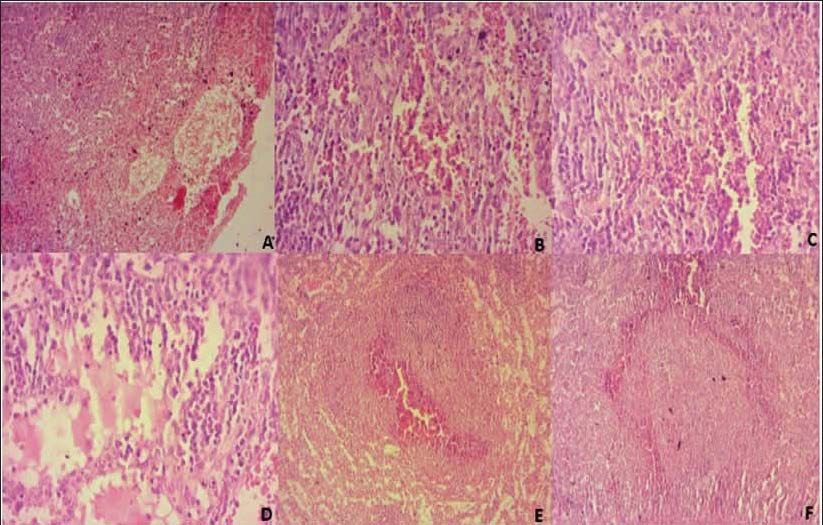
Histological findingsThe splenic tissue still exhibited pronounced inflammatory and degenerative changes 15 days after partial splenectomy. Low-magnification examination revealed disruption of the normal splenic architecture with extensive hemorrhage and vascular congestion (Fig. 2A). Higher magnification revealed marked edema within the red pulp, accompanied by dense infiltration of inflammatory cells, predominantly neutrophils and lymphocytes (Fig. 2B and C).
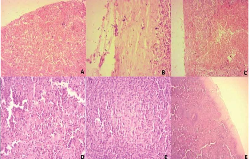
Fig. 2. Histopathological changes in the splenic tissue at day 15. (A) Peripheral region of the spleen showing marked hemorrhage and vascular congestion within the red pulp sinusoids (H&E, × 10). (B) Splenic parenchyma showing moderate congestion and inflammatory cell infiltration, mainly neutrophils and lymphocytes (H&E, × 40). (C) High magnification of the red pulp demonstrating dense neutrophilic infiltration and dilated sinusoids (H&E, × 40). (D) Edematous red pulp with widened intercellular spaces and clusters of inflammatory cells (H&E, × 40). (E) White pulp showing central hemorrhage surrounded by lymphocytic infiltration (H&E, × 10). (F) Organized lymphoid nodule with a well-defined hyperplastic follicle and developing germinal center (H&E, × 10). Dilated and congested sinusoids were evident in several areas, along with focal tissue disorganization and early cellular debris, reflecting ongoing tissue injury and repair (Fig. 2D). Although early signs of regeneration were present, lymphoid follicle formation remained poorly organized at this stage, and inflammatory processes dominated the splenic parenchyma (Fig. 2E and F). On the 30th day after partial splenectomy, the splenic tissue showed marked histological improvement. The overall splenic architecture appeared to be better organized, with a clear distinction between the red and white pulp regions (Fig. 3A). Congestion and hemorrhage were markedly reduced, and the red pulp displayed clearer sinusoidal spaces with minimal residual edema, indicating normal blood circulation was restored Fig. 3B. Inflammatory cell infiltration was significantly decreased compared with earlier stages, with only scattered inflammatory cells remaining within the parenchyma (Fig. 3C). Newly formed lymphoid follicles were clearly observed within the white pulp, several of which exhibited developing germinal centers, reflecting active lymphoid regeneration (Fig. 3D and E). The splenic capsule appeared slightly thickened, and thin fibrous bands were evident along its surface, consistent with tissue remodeling and splenic structure stabilization (Fig. 3F). Overall, these findings indicate that the spleen had undergone substantial structural and cellular regeneration by day 30, approaching a near-normal histological appearance following partial splenectomy.
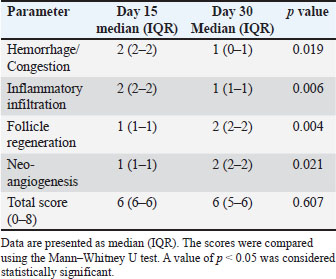
Fig. 3. Histopathological changes in the splenic tissue at day 30. (A) Diffuse capsular thickening and fibrosis with expanded lymphoid areas beneath the capsule (H&E, × 10). (B) High magnification of the splenic capsule showing proliferating fibroblasts and mild inflammatory cell infiltration (H&E, × 40). (C) Splenic parenchyma showing lymphoid hyperplasia and partial reconstruction of the white pulp (H&E, × 10). (D) Red pulp area with dilated sinusoids and scattered lymphocytes (×40, H&E). (E) Interface between the capsule and parenchyma showing chronic mononuclear infiltrate and fibroblastic response (×40, H&E). (F) Mature splenic architecture with well-defined white and red pulp zones and intact capsule (×10, H&E). Histological scoring results supported these microscopic observations. Hemorrhage/congestion scores decreased from day 15 [median (IQR): 2 (2–2)] to day 30 [1 (0–1)] (p=0.019). Inflammatory infiltration scores decreased significantly from 2 (2–2) on day 15 to 1 (1–1) at day 30 (p=0.006). In contrast, follicle regeneration increased from 1 (1–1) at day 15 to 2 (2–2) at day 30 (p=0.004), and neoangiogenesis increased from 1 (1–1) to 2 (2–2) at day 30 (p=0.021). The total score did not significantly differ between day 15 [6 (6–6) and day 30 [6 (5–6)] (p=0.607) (Table 2). Table 2. Histological regeneration score after partial splenectomy.
DiscussionThis study showed that partial splenectomy in rabbits triggers an active, well-organized pattern of regeneration that can be followed both clinically and microscopically. All animals recovered smoothly, and none developed infection or persistent anemia after surgery. In practical terms, this means that keeping even a part of the spleen is sufficient to preserve its main filtering and immune functions. This observation agrees with the earlier work of Holdsworth (1991) and Tavassoli et al. (1973) who described the main stages of splenic repair after transplantation, such as the regrowth of blood vessels and the formation of white pulp. Interestingly, Riera et al. (2009) pointed out that small fragments of splenic tissue known as splenosis can still act as functional spleen, lowering the risk of post-splenectomy sepsis. However, they also noted that it remains uncertain how much tissue is actually needed to provide full protection. Our hematologic findings followed a pattern typical of tissue injury and recovery. On day 15, we noticed a slight drop in red cell and hemoglobin levels, together with a modest increase in white cells and neutrophils. Short-lived anemia and inflammatory response are normal after surgery. By day 30, all values had returned to baseline, indicating that inflammation had subsided and blood production had resumed. Similar temporary changes were reported by Chiu et al. (1970) and Park et al. (2015) who observed comparable fluctuations in splenic regeneration during the transition from inflammation to tissue repair. The return of normal counts in our rabbits by week 3 confirms that the remaining spleen had regained its functional balance. Costi et al. (2019) also emphasized that partial splenectomy tends to carry lower morbidity and mortality than total removal, especially when the remaining portion retains its blood supply. These findings are consistent with rabbit-specific splenic regeneration patterns described by Rahmoun et al. (2019) and with the lower complication rates reported after partial compared to total splenectomy (Buzelé et al., 2016). Our results match those findings. All animals recovered well. These findings indicate that preservation of viable splenic tissue supports both immune and hematologic stability while reducing the risk of complications. In line with this, Khan and Dikki (2004) showed that splenic fragments can survive and recover normal activity after traumatic splenectomy. This is consistent with our observations: the remaining tissue in rabbits not only survived but gradually rebuilt its internal structure and maintained stable blood parameters. The healing process was clear and time-dependent. By the 15th day, the spleen showed congestion, mild bleeding, and a strong inflammatory cell presence, which is a classic feature of early repair. Early signs of new vessel formation and fibroblast activity were observed, although the lymphoid follicles were still poorly developed. By day 30, the picture had changed dramatically: the capsule was thicker, new vessels had spread across the tissue, and well-formed lymphoid follicles with active germinal centers were present. The median histologic score increased from day 15 to day 30, indicating substantial splenic tissue structural recovery. Ishii et al. (2012) and Park et al. (2015) described similar processes and highlighted the role of angiogenesis in restoring normal splenic architecture. Our findings also agree with those of Bradshaw and Thomas (1982) who studied rats after removing different portions of the spleen. They found that the remaining part grew in size over time and that regrowth was roughly proportional to the amount of remaining tissue. Even though our model used rabbits, the pattern was similar: structural regeneration, lymphoid hyperplasia, and clearer tissue organization by the end of the first month. More recently, Elchaninov et al. (2022) confirmed that the spleen has a strong capacity for both structural and functional recovery when some viable tissue remains, despite differences between species. This explains why partial splenectomy helps prevent overwhelming post-splenectomy infection and allows normal blood and immune regulation to continue. Spangler and Kass (1997) observed that the outcome of splenectomy in dogs depended on both the underlying condition and the amount of spleen removed. Animals who underwent total splenectomy had more complications and poorer survival. In contrast, the rabbits in our experiment undergoing partial removal recovered completely. This difference underlines how even a small splenic remnant can be preserved to maintain essential hematologic and immune functions and reduce postoperative risk. The histologic pattern observed over time closely matches the regeneration sequence described by Holdsworth (1991) and Tavassoli et al. (1973). According to their classic model, repair begins with tissue necrosis and capsule formation, followed by vascular regrowth, connective tissue remodeling, and the eventual development of distinct white-pulp regions. The spleen regains normal architecture with clearly defined red and white pulp and a restored vascular system after approximately 5 weeks. ConclusionOur findings demonstrate that partial splenectomy allows both structural and functional spleen recovery. The healing process naturally moves from inflammation to angiogenesis and then to lymphoid reorganization. As a result, the splenic architecture and blood parameters returned to normal. Preserving part of the spleen therefore offers real physiological benefits in maintaining immune function and blood balance while avoiding the complications that often follow total splenectomy. AcknowledgmentsThe authors sincerely thank Dr Mira Chiraoui for her guidance and all the contributors for their valuable support. Conflict of interestThe authors have no conflicts of interest to declare. FundingThis study was conducted without external funding. All research activities were funded by the author’s personal resources. Authors' contributionsAll authors contributed to study conception, design, data collection, and manuscript preparation. All authors have approved the final version. Data availabilityAll data supporting this study’s findings are available within the manuscript. ReferencesAndrey, L., Dor, F.J., Gollackner, B. and Guzzetta, P.C. 2005. Histopathology of spleen allograft rejection in miniature swine. Transplantation 79(1), 29–35. Bradshaw, P.H. and Thomas CG Jr. 1982. Splenic remnant regeneration after partial splenectomy in rats. J. Surg. Res. 32, 176–181. Buzelé, R., Barbier, L., Sauvanet, A. and Fantin, B. 2016. Medical complications following splenectomy. J. Visc. Surg. 153, 277–286. Chiba, T., Boles ET Jr. and Staples, G. 1984. Experimental studies on the rat model for partial splenectomy and spleen regeneration. J. Jpn. Surg. Soc. 85(1), 49–55. Chiu, C.J., McArdle, A.H., Brown, R., Scott, H.J. and Gurd, F.N. 1970. Intestinal mucosal lesion in low-flow states. Arch. Surg. 101(4), 478–483. Costi, R., Castro Ruiz, C., Romboli, A., Wind, P., Violi, V. and Zarzavadjian Le Bian, A. 2019. Partial splenectomy: who, when and how. A systematic review of 2130 published cases. J. Pediatr. Surg. 54(8), 1527–1538. Duncan, J.R. and Prasse, K.W. 2011. Veterinary laboratory medicine: clinical pathology. 5th ed. Ames, IA: Wiley-Blackwell. Elchaninov, A., Vishnyakova, P., Sukhikh, G. and Fatkhudinov, T. 2022. Spleen: reparative regeneration and influence on liver. Life 12(5), 626. Holdsworth, R.J. 1991. Regeneration of the spleen and splenic auto-transplantation. Br. J. Surg. 78, 270–278. Ishii M, Takahashi T, Oka M, Matsushita K, Tanaka C, Miyamae M, Mizukawa N, Nakamura M, Shimohata N and Shimizu Y. 2012. Histological evaluation of mucosal regeneration following tissue injury. J. Histochem. Cytochem. 60(8), 623–634. Khan, Z.A. and Dikki, P.E. 2004. Return of a normal functioning spleen after traumatic splenectomy. J. R. Soc. Med. 97, 391–392. Knowles, D.P., Sears K.P. and Fry, L.M. 2022. The clinical progression of Theileria haneyi in splenectomized horses reveals decreased virulence compared to Theileria equi. Pathogens 11(2), 254. Lewis, S.M., Williams, A. and Eisenbarth, S.C. 2019. Structure and function of the splenic immune system. Sci. Immunol. 4, 6085. Malagó, R., Yamasaki, S., Grubor, B., Carvalho, S.L., Melo, M.N., Campelo, M.J., Lima, M.S., Santana, D.M., Gontijo, C.M. and Santos, R.L. 2008. Inflammation and structural changes of splenic lymphoid tissue in visceral leishmaniasis: a study on naturally infected dogs. Parasite Immunol. 30(12), 721–731. Park, J.H., Kim, D.J. and Park, S.H. 2015. Evaluation of mucosal healing by histologic scoring in experimental models. PLos One 10(8), 135641. Rahmoun, D.E., Fares, M.A., Bouzebda-Afri, F. and Ben Driss, K. 2019. Anatomical and histological study of the rabbit spleen development in the postnatal period in Algeria. Online J. Anim. Feed Res.. Riera, M., Buczacki, S. and Khan, Z.A.J. 2009. Splenic regeneration following splenectomy and impact on sepsis: a clinical review. J. R. Soc. Med. 102(4), 139–142. Slatter, D.H. 2003. Small animal surgery. 3rd ed. Philadelphia, PA: Saunders. Spangler, W.L. and Kass, P.H. 1997. Pathological factors affecting post-splenectomy survival in dogs. J. Vet. Intern. Med. 11, 166–171. Tavassoli, M., Ratzan, R.J. and Crosby, W.H. 1973. Regeneration of heterotopic splenic autotransplants. Blood 41, 701–709. | ||
| How to Cite this Article |
| Pubmed Style Bouknine A, Chikhaoui M, Bouknine A, Houari H, Selmani SA. Does the spleen regenerate in rabbits after partial splenectomy?. Open Vet. J.. 2026; 16(5): 2922-2928. doi:10.5455/OVJ.2026.v16.i5.34 Web Style Bouknine A, Chikhaoui M, Bouknine A, Houari H, Selmani SA. Does the spleen regenerate in rabbits after partial splenectomy?. https://www.openveterinaryjournal.com/?mno=295212 [Access: June 26, 2026]. doi:10.5455/OVJ.2026.v16.i5.34 AMA (American Medical Association) Style Bouknine A, Chikhaoui M, Bouknine A, Houari H, Selmani SA. Does the spleen regenerate in rabbits after partial splenectomy?. Open Vet. J.. 2026; 16(5): 2922-2928. doi:10.5455/OVJ.2026.v16.i5.34 Vancouver/ICMJE Style Bouknine A, Chikhaoui M, Bouknine A, Houari H, Selmani SA. Does the spleen regenerate in rabbits after partial splenectomy?. Open Vet. J.. (2026), [cited June 26, 2026]; 16(5): 2922-2928. doi:10.5455/OVJ.2026.v16.i5.34 Harvard Style Bouknine, A., Chikhaoui, . M., Bouknine, . A., Houari, . H. & Selmani, . S. A. (2026) Does the spleen regenerate in rabbits after partial splenectomy?. Open Vet. J., 16 (5), 2922-2928. doi:10.5455/OVJ.2026.v16.i5.34 Turabian Style Bouknine, Asmaa, Mira Chikhaoui, Abdelhak Bouknine, Hemida Houari, and Sidi Ali Selmani. 2026. Does the spleen regenerate in rabbits after partial splenectomy?. Open Veterinary Journal, 16 (5), 2922-2928. doi:10.5455/OVJ.2026.v16.i5.34 Chicago Style Bouknine, Asmaa, Mira Chikhaoui, Abdelhak Bouknine, Hemida Houari, and Sidi Ali Selmani. "Does the spleen regenerate in rabbits after partial splenectomy?." Open Veterinary Journal 16 (2026), 2922-2928. doi:10.5455/OVJ.2026.v16.i5.34 MLA (The Modern Language Association) Style Bouknine, Asmaa, Mira Chikhaoui, Abdelhak Bouknine, Hemida Houari, and Sidi Ali Selmani. "Does the spleen regenerate in rabbits after partial splenectomy?." Open Veterinary Journal 16.5 (2026), 2922-2928. Print. doi:10.5455/OVJ.2026.v16.i5.34 APA (American Psychological Association) Style Bouknine, A., Chikhaoui, . M., Bouknine, . A., Houari, . H. & Selmani, . S. A. (2026) Does the spleen regenerate in rabbits after partial splenectomy?. Open Veterinary Journal, 16 (5), 2922-2928. doi:10.5455/OVJ.2026.v16.i5.34 |


