





| Research Article | ||
Open Vet. J.. 2026; 16(8): 5397-5411
Open Veterinary Journal, (2026), Vol. 16(8): 5397–5411 Research Article Microbial profile and antimicrobial resistance patterns of traumatic equine wounds in MalaysiaZulfitri Naim Abdul Rahim1, Zunita Zakaria2, Intan Shameha Abdul Razak3, Puteri Rose Camelia Roselan1 and Noraniza Mohd Adzahan1*1Department of Farm and Exotic Animal Medicine and Surgery, Faculty of Veterinary Medicine, Universiti Putra Malaysia, Serdang, Malaysia 2Department of Veterinary Pathology and Microbiology, Faculty of Veterinary Medicine, Universiti Putra Malaysia, Serdang, Malaysia 3Department of Veterinary Preclinical Sciences, Faculty of Veterinary Medicine, Universiti Putra Malaysia, Serdang, Malaysia *Corresponding Author: Noraniza Mohd Adzahan. Department of Farm and Exotic Animal Medicine and Surgery, Faculty of Veterinary Medicine, Universiti Putra Malaysia, Serdang, Malaysia. Email: noraniza [at] upm.edu.my Submitted: 07/11/2025 Revised: 12/06/2026 Accepted: 28/06/2026 Published: 08/08/2026 © 2025 Open Veterinary Journal
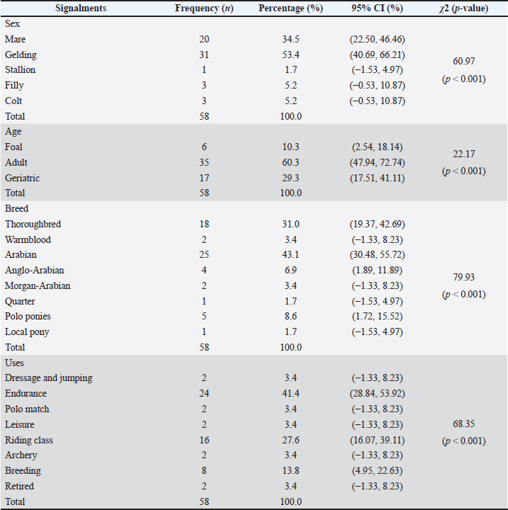
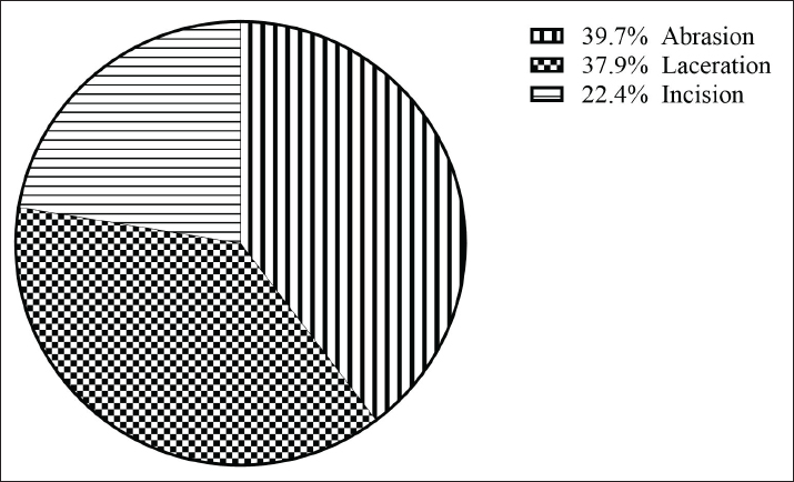
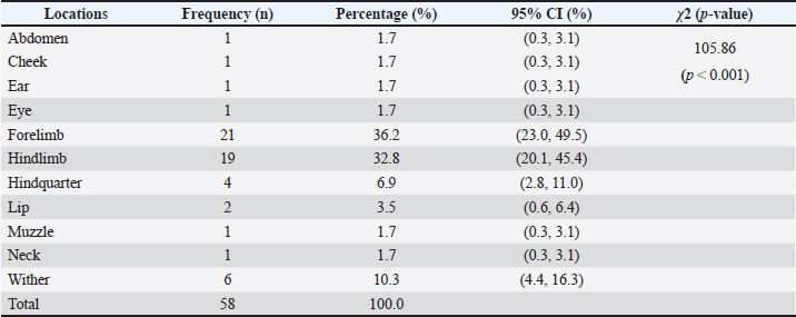
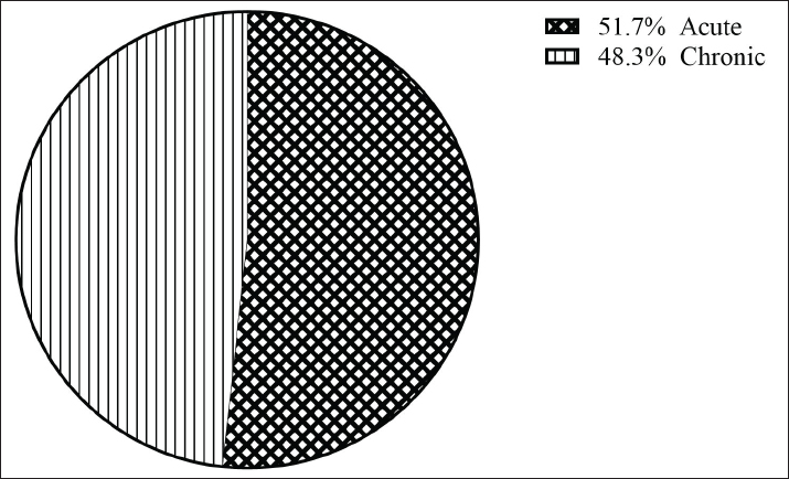
AbstractBackground: Traumatic wounds are a common condition in horses, posing significant challenges to both animal welfare and the equine industry’s economic sustainability. Subcutaneous tissue exposure creates a favorable environment for bacterial colonization and proliferation, which complicates wound management and delays healing. This clinical challenge is often exacerbated by empirical antimicrobial use, which drives the emergence of antimicrobial-resistant (AMR) organisms. Aim: This study aimed to identify bacterial species associated with traumatic wounds in horses in Peninsular Malaysia and to determine their antimicrobial susceptibility profiles. Methods: A cross-sectional study was conducted from February to August 2024 involving 58 horses with traumatic wounds sampled across seven Malaysian states. Wound swabs were collected using the Levine technique and cultured on blood and MacConkey agar. Bacterial isolates were identified using phenotypic and biochemical characterization. Antimicrobial susceptibility testing of the predominant isolates from chronic wounds was performed using the Kirby–Bauer disk diffusion method. Results: A total of 205 bacterial isolates were recovered, comprising 56.6% Gram-positive and 43.4% Gram-negative species. Chronic wounds (56.1%) yielded more isolates than acute wounds (43.9%). The most frequently isolated bacteria were Staphylococcus aureus (24.9%), Escherichia coli (15.6%), Streptococcus sp. (7.8%), Corynebacterium sp. (6.8%), and Klebsiella pneumoniae (6.3%). Penicillin resistance was identified in S. aureus (92.3%) and Corynebacterium sp. (71.4%). In contrast, 100% susceptibility to imipenem was preserved across all tested species. Notably, 23.0% of the tested isolates were classified as multidrug-resistant (MDR). Conclusion: These findings reveal the polymicrobial nature of traumatic wounds in the sampled horses in Malaysia, with S. aureus and E. coli as the predominant pathogens. The detection of MDR isolates emphasizes the need for culture-based diagnosis and prudent antibiotic use. This study provides essential insights to guide veterinarians and horse owners in improving wound management and promoting equine health and welfare. Keywords: Antimicrobial susceptibility, Bacterial colonization, Chronic wound infection, Equine wounds, Multidrug resistance. IntroductionThe occurrence of traumatic wounds in horses remains high due to their behavioral characteristics, environmental exposure, and the physical demands associated with their work (Viljoen et al., 2009; Bowden et al., 2020). Horses are naturally active animals that frequently engage in running, jumping, and social interactions, which predispose them to accidents, falls, or collisions (Owen et al., 2011). Instinctive behaviors such as kicking and fighting also contribute to wound occurrence (Knubben et al., 2008). Environmental factors, including rough terrain, sharp objects, and unsanitary stable conditions, further increase the risk. Once skin integrity is compromised, subcutaneous tissue exposure facilitates microbial colonization from the environment, skin flora, or endogenous sources (Bowler et al., 2001). The microbial population within a wound depends on various factors, including type, depth, location, tissue perfusion, and immune response (Hendrickson and Virgin, 2005). Wound colonization is often polymicrobial, involving both opportunistic and pathogenic microorganisms. Studies have reported that Pseudomonas aeruginosa, Escherichia coli, Enterobacter spp., Acinetobacter spp., Staphylococcus spp., and Streptococcus spp. are the most frequently isolated bacteria from equine wounds (Westgate et al., 2011; Ahmad Nadzir et al., 2020). Infected wounds that fail to respond to therapy can progress to chronic, non-healing states, increasing treatment costs and impairing equine performance (Hendrickson and Virgin, 2005). The current treatment for equine wound infections involves debridement, appropriate dressing, and antibiotic administration (Freeman et al., 2020). However, the use of empirical antibiotics without prior microbiological testing is common in equine practice, increasing the risk of antimicrobial-resistant (AMR) (Ross et al., 2015). Globally, AMR accounts for more than 1.2 million deaths annually, with projections estimating up to 10 million deaths annually by 2050 if left unaddressed (O’Neill, 2016). Although these statistics mirror a devastating trajectory in human medicine, widespread empirical antibiotic use in equine practice heavily accelerates this issue, driving the emergence of resistant pathogens, including extended-spectrum beta-lactamase (ESBL)-producing E. coli, methicillin-resistant Staphylococcus aureus (MRSA), resistant Rhodococcus equi, and multidrug-resistant Salmonella spp. (Weese et al., 2005; Soza-Ossandón et al., 2020; Kauter et al., 2021). These organisms pose zoonotic threats and complicate infection management. The identification of microbial species and the determination of antimicrobial susceptibility patterns are crucial for controlling the spread of AMR strains. This also enables more targeted and effective therapy, ultimately improving clinical outcomes. Moreover, AMR in equines can have profound financial consequences, as it often leads to prolonged treatment periods and increases the management cost of infected horses. In addition, the presence of resistant bacteria in horses can indirectly impact human health through direct contact or environmental transmission, underscoring the importance of antimicrobial stewardship (Kabir et al., 2024). Considering these concerns, this study aimed to identify the bacterial species associated with wound infections in horses in Malaysia and to determine their antimicrobial susceptibility patterns. The findings of this study will provide baseline data for improving wound management strategies, supporting antimicrobial stewardship, and strengthening AMR surveillance in the equine population. Materials and MethodsStudy design and sample sizeThis cross-sectional study was conducted from February to August 2024 to determine the microbial and antibiotic susceptibility profiles of traumatic wounds in horses. A total of 58 horses presenting with traumatic wounds were selected through a non-probability convenience sampling method across seven states in Peninsular Malaysia, including Selangor, Negeri Sembilan, Kedah, Penang, Pahang, Terengganu, and Kelantan. Horses were considered eligible for inclusion if they presented with at least one clinically traumatic wound resulting from an external physical injury. Informed consent was obtained from all owners, and the age, sex, breed, and use of the selected horses were recorded. The traumatic wound assessment procedureThe wound type, wound location, and healing time frame were systematically recorded. Wounds were classified into distinct types, including abrasions, punctures, incisions, and lacerations (Nejash et al., 2017). The anatomical locations of the wounds were categorized into specific regions, including the head, neck, forelimbs, hindlimbs, thorax, abdomen, hindquarters, and inguinal area (Mekuria and Tesfaye, 2017). The healing time frame was categorized into the acute and chronic stages. Acute wounds were defined as those that adhered to the normal healing process, whereas chronic wounds were defined as those that persisted for more than one month without evidence of healing or progression (Bertozzi et al., 2017). Wound size was excluded from the formal analysis because of the high geometric irregularity of the traumatic lesions. Before sampling, clinical histories were verified to confirm that the horses had not received any topical or systemic antimicrobial treatments for at least 7 days. None of the sampled wounds were bandaged before or during the collection period; all wounds were managed as open wounds. Sample collectionBefore sample collection, the wound surfaces were thoroughly irrigated with sterile normal saline to remove debris and surface contaminants. Sterile swab sticks were used, and sampling was performed using the Levine technique, which involves rotating a sterile swab firmly over a 1‐cm2 area of the wound surface in a circular motion to obtain representative samples from both surface and deeper tissues (Haalboom et al., 2019). The swabs were placed in Cary Blair transport medium (Labchem, Malaysia) and transported in an ice-cooled container (~4°C) to the Bacteriology Laboratory, Faculty of Veterinary Medicine, Universiti Putra Malaysia, for immediate processing. Isolation and identification of bacteriaBlood agar (Oxoid, Thermo Fisher Scientific, United Kingdom) supplemented with 5% horse blood and MacConkey agar (Oxoid, Thermo Fisher Scientific, United Kingdom) were used as the culture media for inoculating all collected samples. Plates were incubated at 37°C for 24–48 hours in an inverted position to prevent condensation and minimize the risk of contamination. Bacterial growth on culture plates was carefully examined for macroscopic colony morphology, including size, shape, color and pigmentation, elevation, margin, texture, and hemolytic pattern, after incubation. Each isolated colony was then subcultured on tryptic soy agar (Liofilchem, Italy) for purification. Pure isolates were then Gram-stained, and their Gram reaction, cell shape, and arrangement were examined by microscopic examination. Subsequently, a series of biochemical tests was performed to identify the genus and species of the bacteria. These included catalase, oxidase, coagulase, indole, methyl red (MR), Voges–Proskauer (VP), citrate utilization, sugar fermentation, triple sugar iron (TSI), and urease tests. The results of these tests, combined with the colony and cellular morphology and Gram reaction, aided in their accurate identification. Antimicrobial susceptibility testingAntimicrobial susceptibility testing was performed on the 74 bacterial isolates derived from chronic wounds using the Kirby–Bauer disk diffusion method, following the Clinical and Laboratory Standards Institute (CLSI, 2023) guidelines. These isolates represented the five most predominant bacterial species recovered across the entire sample, selected strictly based on their high isolation frequency. This targeted approach was adopted because chronic wounds serve as the primary reservoir for persistent, treatment-refractory pathogens, making these isolates the most clinically relevant targets for evaluating localized resistance trends. Bacterial suspensions were prepared by transferring colonies into sterile distilled water and adjusting turbidity to match a 0.5 McFarland standard (approximately 1.5 × 10⁸ CFU/mL). Sterile cotton swabs were dipped into the suspension and spread evenly on Mueller–Hinton agar (Oxoid, Thermo Fisher Scientific, United Kingdom) using the lawn technique. Antibiotic-impregnated paper discs were applied to the plates using a disc dispenser (Oxoid, Thermo Fisher Scientific, United Kingdom), and the plates were incubated at 37°C for 18–24 hours. The diameters of inhibition zones were measured in millimeters using a digital vernier caliper (Mitutoyo, Japan) and interpreted as susceptible, intermediate, or resistant according to CLSI (2023) breakpoints. Where CLSI data were unavailable, the guidelines of the European Committee on Antimicrobial Susceptibility Testing (EUCAST, 2023) or published reference data were used. Antibiotic agents testedThe antibiotics tested were selected based on the World Organization for Animal Health (OIE, 2021) classification and the prescription practices for horses in Malaysia (Department of Veterinary Services Malaysia, 2021). The susceptibility of Gram-positive bacteria was assessed against 12 antibiotic agents (Oxoid, Thermo Fisher Scientific, United Kingdom) from nine different classes, while Gram-negative bacteria were tested against 10 antibiotic agents from eight different classes. These antibiotic agents were ampicillin (AMP), penicillin (P), imipenem (IPM), amoxicillin– clavulanic acid (AMC), enrofloxacin (ENR), erythromycin (E), trimethoprim–sulfamethoxazole (SXT), chloramphenicol (C), doxycycline (DO), tetracycline (TE), gentamicin (CN), and streptomycin (STR). Reference strains of S. aureus (ATCC 25923) and E. coli (ATCC 25922) were included for quality control to ensure the reliability, reproducibility, and consistency of the testing process. The isolates tested that demonstrated non-susceptibility to at least one agent in three or more antimicrobial categories were considered multidrug-resistant (MDR) bacteria. Data management and statistical analysisAll data were recorded in Microsoft Excel 2021 (Microsoft Corporation) and analyses using GraphPad Prism version 10 (GraphPad Software, California, USA). Descriptive statistics, including frequency distributions, were used to summarize the bacterial isolates and the antimicrobial susceptibility patterns. Chi-square tests were performed to evaluate the distribution patterns of demographic variables within the studied cohort; Fisher’s exact test was applied when sample sizes were small. A p-value < 0.05 was considered statistically significant. Ethical approvalThe Institutional Animal Care and Use Committee, Universiti Putra Malaysia, approved all procedures (AUP Number R02/2024). ResultsA total of 58 horses with traumatic wounds were included in this study. Table 1 summarizes the distribution of horses sustaining traumatic wounds according to sex, age, breed, and use. Statistically significant differences (p < 0.05) were found in the sex, age, breed, and use distribution among the sampled horses. Geldings sustained the highest proportion of injuries (53.4%), followed by mares (34.5%), while stallions, colts, and fillies were the least represented. Adult horses (60.3%) were more frequently affected than geriatric horses (29.3%) and foals (10.3%). Arabian (43.1%) and Thoroughbred (31.0%) horses were more represented than other breeds. Horses used for endurance (41.4%) and riding (27.6%) activities were more frequently represented with traumatic wounds than those used for breeding, leisure, or other purposes. The distribution of wound types (Fig. 1) showed that abrasions were the most common (39.7%), followed by lacerations (37.9%) and incisions (22.4%). The chi-square test yielded no statistically significant difference in the distribution of these wound types within the sample (χ2=3.14, p=0.208). Based on anatomical distribution (Table 2), the forelimb (36.2%) and hindlimb (32.8%) were the most frequently affected areas, followed by the withers (10.3%). Less frequent injury sites included the hindquarter (6.9%) and the lip (3.5%), while the abdomen, cheek, ear, eye, muzzle, and neck each accounted for a minor proportion (1.72% each) of the recorded wounds. The Chi-square test demonstrated a statistically significant difference in wound localization across the different anatomical regions (χ2=105.86, p < 0.001). The healing time of the traumatic wounds was categorized into acute and chronic stages (Fig. 2). Acute wounds accounted for 51.7% of cases, whereas chronic wounds accounted for 48.3%, with no significant difference in the distribution of healing stages (p > 0.05). Table 1. Distribution of horses with traumatic wounds according to sex, age, breed, and use.
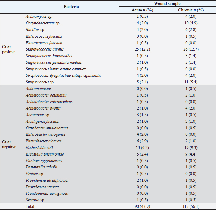
The collected wound swab samples yielded 205 distinct bacterial isolates. Table 3 illustrates the frequency distribution of 116 (56.6%) Gram-positive and 89 (43.4%) Gram-negative bacterial isolates. Of these bacteria, 90 (43.9%) originated from acute wounds, whereas 115 (56.1%) originated from chronic wounds. Among these isolates, S. aureus (24.9%), E. coli (15.6%), Streptococcus sp. (7.8%), Corynebacterium sp. (6.8%), and Klebsiella pneumoniae (6.3%) were the most common bacteria. Moreover, certain bacterial species were isolated exclusively from either acute or chronic wounds. Specifically, Achromobacter, Citrobacter amalonaticus, Enterococcus faecalis, Pasteurella caballi, Providencia stuartii, and P. aeruginosa were found only in chronic wounds. Conversely, Acinetobacter calcoaceticus, Enterococcus faecium, Proteus sp., and Streptococcus bovis-equine complex were isolated exclusively from acute wounds. These bacteria were also the least frequently isolated species from these traumatic wounds.
Fig. 1. Distribution of traumatic wounds sustained by horses according to the types of the wounds. Table 2. Distribution of traumatic wounds sustained by horses across different anatomical body regions.
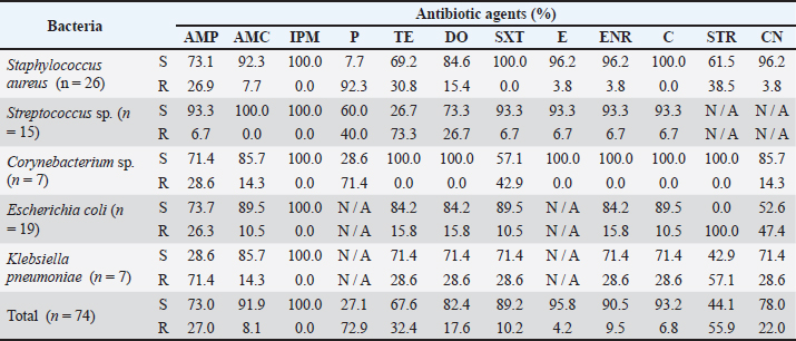
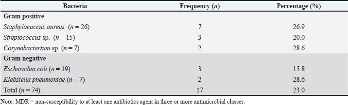
Table 4 summarizes the antimicrobial susceptibility of 74 bacterial isolates, including both Gram-positive and Gram-negative species. These isolates were the five predominant bacterial species selected based on the isolation frequency from pooled isolates obtained from 30 chronic wounds. Staphylococcus aureus was highly susceptible to most antibiotics, with complete susceptibility to trimethoprim–sulfamethoxazole, imipenem, and chloramphenicol. However, it showed significant resistance to penicillin (92.3%), streptomycin (38.5%), tetracycline (30.8%), and ampicillin (26.9%). Streptococcus sp. exhibited 100% susceptibility to amoxicillin–clavulanic acid and imipenem, but reduced susceptibility to tetracycline (26.7%) and penicillin (60.0%). Corynebacterium sp. also exhibited complete susceptibility to most antibiotics, although resistance to penicillin (71.4%) and trimethoprim-sulfamethoxazole (42.9%) was observed. In addition, E. coli showed high susceptibility to 9 out of 10 antibiotics tested, with 100% susceptibility to imipenem, but complete resistance to streptomycin, and reduced susceptibility to gentamicin (52.6%). Klebsiella pneumoniae exhibited broad susceptibility, with complete susceptibility to imipenem, moderate susceptibility to most antibiotics, and the lowest susceptibility to ampicillin (28.6%) and streptomycin (42.9%). Table 5 summarizes the frequency of MDR bacteria. Of the 74 bacterial isolates tested, 17 (23.0%) were identified as MDR, where bacteria become non-susceptible to at least one antibiotic agent in three or more antimicrobial classes.
Fig. 2. Distribution of traumatic wounds sustained by horses according to the acute and chronic time frames. DiscussionThis study describes the distribution of sex, age, breed, and use among horses presenting with traumatic wounds. Although the study design does not allow for risk or incidence assessment, the observed patterns are consistent with previously reported risk factors in the literature (Mekuria and Tesfaye, 2017; Nejash et al., 2017; Mekete, 2022; Kompi et al., 2023). Adult horses, particularly geldings and mares, accounted for a higher proportion of the sampled population, likely due to their frequent use in work and sport. Geldings are commonly selected for sport due to their manageable temperament, while mares may experience injury risk due to excitable behavior (Hennessy et al., 2008; Duberstein and Gilkeson, 2010). Geriatric horses also sustain injuries, especially in riding schools, often due to ill-fitting saddles and mishandling. Age-related decline in physical resilience and prolonged workload also contribute to susceptibility to injury. Foals were underrepresented in this study because most sampling occurred in Kelantan, one of the few states with active breeding programs, whereas adult horses are more commonly found in other states. Breed distribution variation was observed within the study sample, particularly in competitive settings. Arabians and Thoroughbreds, which are commonly used in endurance racing, were more frequently affected, likely due to the intense demands of the sport (Ropka-Molik et al., 2019). Similarly, Polo ponies and Warmbloods face discipline-specific risks in polo, jumping, and dressage, respectively (Rovere et al., 2016; Martinez et al., 2021). Retired Thoroughbreds repurposed as riding school horses also face increased injury risks due to inexperienced riders, shared tack, and limited rest (Greve and Dyson, 2014; Dyson et al., 2015). Hence, signalment characteristics showed differences in distribution within the study population, reflecting the combined effects of physiological traits and the roles that horses fulfill in various equine disciplines. Table 3. Distribution of bacteria isolated from acute and chronic traumatic equine wounds.
The most common wound types observed were abrasions and lacerations, consistent with previous reports in horses and donkeys (Fikru et al., 2015; Nejash et al., 2017). Mechanical trauma, including friction, falls, collisions, and contact with environmental hazards such as rough surfaces or sharp objects encountered during training, handling, transport, and competition, is typically associated with these lesions. However, no significant association was found between wound type and frequency, suggesting that traumatic injuries are multifactorial in origin. The distal regions of the forelimbs and hindlimbs were the most commonly affected sites in the sampled horses, differing from previous studies that reported the back, withers, and prescapular regions as common injury locations related to poorly fitted tack (Fikru et al., 2015; Nejash et al., 2017; Chala et al., 2017; Mekete, 2022). Distal limbs are particularly vulnerable because of limited soft tissue coverage, high contamination risk, reduced vascularity, and constant joint motion, all of which impair healing (Kayode, 2017; Jørgensen et al., 2021). The carpal, tarsal, and fetlock joints were commonly injured, likely due to the high biomechanical stress associated with training and competition (Paris et al., 2021). Wounds on the withers and hindquarters were likely associated with tack-related pressure injuries, particularly from improper saddle fit. Increased saddle pressure has been linked to the development of saddle sores and dry spots, whereas factors such as girth straps and saddle flocking may influence pressure distribution (Von Peinen et al., 2010; Byström et al., 2010). Hindquarter pressure sores were also observed in horses with prolonged confinement or reduced mobility, emphasising the importance of proper flooring and adequate bedding. Table 4. Antibiotic susceptibility and resistance of bacteria isolated from equine chronic traumatic wounds using the disk diffusion method.
Table 5. Multidrug resistance bacteria identified from equine chronic traumatic wounds.
Acute and chronic wounds were represented at comparable frequencies within the sampled horses. However, chronic wounds yielded a higher number of bacterial isolates than acute wounds, suggesting greater microbial colonization in delayed-healing lesions. Chronic wounds are often associated with persistent inflammation, contamination, repeated trauma, and prolonged exposure to the environment, all of which may favor bacterial colonization and persistence (Knottenbelt, 2003). In contrast, acute wounds generally benefit from more effective vascular and immune responses during the early stages of healing (Raziyeva et al., 2021). These findings support the observation that chronic equine wounds may provide a more favorable environment for persistent bacterial colonization. The recovered bacterial isolates from traumatic wounds demonstrated the polymicrobial nature of wound infections. The frequent isolation of S. aureus, E. coli, Streptococcus sp., Corynebacterium sp., and K. pneumoniae from both acute and chronic wounds aligns with previous reports in Malaysia and other countries (Westgate et al., 2011; Ahmad Nadzir et al., 2020; Abdul Rahman et al., 2021). Their prevalence suggests that these bacteria play a central role in equine wound infections. Staphylococcus aureus, a part of the normal flora of the skin, may contribute to wound persistence through its ability to form biofilms, particularly in chronic wounds (Wu et al., 2024). The emergence of MRSA strains, as reported in other studies, could further complicate treatment and infection control, although MRSA was not specifically assessed in the present study (Uchida-Fujii et al., 2022; Kannekens-Jager et al., 2024). Similarly, E. coli, which commonly originates from gastrointestinal or environmental contamination, may persist under both aerobic and anaerobic conditions and has been reported to exhibit antibiotic resistance, which could influence treatment outcomes (Basavaraju and Gunashree, 2022; Ballén et al., 2022). Meanwhile, Streptococcus sp. may contribute to infection via virulence factors, such as streptokinase and streptolysin S, which promote tissue invasion and inflammation (Barnett et al., 2015; Bekele et al., 2024). Corynebacterium sp. have been associated with abscesses and lymphangitis and may establish chronic infections in damaged tissues (Pratt et al., 2005; Kilcoyne et al., 2014). Although K. pneumoniae is commonly linked to respiratory and gastrointestinal infections, it has also been reported in wound infections, particularly due to its ability to produce ESBL, which may contribute to multidrug resistance (Trigo da Roza et al., 2019; Gravey et al., 2024). Its environmental resilience could further enable persistent wound colonization (Riwu et al., 2022). In addition, microbial profiles show compositional divergence between acute and chronic wounds. Acute wounds that are early in the healing process tend to host transient bacteria in oxygen-rich environments where immune defenses are more effective (Raziyeva et al., 2021). However, chronic wounds support more resilient, often biofilm-forming species that thrive in hypoxic and inflamed tissue, which may impair healing and complicate treatment (Schilrreff and Alexiev, 2022; Sahoo and Meshram, 2024). Conventional culture and phenotypic identification remain the gold standard for isolating viable, clinically significant bacteria; however, they possess an inherent bias toward fast-growing aerobic and facultative anaerobic species. Fastidious or strict anaerobic bacteria embedded within mature biofilms may be underrepresented. Future surveillance could build upon these foundational culture baselines by incorporating complementary molecular tools, such as 16S rRNA gene sequencing or rapid MALDI-TOF MS platforms, to comprehensively profile the wider equine wound microbiome. Expanding upon these cultural baselines, analysis of the isolates’ specific phenotypic profiles revealed significant AMR patterns that directly compromise routine wound therapy. Antimicrobial resistance within these wounds can arise intrinsically through natural genetic recombination or be acquired via horizontal gene transfer mechanisms, such as conjugation, transformation, and transduction (Martínez, 2008). Therefore, it is essential to assess bacterial susceptibility to antibiotics before treatment. In this study, S. aureus showed the highest resistance to penicillin, followed by streptomycin, tetracycline, and ampicillin, which aligns with the findings of Robinson et al. (2016). Penicillin is a beta-lactam antibiotic that inhibits bacterial cell wall synthesis by targeting penicillin-binding proteins (PBPs), which are critical for peptidoglycan cross-linking (Bush and Bradford, 2016). Resistance occurs when S. aureus produces beta-lactamase or penicillinase, enzymes that degrade the beta-lactam ring. Penicillin and streptomycin are frequently used as first-line treatments for equine infections in Malaysia, increasing the risk of resistance development (Department of Veterinary Services Malaysia, 2021). The overwhelming penicillin resistance observed in our study provides clear evidence that standard husbandry practices and unregulated empirical selection are directly exhausting the utility of these foundational frontline drugs. Streptomycin resistance typically results from enzymatic modification, which impairs its ability to inhibit protein synthesis (Schmitz, 1999; Fathi et al., 2022). Additionally, the high susceptibility of S. aureus to trimethoprim-sulfamethoxazole, imipenem, and chloramphenicol in the current study agrees with previous equine studies, likely due to the limited use of these agents (Tahoun et al., 2022; Marshall and Marsella, 2023). In this study, Streptococcus sp. showed the highest resistance to tetracycline, followed by penicillin and doxycycline, while remaining fully susceptible to amoxicillin–clavulanic acid and imipenem. Only one isolate demonstrated resistance to other antibiotics, indicating that most Streptococcus sp. isolates remain highly susceptible to antibiotics. Clark et al. (2008) and Erol et al. (2012) reported similar susceptibility trends, who noted resistance primarily to tetracycline, gentamicin, and trimethoprim–sulphonamide. Duchesne et al. (2019) also observed a significant increase in the number of tetracycline-resistant Streptococcus isolates over a decade. The frequent empirical use of broad-spectrum antibiotics may contribute to selective resistance, whereas limited exposure to less commonly used agents likely helps preserve susceptibility. Consistent with previous findings, the extensive use of tetracycline and penicillin in the Malaysian equine population may have facilitated the persistence of resistant isolates (Wanninger et al., 2016; Arredondo et al., 2020). Biofilm formation may further promote resistance, as tetracycline accumulation within biofilms increases the selection pressure for resistance gene expression (Hu et al., 2023). Furthermore, tetracycline resistance is primarily mediated by active efflux pumps and ribosomal protection, both of which are regulated by specific resistance genes (Grossman, 2016). Meanwhile, penicillin resistance in Streptococcus sp. can develop gradually through the accumulation of amino acid changes in PBPs, rather than through horizontal gene transfer of beta-lactamase or low-affinity PBPs (Yu et al., 2023). Despite the high susceptibility of Streptococcus sp. to most antibiotics, resistance to tetracycline and penicillin is still increasing, which underscores the need for careful antibiotic use to prevent further resistance. In the current study, Corynebacterium sp. demonstrated complete susceptibility to seven out of 12 antibiotics tested, consistent with reports of high susceptibility in isolates recovered from the conjunctiva of horses (Raquel et al., 2017; Fernández-Garayzábal et al., 2022). This broad-spectrum susceptibility suggests that multiple therapeutic options remain effective for managing Corynebacterium-associated wound infections, reducing the need for empirical use of broad-spectrum antibiotics. However, resistance to penicillin, trimethoprim-sulfamethoxazole, ampicillin, amoxicillin–clavulanic acid, and gentamicin was also detected, aligning with the findings of Saleh et al. (2023) and Elwalaly et al. (2024). The resistance to beta-lactam antibiotics is likely due to the production of beta-lactamases and altered PBPs. Resistance to trimethoprim-sulfamethoxazole often arises from the acquisition of drug-resistant dihydrofolate reductase variants encoded by dfr genes, which are frequently found within integrons or associated with insertion sequence common region-1 (ISCR1) (Shin et al., 2015). Moreover, gentamicin resistance can arise through aminoglycoside-modifying enzymes, which hinder antimicrobial binding and activity (Garneau-Tsodikova and Labby, 2016). Therefore, although Corynebacterium sp. exhibits generally favorable susceptibility patterns, the emergence of resistance to specific antibiotics highlights the need for targeted antimicrobial selection. E. coli exhibits complete susceptibility to imipenem but significant resistance to streptomycin and gentamicin, with moderate resistance to ampicillin and low resistance to other tested antibiotics. These findings are consistent with Chipangura et al. (2017), who also observed high susceptibility of E. coli from horses to imipenem and marked resistance to ampicillin, doxycycline, oxytetracycline, and gentamicin. Similarly, studies in Canada and the United Kingdom found that a substantial proportion of horses shed E. coli resistant to tetracyclines, folate pathway inhibitors, aminoglycosides, and aminopenicillins (de Lagarde et al., 2020; Isgren et al., 2021). The notable resistance to aminoglycosides observed in this study is likely due to aminoglycoside-modifying enzymes that inhibit ribosomal binding in E. coli (Schmitz, 1999; Fathi et al., 2022). Comparable resistance patterns have also been reported in Malaysian ruminants, suggesting that cross-species trends are potentially influenced by shared antibiotic use and reservoirs of resistant bacteria (Haulisah et al., 2021). Hence, this suggests the critical impact of antibiotic overuse in animals and the potential for cross-species transmission of resistant strains. Klebsiella pneumoniae is commonly found in feces and has the potential to contaminate and colonize wounds. The increasing prevalence of AMR has raised significant concerns within the medical and veterinary fields (Estell et al., 2015; Trigo da Roza et al., 2019). In this study, K. pneumoniae exhibited high resistance to almost all tested antibiotics, except imipenem, to which all isolates remained fully susceptible. Resistance to penicillin-class antibiotics, particularly ampicillin, is a characteristic feature of this species and is attributed to the chromosomal SHV beta-lactamase gene, which confers intrinsic resistance to several beta-lactam agents (Li et al., 2023). In addition, aminoglycoside resistance in K. pneumoniae is partly due to the acquisition of the armA gene, which encodes a 16S rRNA methylase located on plasmids, which protects ribosomal binding sites and confers high-level resistance to nearly all aminoglycosides, including newer-generation compounds (Galimand et al., 2003; Doi et al., 2016). Chromosomal genes also contribute to the resistance of K. pneumoniae to aminoglycosides by altering cell permeability through modifications in the efflux pump systems and by the loss of porin channels (Li et al., 2023). Although K. pneumoniae is typically a harmless commensal, the emergence of MDR strains poses a significant health risk to horses and carries zoonotic potential. Therefore, routine susceptibility testing and targeted antimicrobial therapy are essential to improve wound management and limit the spread of resistance. High levels of MDR of clinically important bacteria isolated from traumatic equine wounds have serious implications. In this study, 17 isolates (23.0%) were identified as MDR, limiting therapeutic options and increasing the risk of resistance dissemination by horizontal gene transfer to the same or different bacterial species (Lerminiaux and Cameron, 2019). Resistant bacteria may also pose a zoonotic risk, as transmission to humans can occur through direct contact or environmental exposure. The emergence of MDR bacteria in the equine population is likely driven by empirical use of antibiotics without proper diagnostic confirmation of the causative agent, unregulated sale and trade of drugs without veterinary consultation, incorrect dosage and administration routes of medications, and self-medication (Matheou et al., 2025). These factors collectively contribute to resistance selection pressure and the high prevalence of MDR isolates in equine wound infections. Therefore, obtaining data on the bacterial species present in wounds and their resistance patterns are essential for mitigating the emergence of MDR bacteria in horses. This information is crucial for effective wound management and the selection of appropriate antibiotic treatment, while also facilitating the ongoing monitoring and surveillance of AMR in horses in Malaysia. ConclusionThis study describes the characteristics of horses presenting with traumatic wounds in Peninsular Malaysia, with higher representation observed among geldings, adult horses, and horses involved in competitive activities. Traumatic wounds were observed across horses of different signalments within the sampled population. The wounds were polymicrobial, with a higher number of bacterial isolates recovered from chronic wounds than from acute wounds. Staphylococcus aureus was the most frequently isolated organism, followed by E. coli, Streptococcus sp., Corynebacterium sp., and K. pneumoniae. Antimicrobial susceptibility profiling revealed variable resistance patterns among isolates. Amoxicillin–clavulanic acid and trimethoprim–sulfamethoxazole demonstrated good in vitro activity against several isolates, suggesting their potential utility in empirical therapy. However, the detection of MDR organisms indicates AMR within the studied population. Although imipenem showed high in vitro efficacy against all isolates, its use should be reserved for severe or refractory cases to minimize the risk of resistance development. Of particular concern, MDR K. pneumoniae is recognized in the literature as a pathogen of both veterinary and public health importance, with documented capacity for opportunistic infection and potential zoonotic transmission in close human–animal contact settings. Therefore, its presence in equine wounds may represent a potential interface for AMR dissemination, particularly in environments where horses, handlers, and veterinary personnel interact closely, although direct transmission was not assessed in this study. Overall, these findings support the importance of culture-based diagnosis and antibiotic susceptibility-guided therapy in equine wound management. Strengthening antimicrobial stewardship, promoting responsible antibiotic use among veterinarians and horse owners, and maintaining ongoing surveillance are important strategies to help limit the spread of MDR bacteria and safeguard equine health within a One Health framework. AcknowledgmentsThe authors would like to thank the veterinary officers and staff of the Farm and Exotic Animal Medicine and Surgery, University Veterinary Hospital, Universiti Putra Malaysia, and the Bacteriology Laboratory, Faculty of Veterinary Medicine, Universiti Putra Malaysia, for their assistance during sample collection and processing. Conflict of interestThe authors declare no conflict of interest. FundingThis study received no specific grant. Authors' contributionsZNAR contributed to the research design, sample collection coordination, laboratory work and data analysis, and manuscript preparation. NMA, ZZ, and ISAR supervised the field sampling and laboratory work, validated the data analysis, and revised and edited the manuscript. PRCR contributed to sample collection and manuscript preparation. All authors have read and approved the final version of the manuscript. Data availabilityAll data supporting the findings of this study are available within the manuscript. ReferencesAbdul Rahman, A.Z, Adzahan, N.M., Zakaria, Z. and Mayaki, A.M. 2021. Antibacterial effect of acidic ionized water on horse wounds bacterial isolates. Vet. World. 14(5), 1128–1132. Ahmad Nadzir, N.A., Zakaria, Z., Mohd Adzahan, N. and Mayaki, A.M. 2020. Antibiotic susceptibilities of biofilm producing bacteria isolated from horse wounds. Explor. Anim. Med. Res. 10(1), 42–49. Arredondo, A., Blanc, V., Mor, C., Nart, J. and León, R. 2020. Tetracycline and multidrug resistance in the oral microbiota: differences between healthy subjects and patients with periodontitis in Spain. J. Oral Microbiol. 13(1), 1847431. Ballén, V., Cepas, V., Ratia, C., Gabasa, Y. and Soto, S.M. 2022. Clinical Escherichia coli: from biofilm formation to new antibiofilm strategies. Microorganisms 10(6), 1103. Barnett, T.C., Cole, J.N., Rivera-Hernandez, T., Henningham, A., Paton, J.C., Nizet, V. and Walker, M.J. 2015. Streptococcal toxins: role in pathogenesis and disease. Cell. Microbiol. 17(12), 1721–1741. Basavaraju , M. and Gunashree, B.S. 2022. Escherichia coli: an overview of main characteristics. In Escherichia coli – old and new insights. Ed., Erjavec, M.S, London, UK: IntechOpen, pp: 3–24. Bekele, D., Dessalegn, B., Tadesse, B. and Abey, S.L. 2024. Streptococcus equi subspecies equi from strangles suspected equines: molecular detection, antibiogram profiles and risk factors. BMC. Vet. Res. 20(1), 377. Bertozzi, N., Simonacci, F., Grieco, M.P., Grignaffini, E. and Raposio, E. 2017. The biological and clinical basis for the use of adipose-derived stem cells in the field of wound healing. Ann. Med. Surg. 20, 41–48. Bowden, A., Boynova, P., Brennan, M.L., England, G.C.W., Mair, T.S., Furness, W.A., Freeman, S.L. and Burford, J.H. 2020. Retrospective case series to identify the most common conditions seen 'out‐of‐hours' by first‐opinion equine veterinary practitioners. Vet. Rec. 187(10), 404. Bowler, P.G., Duerden, B.I. and Armstrong, D.G. 2001. Wound microbiology and associated approaches to wound management. Clin. Microbiol. Rev. 14(2), 244–269. Bush, K. and Bradford, P.A. 2016. Β-lactams and β-lactamase inhibitors: an overview. Cold. Spring. Harb. Perspect. Med. 6(8), 25247. Byström, A., Stalfelt, A., Egenvall, A., Von Peinen, K., Morgan, K. and Roepstorff, L. 2010. Influence of girth strap placement and panel flocking material on the saddle pressure pattern during riding of horses. Equine Vet. J. 42(38), 502–509. Chala, F., Ayele, B. and Tariku, T. 2017. Prevalence of work-related wound and the associated risk factors in cart horses in Bishoftu Town, Central Ethiopia. J. Biol. Agric. Healthcare. 7(17), 80–86. Chipangura, J.K., Chetty, T., Kgoete, M. and Naidoo, V. 2017. Prevalence of antimicrobial resistance from bacterial culture and susceptibility records from horse samples in South Africa. Prev. Vet. Med. 148, 37–43. Clark, C., Greenwood, S., Boison, J.O., Chirino-Trejo, M. and Dowling, P.M. 2008. Bacterial isolates from equine infections in western Canada (1998-2003). Can. Vet. J. 49(2), 153–160. Clinical and Laboratory Standards Institute (CLSI). 2023. Performance standards for antimicrobial susceptibility testing: CLSI supplement M100. 33rd ed. Wayne, PA: Clinical and Laboratory Standards Institute, pp: 58–146. De Lagarde, M., Fairbrother, J.M. and Arsenault, J. 2020. Prevalence, risk factors, and characterization of multidrug resistant and ESBL/AmpC producing Escherichia coli in healthy horses in Quebec, Canada, in 2015–2016. Animals 10(3), 523. Department of Veterinary Services Malaysia. 2021. Malaysian Veterinary Antimicrobials Guidelines. 1st ed. Available via: https://www.dvs.gov.my/dvs/resources/user_1/2021/BKAV/MVAG_2021.pdf. Doi, Y., Wachino, J. and Arakawa, Y. 2016. Aminoglycoside resistance. Infect. Dis. Clin. N. Am. 30(2), 523–537. Duberstein, K.J. and Gilkeson, J.A. 2010. Determination of sex differences in personality and trainability of yearling horses utilizing a handler questionnaire. Appl. Anim. Behav. Sci. 128(1–4), 57–63. Duchesne, R., Castagnet, S., Maillard, K., Petry, S., Cattoir, V., Giard, J.C. and Leon, A. 2019. In vitro antimicrobial susceptibility of equine clinical isolates from France, 2006–2016. J. Glob. Antimicrob. Resist. 19, 144–153. Dyson, S., Carson, S. and Fisher, M. 2015. Saddle fitting, recognising an ill-fitting saddle and the consequences of an ill-fitting saddle to horse and rider. Equine Vet. Educ. 27(10), 533–543. Elwalaly, N., Abd El Alim, M., Saber, S., El Masry, S. and Fahim, N. 2024. Identification and antibiogram of clinically relevant non-diphtheriae Corynebacterium species. Microbes Infect. Dis. 5(2), 682–689. Erol, E., Locke, S.J., Donahoe, J.K., Mackin, M.A. and Carter, C.N. 2012. Beta-hemolytic Streptococcus spp. from horses. J. Vet. Diagn. Invest. 24(1), 142–147. Estell, K.E., Young, A., Kozikowski, T., Swain, E.A., Byrne, B.A., Reilly, C.M., Kass, P.H. and Aleman, M. 2015. Pneumonia caused by Klebsiella spp. in 46 Horses. J. Vet. Intern. Med. 30(1), 314–321. European Committee on Antimicrobial Susceptibility Testing (EUCAST). 2023. Breakpoints table for interpretation of MICs and zone diameters. 13th ed. Sweden: European Committee on Antimicrobial Susceptibility Testing. Fathi, J., Hashemizadeh, Z., Dehkordi, R.S., Bazargani, A., Javadi, K., Hosseini-Nave, H. and Hadadi, M. 2022. Evaluation of aminoglycoside modifying enzymes, SCCmec, coagulase gene and PCR-RFLP coagulase gene typing of Staphylococcus aureus isolates from hospitals in Shiraz, southwest of Iran. Heliyon 8(8), e10230. Fernández-Garayzábal, J., Lafrentz, S., Casamayor, A., Abarca, E., Mohammed, H., Cuming, R., Arias, C., Domínguez, L. and Vela, A. 2022. Corynebacterium conjunctivae: a new Corynebacterium species isolated from the ocular surface of healthy horses. Animals 12(14), 1827. Fikru, A., Tadase, A. and Gebreegziabher, Z. 2015. Prevalence of equine wound and associated risk factors in and around Kombolcha Town, North Ethiopia. J. Vet. Sci. Technol. 6(4), 234. Freeman, S.L., Ashton, N.M., Elce, Y.A., Hammond, A., Hollis, A.R. and Quinn, G. 2020. BEVA primary care clinical guidelines: wound management in the horse. Equine Vet. J. 53(1), 18–29. Galimand, M., Courvalin, P. and Lambert, T. 2003. Plasmid-mediated high-level resistance to aminoglycosides in Enterobacteriaceae due to 16S rRNA methylation. Antimicrob. Agents Chemother. 47(8), 2565–2571. Garneau-Tsodikova, S. and Labby, K.J. 2016. Mechanisms of resistance to aminoglycoside antibiotics: overview and perspectives. MedChemComm 7(1), 11–27. Gravey, F., Sevin, C., Castagnet, S., Foucher, N., Maillard, K., Trapprest, J., Léon, A., Langlois, B., Hello, S.L. and Petry, S. 2024. Antimicrobial resistance and genetic diversity of Klebsiella pneumoniae strains from different clinical sources in horses. Front. Microbiol. 14, 1334555. Greve, L. and Dyson, S. 2014. Saddle fit and management: an investigation of the association with equine thoracolumbar asymmetries, horse and rider health. Equine. Vet. J. 47(4), 415–421. Grossman, T.H. 2016. Tetracycline antibiotics and resistance. Cold Spring Harbor Perspect. Med. 6(4), 25387. Haalboom, M., Blokhuis-Arkes, M.H.E., Beuk, R.J., Meerwaldt, R., Klont, R., Schijffelen, M.J., Bowler, P.B., Burnet, M., Sigl, E. and Van Der Palen, J.A.M. 2019. Culture results from wound biopsy versus wound swab: does it matter for the assessment of wound infection?. Clin. Microbiol. Infect. 25(5), 629.e7–629.e12. Haulisah, N.A., Hassan, L., Bejo, S.K., Jajere, S.M. and Ahmad, N.I. 2021. High levels of antibiotic resistance in isolates from diseased livestock. Front. Vet. Sci. 8, 652351. Hendrickson, D. and Virgin, J. 2005. Factors that affect equine wound repair. Vet. Clin. North Am. Equine Pract. 21(1), 33–44. Hennessy, K.D., Quinn, K.M. and Murphy, J. 2008. Producer or purchaser: different expectations may lead to equine wastage and welfare concerns. J. Appl. Anim. Welf. Sci. 11(3), 232–235. Hu, X., Zhang, Y., Chen, Z., Gao, Y., Teppen, B., Boyd, S.A., Zhang, W., Tiedje, J.M. and Li, H. 2023. Tetracycline accumulation in biofilms enhances the selection pressure on Escherichia coli for expression of antibiotic resistance. Sci. Total Environ. 857(2), 159441. Isgren, C.M., Williams, N.J., Fletcher, O.D., Timofte, D., Newton, R.J., Maddox, T.W., Clegg, P.D. and Pinchbeck, G.L. 2021. Antimicrobial resistance in clinical bacterial isolates from horses in the UK. Equine Vet. J. 54(2), 390–414. Jørgensen, E., Bjarnsholt, T. and Jacobsen, S. 2021. Biofilm and equine limb wounds. Animals 11, 2825. Kabir, A., Lamichhane, B., Habib, T., Adams, A., El-sheikh Ali, H., Slovis, N.M., Troedsson, M.H.T. and Helmy, Y.A. 2024. Antimicrobial resistance in equines: a growing threat to horse health and beyond—a comprehensive review. Antibiotics 13(8), 713. Kannekens-Jager, M.M., Duim, B., Der Graaf-van Bloois, L.V., Zomer, A.L., Broekhuizen-Stins, M.J., Boswinkel, M., Wagenaar, J.A. and Broens, E.M. 2024. Methicillin-resistant Staphylococcus aureus outbreak in a Dutch equine referral clinic. J. Med. Microbiol. 73(8), 1873. Kauter, A., Epping, L., Ghazisaeedi, F., Lübke-Becker, A., Wolf, S.A., Kannapin, D., Stoeckle, S.D., Semmler, T., Günther, S., Gehlen, H. and Walther, B. 2021. Frequency, local dynamics, and genomic characteristics of ESBL-Producing Escherichia coli isolated from specimens of hospitalized horses. Front. Microbiol. 12, 671676. Kayode, OA. 2017. Epidemiological study on wound distribution pattern in horses presented at two veterinary clinics in South West, Nigeria between 2007-2010. J. Dairy Vet. Anim. Res. 5(4), 127–129. Kilcoyne, I., Spier, S.J., Carter, C.N., Smith, J.L., Swinford, A.K. and Cohen, N.D. 2014. Frequency of Corynebacterium pseudotuberculosis infection in horses across the United States during a 10-year period. J. Am. Vet. Med. Assoc. 245(3), 309–314. Knottenbelt. 2003. Handbook of equine wound management. London, UK: Saunders. Knubben, J.M., Fürst, A., Gygax, L. and Stauffacher, M. 2008. Bite and kick injuries in horses: prevalence, risk factors and prevention. Equine Vet. J. 40(3), 219–223. Kompi, P.P., Molapo, S. and Mosebo, K.A. 2023. Prevalence of equine wounds and associated risk factors in different agro-ecological zones of Lesotho. J. Adv. Vet. Res. 13(8), 1664–1667. Lerminiaux, N.A. and Cameron, A.D.S. 2019. Horizontal transfer of antibiotic resistance genes in clinical environments. Can. J. Microbiol. 65(1), 34–44. Li, Y., Kumar, S., Zhang, L., Wu, H. and Wu, H. 2023. Characteristics of antibiotic resistance mechanisms and genes of Klebsiella pneumoniae. Open Med. 18(1), 20230707. Marshall, K. and Marsella, R. 2023. Evolution of the prevalence of antibiotic resistance to Staphylococcus spp. Isolated from horses in Florida over a 10-year period. Vet. Sci. 10(2), 71. Martínez, J.L. 2008. Antibiotics and antibiotic resistance genes in natural environments. Science 321(5887), 365–367. Martinez, M.M., Costa, M. and Corva, P.M. 2021. Analysis of genetic variability in the Argentine Polo Horse with a panel of microsatellite markers. J. Equine Vet. Sci. 96, 103320. Matheou, A., Abousetta, A., Pascoe, A.P., Papakostopoulos, D., Charalambous, L., Panagi, S., Panagiotou, S., Yiallouris, A., Filippou, C. and Johnson, E.O. 2025. Antibiotic use in livestock farming: a driver of multidrug resistance?. Microorganisms 13(4), 779. Mekete, A.H. 2022. The Prevalence of work related wound and associated risk factors in working equines. J. Med. Healthc. 4(3), 1–7. Mekuria, K. and Tesfaye, W. 2017. Prevalence of wound and associated risk factors in working donkeys in and around Wolaita Sodo Town, Southern Ethiopia. Int. J. Agri. Sci. Vet. Med. 7(9), 77–81. Nejash, A., Endale, T., Abdi, H. and Feyissa, B.D. 2017. Prevalence and associated risk factors of equine wound in and around Asella town, South Eastern Ethiopia. J. Vet. Med. Anim. Health 9(4), 63–71. World Organisation for Animal Health. 2021. OIE list of antimicrobials of veterinary importance. Available via https://www.woah.org/app/uploads/2021/03/ oie-list-antimicrobials.pdf O’Neill, J. 2016. Tackling drug-resistant infections globally: Final report and recommendations. Available via: https://amr-review.org/sites/default/files/160525_Final%20paper_with%20cover.pdf. Owen, K.R., Singer, E.R., Clegg, P.D., Ireland, J.L. and Pinchbeck, G.L. 2011. Identification of risk factors for traumatic injury in the general horse population of north-west England, Midlands and north Wales. Equine Vet. J. 44(2), 143–148. Paris, A., Beccati, F. and Pepe, M. 2021. Type, prevalence, and risk factors for the development of orthopedic injuries in endurance horses during training and competition. J. Am. Vet. Med. Assoc. 258(10), 1109–1118. Pratt, S.M., Spier, S.J., Carroll, S.P., Vaughan, B., Whitcomb, M.B. and Wilson, W.D. 2005. Evaluation of clinical characteristics, diagnostic test results, and outcome in horses with internal infection caused by Corynebacterium pseudotuberculosis: 30 cases (1995-2003). J. Am. Vet. Med. Assoc. 227(3), 441–449. Raquel, A., Santana, A.F., Almeida, A.C.D.V.R.D., Sousa, R.F., Perecmanis, S. and Galera, P.D. 2017. Bacterial culture and antibiotic sensitivity from the ocular conjunctiva of horses. Ciênc. Rural 47(6), e20160753. Raziyeva, K., Kim, Y., Zharkinbekov, Z., Kassymbek, K., Jimi, S. and Saparov, A. 2021. Immunology of acute and chronic wound healing. Biomolecules 11(5), 700. Riwu, K.H.P., Effendi, M.H., Rantam, F.A., Khairullah, A.R. and Widodo, A. 2022. A review: virulence factors of Klebsiella pneumonia as emerging infection on the food chain. Vet. World 15(9), 2172–2179. Robinson, C.S., Timofte, D., Singer, E.R., Rimmington, L. and Rubio-Martínez, L.M. 2016. Prevalence and antimicrobial susceptibility of bacterial isolates from horses with synovial sepsis: a cross-sectional study of 95 cases. Vet. J. 216, 117–121. Ropka-Molik, K., Stefaniuk-Szmukier, M., Musiał, A.D. and Velie, B.D. 2019. The genetics of racing performance in Arabian horses. Int. J. Genomics 2019, 9013239. Ross, S.E., Duz, M. and Rendle, D.I. 2015. Antimicrobial selection and dosing in the treatment of wounds in the United Kingdom. Equine. Vet. J. 48(6), 676–680. Rovere, G., Ducro, B.J., Van Arendonk, J.A.M., Norberg, E. and Madsen, P. 2016. Analysis of competition performance in dressage and show jumping of Dutch Warmblood horses. J. Anim. Breed. Genet. 133(6), 503–512. Sahoo, K. and Meshram, S. 2024. Biofilm formation in chronic infections: a comprehensive review of pathogenesis, clinical implications, and novel therapeutic approaches. Cureus 16(10), e70629. Saleh, O.M.A., Zein, S.E., Rivera, C.G., Stevens, R.W.W., Schuetz, A.N. and Fida, M. 2023. 2768. In vitro antibiotic susceptibility pattern of non-diphtheriae Corynebacterium isolates 2012-2023, reference lab experience. Open Forum Infect. Dis. 10(2), ofad500.2379. Schilrreff, P. and Alexiev, U. 2022. Chronic inflammation in non-healing skin wounds and promising natural bioactive compounds treatment. Int. J. Mol. Sci. 23(9), 4928. Schmitz, F.J. 1999. The prevalence of aminoglycoside resistance and corresponding resistance genes in clinical isolates of staphylococci from 19 European hospitals. J. Antimicrob. Chemother. 43(2), 253–259. Shin, H.W., Lim, J., Kim, S., Kim, J., Kwon, G.C. and Koo, S.H. 2015. Characterization of Trimethoprim-Sulfamethoxazole resistance genes and their relatedness to Class 1 Integron and Insertion Sequence Common Region in Gram-Negative Bacilli. J. Microbiol. Biotechnol. 25(1), 137–142. Soza-Ossandón, P., Rivera, D., Tardone, R., Riquelme-Neira, R., García, P., Hamilton-West, C., Adell, A.D., González-Rocha, G. and Moreno-Switt, A.I. 2020. Widespread environmental presence of multidrug-resistant Salmonella in an equine veterinary hospital that received local and international horses. Front. Vet. Sci. 7, 346. Tahoun, A., Elnafarawy, H.K., El-Sharkawy, H., Rizk, A.M., Alorabi, M., El-Shehawi, A.M., Youssef, M.A., Ibrahim, H.M.M. and El-Khodery, S. 2022. The prevalence and molecular biology of Staphylococcus aureus isolated from healthy and diseased equine eyes in Egypt. Antibiotics 11(2), 221. Trigo Da Roza, F., Couto, N., Carneiro, C., Cunha, E., Cristina, T., Magalhães, M., Tavares, L., Novais, A., Peixe, L., Rossen, J., Lamas, L.P. and Oliveira, M. 2019. Commonality of multidrug-resistant Klebsiella pneumoniae ST348 isolates in horses and humans in Portugal. Front. Microbiol. 10, 1657. Uchida-Fujii, E., Niwa, H., Kanai, K., Kinoshita, Y., Kuroda, T., Nukada, T. and Ueno, T. 2022. Outbreak of methicillin-resistant Staphylococcus aureus sequence type 1, spa type t1784, in an equine hospital in Japan. Vet. Anim. Sci. 17, 100259. Viljoen, A., Saulez, M.N., Donnellan, C.M., Bester, L. and Gummow, B. 2009. After-hours equine emergency admissions at a university referral hospital (1998 - 2007): causes and interventions. J. S. Afr. Vet. Assoc. 80(3), 169–173. Von Peinen, K., Wiestner, T., Von Rechenberg, B. and Weishaupt, M.A. 2010. Relationship between saddle pressure measurements and clinical signs of saddle soreness at the withers. Equine Vet. J. 42(38), 650–653. Wanninger, S., Donati, M., Di Francesco, A., Hässig, M., Hoffmann, K., Seth-Smith, H.M.B., Marti, H. and Borel, N. 2016. Selective pressure promotes tetracycline resistance of Chlamydia Suis in fattening pigs. PLos One 11(11), 166917. Weese, J.S., Rousseau, J., Traub-Dargatz, J.L., Willey, B.M., McGeer, A.J. and Low, D.E. 2005. Community-associated methicillin-resistant Staphylococcus aureus in horses and humans who work with horses. J. Am. Vet. Med. Assoc. 226(4), 580–583. Westgate, S.J., Percival, S.L., Knottenbelt, D.C., Clegg, P.D. and Cochrane, C.A. 2011. Microbiology of equine wounds and evidence of bacterial biofilms. Vet. Microbiol. 150(1–2), 152–159. Wu, X., Wang, H., Xiong, J., Yang, G.X., Hu, J.F., Zhu, Q. and Chen, Z. 2024. Staphylococcus aureus biofilm: formulation, regulatory, and emerging natural products-derived therapeutics. Biofilm 7, 100175. Yu, D., Guo, D., Zheng, Y. and Yang, Y. 2023. A review of penicillin binding protein and group A Streptococcus with reduced-β-lactam susceptibility. Front. Cell. Infect. Microbiol.13, 1117160. | ||
| How to Cite this Article |
| Pubmed Style Rahim ZNA, Zakaria Z, Razak ISA, Roselan PRC, Adzahan NM. Microbial profile and antimicrobial resistance patterns of traumatic equine wounds in Malaysia. Open Vet. J.. 2026; 16(8): 5397-5411. doi:10.5455/OVJ.2026.v16.i8.33 Web Style Rahim ZNA, Zakaria Z, Razak ISA, Roselan PRC, Adzahan NM. Microbial profile and antimicrobial resistance patterns of traumatic equine wounds in Malaysia. https://www.openveterinaryjournal.com/?mno=295110 [Access: August 08, 2026]. doi:10.5455/OVJ.2026.v16.i8.33 AMA (American Medical Association) Style Rahim ZNA, Zakaria Z, Razak ISA, Roselan PRC, Adzahan NM. Microbial profile and antimicrobial resistance patterns of traumatic equine wounds in Malaysia. Open Vet. J.. 2026; 16(8): 5397-5411. doi:10.5455/OVJ.2026.v16.i8.33 Vancouver/ICMJE Style Rahim ZNA, Zakaria Z, Razak ISA, Roselan PRC, Adzahan NM. Microbial profile and antimicrobial resistance patterns of traumatic equine wounds in Malaysia. Open Vet. J.. (2026), [cited August 08, 2026]; 16(8): 5397-5411. doi:10.5455/OVJ.2026.v16.i8.33 Harvard Style Rahim, Z. N. A., Zakaria, . Z., Razak, . I. S. A., Roselan, . P. R. C. & Adzahan, . N. M. (2026) Microbial profile and antimicrobial resistance patterns of traumatic equine wounds in Malaysia. Open Vet. J., 16 (8), 5397-5411. doi:10.5455/OVJ.2026.v16.i8.33 Turabian Style Rahim, Zulfitri Naim Abdul, Zunita Zakaria, Intan Shameha Abdul Razak, Puteri Rose Camelia Roselan, and Noraniza Mohd Adzahan. 2026. Microbial profile and antimicrobial resistance patterns of traumatic equine wounds in Malaysia. Open Veterinary Journal, 16 (8), 5397-5411. doi:10.5455/OVJ.2026.v16.i8.33 Chicago Style Rahim, Zulfitri Naim Abdul, Zunita Zakaria, Intan Shameha Abdul Razak, Puteri Rose Camelia Roselan, and Noraniza Mohd Adzahan. "Microbial profile and antimicrobial resistance patterns of traumatic equine wounds in Malaysia." Open Veterinary Journal 16 (2026), 5397-5411. doi:10.5455/OVJ.2026.v16.i8.33 MLA (The Modern Language Association) Style Rahim, Zulfitri Naim Abdul, Zunita Zakaria, Intan Shameha Abdul Razak, Puteri Rose Camelia Roselan, and Noraniza Mohd Adzahan. "Microbial profile and antimicrobial resistance patterns of traumatic equine wounds in Malaysia." Open Veterinary Journal 16.8 (2026), 5397-5411. Print. doi:10.5455/OVJ.2026.v16.i8.33 APA (American Psychological Association) Style Rahim, Z. N. A., Zakaria, . Z., Razak, . I. S. A., Roselan, . P. R. C. & Adzahan, . N. M. (2026) Microbial profile and antimicrobial resistance patterns of traumatic equine wounds in Malaysia. Open Veterinary Journal, 16 (8), 5397-5411. doi:10.5455/OVJ.2026.v16.i8.33 |


