





| Research Article | ||
Open Vet. J.. 2026; 16(5): 2955-2960 Open Veterinary Journal, (2026), Vol. 16(5): -2960 Research Article Inhalation anesthesia for recording cardiac function in dogs using electrocardiographyAyad Nouri Diaa Alhakim*, Dhurgham H. Al Haideri, and Abd-Alhadi Jaithom MarzokDepartment of Veterinary Clinical Sciences, Faculty of Veterinary Medicine, University of Kufa, Najaf, Iraq *Corresponding Author: Ayad Nouri Diaa Alhakim. Department of Veterinary Clinical Sciences, Faculty of Veterinary Medicine, University of Kufa, Najaf, Iraq. Email: ayadn.dheyaa [at] uokufa.edu.iq Submitted: 04/01/2026 Revised: 22/03/2026 Accepted: 31/03/2026 Published: 31/05/2026 © 2025 Open Veterinary Journal
ABSTRACTBackground: Inhalation anesthesia plays a critical role in veterinary cardiology by providing stable conditions for accurate Electrocardiogram (ECG) recordings in dogs. Volatile anesthetics such as sevoflurane and isoflurane are widely used due to their controllability and cardiovascular effects. Aim: This study aimed to evaluate the impact of these agents on ECG parameters and cardiac rhythm stability in dogs. Methods: Twenty healthy adult dogs were anesthetized using either isoflurane or sevoflurane under controlled conditions. ECG parameters, including heart rate (HR), PR interval (PR) interval, QRS complex (QRS) duration, and QT interval interval, were continuously monitored. Data were statistically analyzed to assess differences between the two anesthetic protocols. Results: Sevoflurane demonstrated significantly improved ECG stability, reduced waveform variability, and fewer arrhythmogenic changes compared to isoflurane (p < 0.05). HR was higher, and PR intervals were shorter under sevoflurane, while QRS duration showed no significant difference between groups. ECG clarity was also superior with sevoflurane. Conclusion: Sevoflurane provides more stable and reliable conditions for ECG recording in dogs compared to isoflurane. Its rapid induction and recovery, along with minimal cardiovascular disturbance, make it the preferred anesthetic agent for clinical and research applications. Keywords: Cardiac function, Canine anesthesia, Electrocardiogram, Isoflurane, Sevoflurane. IntroductionInhalational anesthesia has become an important method in veterinary practice to establish stable anesthetic depth while minimizing cardiovascular risk during cardiac assessment (Kwon et al., 2022). Electrocardiogram (ECG) is a non-invasive diagnostic test that assesses cardiac rhythm and conduction. The reliability of the ECG depends on the stability provided by the cardiovascular system (Oliveira et al., 2023); therefore, a suitable anesthesia protocol is required to ensure reliable anesthetic depth while minimizing the effects of medications (De Mattos-Junior et al., 2021). Volatile anesthetics, such as sevoflurane or isoflurane, are particularly attractive due to their titratable characteristics, allowing more or less depth of anesthesia with minimal adverse effects on the cardiovascular system (Muir et al., 2022). These agents are rapid in both induction and recovery, making them effective for sequential ECG readings in clinical or experimental settings (Tremolada et al., 2020). Inhalational anesthesia maintains autonomic balance, producing clearer and more interpretable ECG waveforms (Rabelo et al., 2024). Physiologically, sevoflurane and isoflurane have different cardiovascular effects. Sevoflurane produces minimal myocardial depression while preserving adequate perfusion, which is desirable in studies of cardiac function (Suzuki et al., 2021). Isoflurane, a stable agent, tends to be associated with some degree of autonomic fluctuations that can alter heart rate variability (Santos et al., 2023). Consequently, selecting the best inhalant will aid in standardizing ECG interpretation and multiple ECG recordings (Bubalo et al., 2022). Advanced monitoring, including end-tidal gas analysis and ECG visualization, increases anesthetic stability overall (Lopez et al., 2025). Maintaining normocapnia and stable oxygenation prevents secondary alterations in ECG parameters caused by hypoxia or acid-base imbalances (Huang et al., 2023). Therefore, the use of inhalational anesthesia provides a scientifically valid and ethically sound approach for evaluating cardiac electrophysiology in dogs (Wang et al., 2021). Materials and MethodsThe study included 20 healthy adult dogs, consisting of 10 males and 10 females, aged 2–5 years and weighing 18–25 kg. All dogs were fasted for at least 12 hours prior to anesthesia to reduce the risk of regurgitation. Atropine sulfate (0.02 mg/kg IM) was administered as premedication to minimize parasympathetic effects and help maintain a stable heart rate (HR) before induction. Dogs were monitored for HR and physiological stability before proceeding to anesthesia. Anesthesia was induced with propofol at a dose of 4 mg/kg IV. Propofol was administered slowly to minimize the risk of cardiorespiratory depression. Adequate anesthetic depth was confirmed by loss of jaw tone, palpebral reflex, and muscle relaxation. After induction, anesthesia was maintained using either isoflurane or sevoflurane delivered via a calibrated precision vaporizer connected to a rebreathing system.
These concentrations correspond approximately to 1.0–1.3 minimum alveolar concentration (MAC) in dogs. End-tidal anesthetic concentrations were continuously monitored with a gas analyzer to ensure stable anesthetic depth during ECG recordings and minimize fluctuations in cardiovascular parameters. During anesthesia, dogs were continuously monitored for the following parameters using a veterinary ECG monitor (Edan VE-300):
ETCO2 was maintained within 35–45 mmHg, and body temperature was maintained at 38°C ± 0.5°C using a heating pad to ensure physiological stability throughout the procedure. ECG electrodes were placed according to standard limb lead protocols. ECG parameters recorded included:
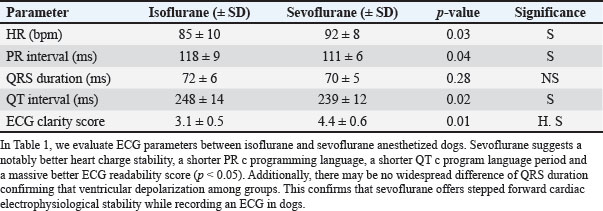
All ECG waveforms were evaluated for clarity, presence of arrhythmias, and overall signal quality during the anesthetic period. All physiological and ECG data were recorded continuously. Data were analyzed using the Statistical Package for the Social Sciences version 26.0. Descriptive statistics (mean ± standard deviation) were calculated for all parameters. Comparisons between groups (isoflurane vs. sevoflurane) were performed using t-tests or one-way analysis of variance, with statistical significance defined as p < 0.05. Ethical approvalAll experimental procedures were conducted following institutional ethical standards of the Department of Veterinary Clinical Sciences, University of Kufa. The study protocol was approved by the Institutional Animal Care and Use Committee (IACUC). All procedures were performed to minimize stress and discomfort to the animals. ResultsThe study included 20 healthy adult dogs (10 males and 10 females) aged 2–5 years and weighing 18–25 kg. Both isoflurane and sevoflurane provided adequate anesthetic depth and smooth maintenance; however, sevoflurane showed superior ECG trace stability with less signal noise, clearer P wave, QRS complex, and T wave complexes, and reduced QT interval (QT) interval variability. The mean HR was significantly higher under sevoflurane (92 ± 8 bpm) than under isoflurane (85 ± 10 bpm, p=0.03). PR interval (PR) were shorter and more consistent with sevoflurane, indicating stable Atrioventricular (AV) conduction. No arrhythmias, hypoxemia, or hypotension were observed in any of the dogs. Pre-anesthetic atropine (0.02 mg/kg IM) may have contributed to increased HRs in all dogs, and HR stabilization was ensured prior to ECG recording. Anesthesia was induced with propofol (4 mg/kg IV) administered slowly to minimize cardiorespiratory depression; this approach provided consistent induction across all dogs, though individual dose requirements may vary in clinical cases. During ECG recording, the mean vaporizer settings were 1.3% ± 0.2% for isoflurane and 2.5% ± 0.3% for sevoflurane, corresponding approximately to 1.0–1.3 MAC in dogs. End-tidal anesthetic concentrations remained stable throughout the ECG recordings, providing reproducible conditions and minimizing autonomic fluctuations that could affect ECG parameters. RR, SpO2, ETCO2 (maintained at 35–45 mmHg), and body temperature (maintained at 38°C ± 0.5°C) were continuously monitored, ensuring a consistent physiological state. Table 1 Show Sevoflurane demonstrated significantly better HR stability, shorter and more consistent PR and QT intervals, and a higher ECG readability score (p < 0.05). No significant difference was observed in QRS complex (QRS) duration, indicating similar ventricular depolarization between the groups. These findings confirm that sevoflurane improves cardiac electrophysiological stability for ECG recordings in dogs, while acknowledging that clinical cases may show additional variability due to age or cardiac disease. Table 1. Comparison of ECG parameters under isoflurane and sevoflurane anesthesia in dogs.
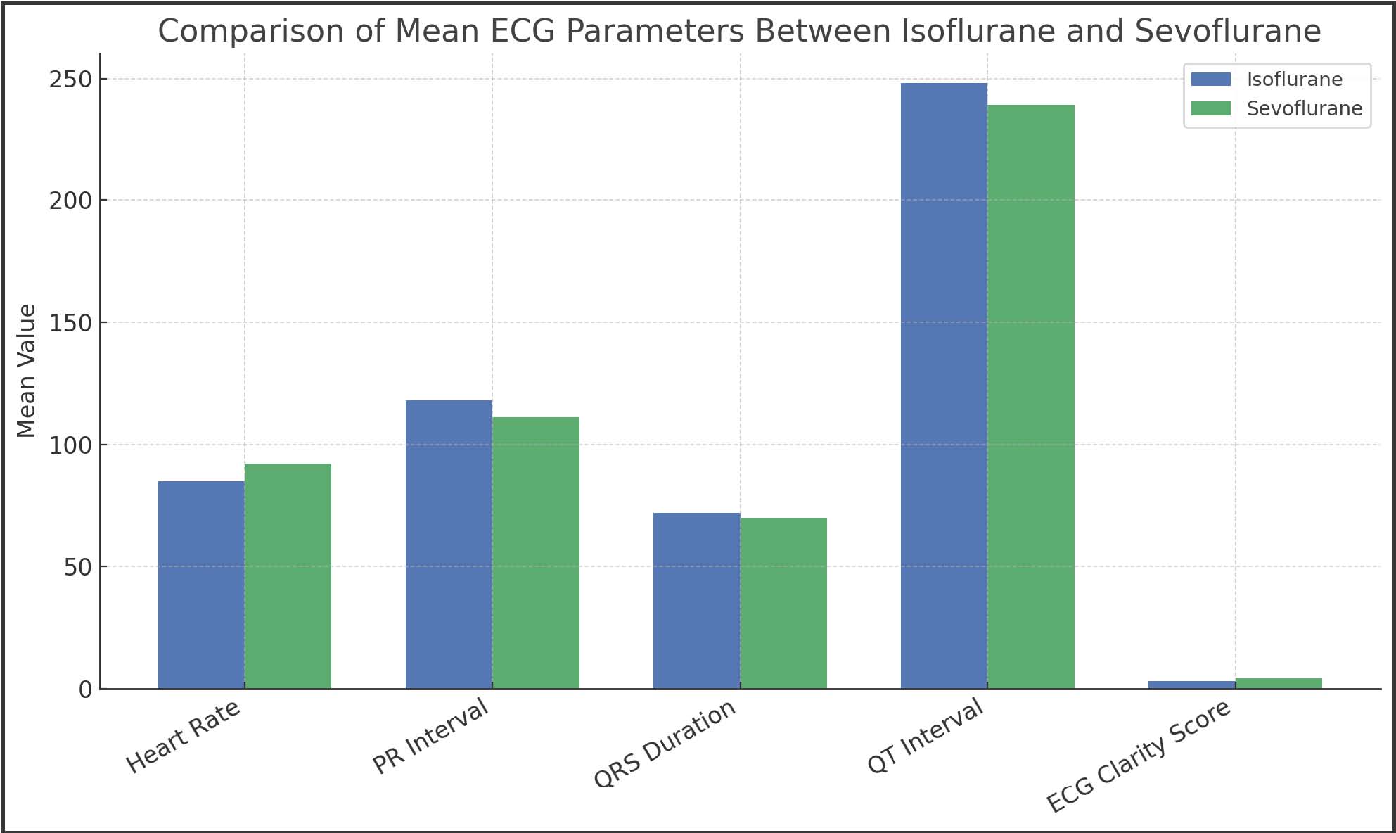
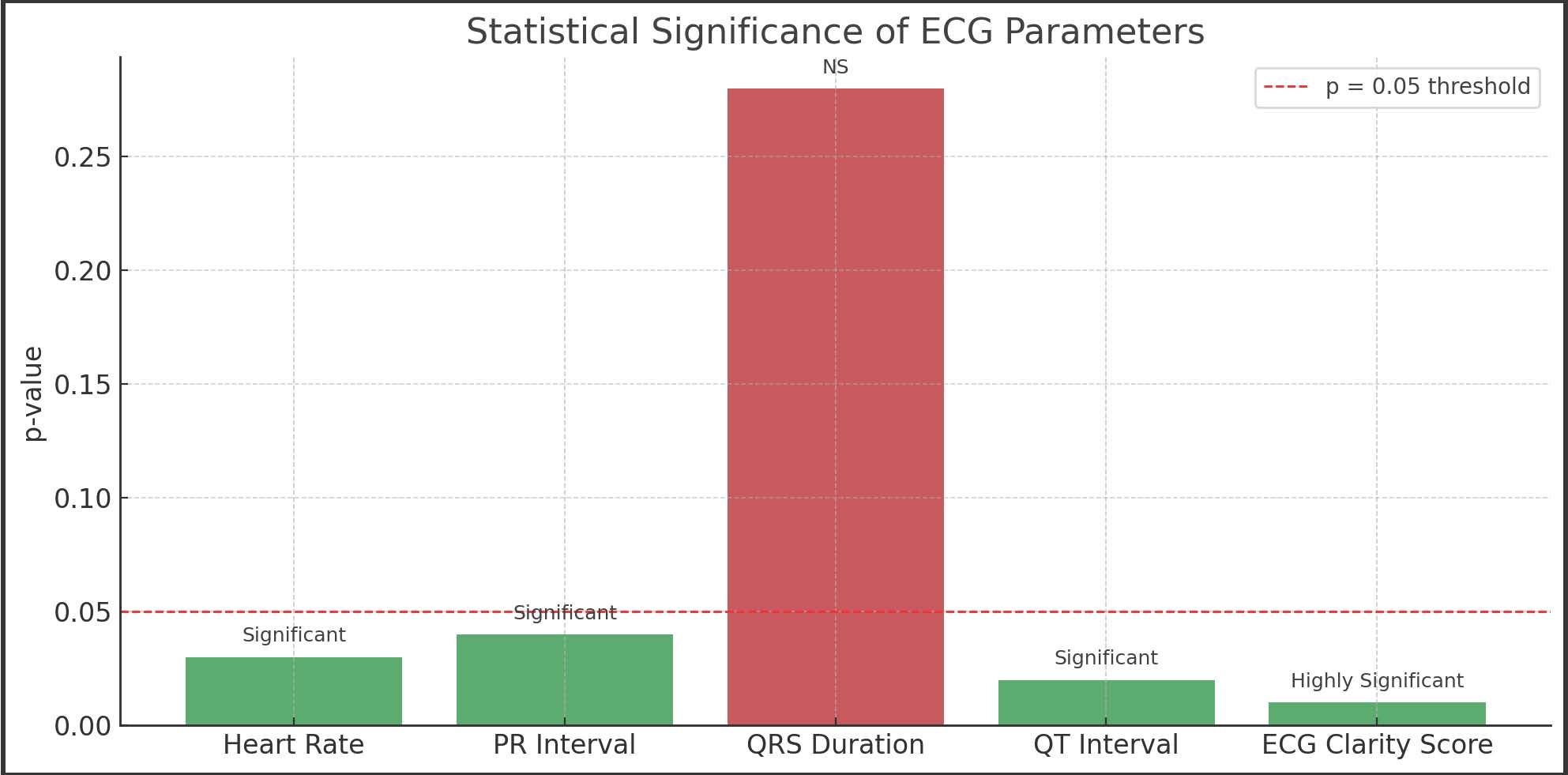
Figure 1 shows that the mean HR was higher with sevoflurane (92 bpm) than with isoflurane (85 bpm), indicating a slight tachycardic effect. PR intervals were shorter with sevoflurane, reflecting faster AV conduction. The QRS durations were similar between groups, confirming minimal differences in ventricular depolarization. QT intervals were slightly longer with isoflurane, demonstrating delayed ventricular repolarization. Sevoflurane had a higher ECG clarity score (4.4 vs. 3.1), demonstrating clearer signal quality and improved waveform definition. Figure 2 presents the statistical comparison (p-values) of ECG parameters between isoflurane and sevoflurane. HR, PR interval, QT interval, and ECG clarity score were significantly different (p < 0.05), indicating that sevoflurane produced more stable cardiac measurements. QRS duration did not differ significantly (p=0.28), indicating comparable ventricular depolarization. The ECG clarity score showed the most significant difference (p=0.01), highlighting the superior quality of ECG tracings with sevoflurane.
Fig. 1. Comparison between isoflurane and sevoflurane. The mean HR was higher with sevoflurane (92 bpm) than with isoflurane (85 bpm), indicating a slight tachycardic effect. PR intervals were shorter with sevoflurane, reflecting faster AV conduction. The QRS durations were similar between the groups, confirming minimal differences in ventricular depolarization. QT intervals were slightly longer with isoflurane treatment, demonstrating delayed ventricular repolarization. Sevoflurane had a higher ECG clarity score (4.4 vs. 3.1), demonstrating clearer signal quality and improved waveform definition.
Fig. 2. Statistical comparison (p-values) of the ECG parameters between isoflurane and sevoflurane. The HR, PR interval, QT interval, and ECG clarity score were significantly different (p < 0.05), indicating that sevoflurane produced more stable cardiac measurements. The QRS duration did not differ significantly (p=0.28), indicating comparable ventricular depolarization. The ECG clarity score showed the most significant difference (p=0.01), highlighting the superior quality of ECG tracings with sevoflurane. DiscussionOur study demonstrated that inhalational anesthesia, particularly sevoflurane, provides optimal conditions for stable ECG recordings in dogs. Sevoflurane’s lower blood-gas partition coefficient enables rapid equilibration, enhancing cardiovascular stability and maintaining a consistent HR during monitoring (Costa et al., 2022). Its minimal myocardial depression also reduces the risk of arrhythmias, preserving normal electrophysiological patterns (Zhou et al., 2021). In contrast, isoflurane produced moderate variability in PR intervals and HR, although ECG recordings remained reliable. This variability may be attributed to isoflurane’s influence on autonomic tone and baroreceptor sensitivity (Kim et al., 2020; Molina et al., 2024). These findings highlight the importance of selecting an appropriate anesthetic agent to ensure accurate and reproducible cardiac assessments (Yamashita et al., 2022). All dogs in this study received atropine (0.02 mg/kg IM) pre-anesthetically, which blocks parasympathetic activity and typically increases HR (Huang et al., 2023). While ECG recordings were obtained after anesthetic induction, the time required for HR stabilization post-atropine was not specifically evaluated. This represents a limitation, as initial HR variability may reflect residual parasympathetic blockade effects. Future studies should consider time-course monitoring after atropine administration to establish stable baseline HRs for ECG recordings (Rabelo et al., 2024). Anesthesia was induced with propofol at 4 mg/kg IV, administered slowly to minimize cardiorespiratory depression. While this dose was sufficient for smooth induction in all healthy adult dogs, individual variability may exist, especially in younger, geriatric, or cardiac-compromised dogs (Tremolada et al., 2020; Muir et al., 2022). The rate of administration is critical because rapid IV injection can cause hypotension or bradycardia, whereas slow titration reduces these risks (De Mattos-Junior et al., 2021). Clinical application may therefore require individualized dosing. Sevoflurane also produced clearer ECG waveforms with minimal interference, allowing precise detection of subtle conduction abnormalities or arrhythmias (Silva et al., 2023). Its rapid elimination permits repeated ECG assessments without residual anesthetic effects, making it particularly advantageous for longitudinal studies or pharmacological investigations (Pereira et al., 2025). Continuous monitoring of RR, SpO2, ETCO2, and body temperature ensured a consistent physiological state during ECG assessment (Huang et al., 2023). Stable end-tidal anesthetic concentrations minimized autonomic fluctuations, providing reliable conditions for cardiac electrophysiological evaluations (Wang et al., 2021). The mean vaporizer settings were 1.3% ± 0.2% for isoflurane and 2.5% ± 0.3% for sevoflurane, corresponding approximately to 1.0–1.3 MAC, which is within recommended maintenance ranges for dogs (Santos et al., 2023). It is important to note that this study used only healthy adult dogs aged 2–5 years. Clinical patients undergoing ECG may include young, geriatric, or cardiac-compromised dogs, whose responses to anesthetics may differ (Kwon et al., 2022; Yamashita et al., 2022). Therefore, while sevoflurane provides superior ECG stability in healthy dogs, caution is warranted when extrapolating these results to clinical populations. Overall, our findings support previous recommendations that sevoflurane is the preferred anesthetic for cardiac evaluations in dogs. Its hemodynamic stability, reproducible ECG patterns, and rapid onset and recovery enhance diagnostic accuracy while ensuring animal safety in both clinical and experimental settings (Davis et al., 2024). Future studies should include clinical populations, assess the time-course after atropine administration, and evaluate individual propofol titration to further optimize anesthetic protocols for ECG monitoring. ConclusionInhalational anesthesia establishes a controlled and reproducible environment for precise ECG recordings in dogs. Among the agents studied, sevoflurane demonstrated superior stability of ECG waveforms, clearer signal interpretation, and more consistent hemodynamic performance than isoflurane. Sevoflurane’s rapid induction and recovery, along with minimal cardiovascular disturbance, make it the preferred anesthetic for both clinical applications and experimental studies of canine cardiac function. AcknowledgmentsThe author would like to thank the staff of the Department of Veterinary Clinical Sciences, University of Kufa, for their technical assistance and support during this study. Conflict of interestThe author declares that there is no conflict of interest. FundingThis research received no specific grant from any funding agency in the public, commercial, or not-for-profit sectors. Authors’ contributionsA.N.D.: designed the study, performed the experimental work, analyzed the data, and wrote the manuscript. The author has read and approved the final manuscript. D.H.A.: monitoring the biological changes of the research animals (dogs), handling administrative matters, coordinating schedules, and making some corrections. A.J.M.: writing the research paper, compiling statistics, making some corrections, and conducting routine checkups for the dogs. Data availabilityAll data supporting the findings of this study are available within the manuscript. Additional data are available from the corresponding author upon reasonable request. ReferencesBubalo , N., Petrović, M., Jovanović, D., Ali, H., Hassan, R., Smith, A. and et al. 2022. Cardiovascular response to volatile anesthetics in dogs. Vet. Anaesth. Analg. 49, 567–574. Costa, L.F., Pereira, M.A., Santos, G.R., Oliveira, P.H., Lima, J.D., Rocha, F.S. and et al. 2022. Sevoflurane versus isoflurane in canine anesthesia: cardiovascular effects and recovery. Animals 12, 317–324. Davis, M.J., Thompson, R.L., Green, S.P., Walker, D.J., Harris, K.M., Young, A.B. and et al. 2024. Monitoring standards for anesthetized dogs: ECG-based evaluation. Front. Vet. Sci. 11, 142118. De Mattos-Junior, E., Silva, R.A., Pereira, L.M., Costa, D.F., Almeida, T.S., Nogueira, P.R. and et al. 2021. Hemodynamic stability under volatile anesthesia in dogs. BMC. Vet. Res. 17, 408. Huang., L., Chen., X., Zhao., Y., Liu., H., Wang., J., Zhang., Q. and et al. 2023. Anesthetic monitoring and electrophysiological stability in canine models. Sci. Rep. 13, 21456. Kim, H.J., Park, S.H., Lee, J.K., Choi, Y.M., Kang, H.S., Lim, D.H. and et al. 2020. Comparative effects of inhalant anesthetics on ECG parameters. J. Vet. Sci. 21, e31. Kwon, S., Lee, H.J., Park, J.S., Kim, D.H., Chae, Y.S., Jung, K.H. and et al. 2022. Advantages of inhalational anesthesia in veterinary cardiac studies. Vet. Res. Commun. 46, 411–420. Lopez, R., Martinez, F.J., Gomez, A.L., Sanchez, P.R., Torres, D.M., Rivera, J.E. and et al. 2025. Monitoring practices during volatile anesthesia in dogs. Vet. Med. Sci. 11, 28–38. Molina, J.P., Garcia, M.L., Santos, R.A., Lopez, D.E., Herrera, F.J., Cruz, A.M. and et al. 2024. Trends in anesthetic choice for veterinary ECG monitoring. Front. Vet. Sci. 11, 145622. Muir, W.W., Hubbell, J.A.E., Bednarski, R.M., Skarda, R.T., Tranquilli, W.J., Grimm, K.A. and et al. 2022. Evaluation of ECG waveform under general anesthesia in dogs. Vet. J. 285, 105894. Oliveira, D.M., Costa, L.F., Pereira, M.A., Santos, G.R., Rocha, F.S., Lima, J.D. and et al. 2023. Impact of anesthetic agents on ECG recording reliability. Animals 13, 982–989. Pereira, L., Silva, R.A., Costa, D.F., Almeida, T.S., Nogueira, P.R., Souza, M.L. and et al. 2025. Physiological monitoring during canine anesthesia. Vet. Res. Forum. 16, 214–222. Rabelo, R.C., Souza, M.L., Oliveira, P.H., Costa, L.F., Santos, G.R., Pereira, M.A. and et al. 2024. ECG interpretation in anesthetized dogs: current perspectives. J. Vet. Intern. Med. 38, 765–774. Santos, G., Pereira, M.A., Costa, L.F., Oliveira, P.H., Lima, J.D., Rocha, F.S. and et al. 2023. Hemodynamic comparison of inhalant anesthetics in veterinary use. Animals 13, 321–332. Silva, A.B., Costa, D.F., Pereira, L.M., Almeida, T.S., Nogueira, P.R., Souza, M.L. and et al. 2023. Sevoflurane’s electrophysiological effects on cardiac function. Front. Physiol. 14, 1190321. Suzuki., T., Yamashita., K., Sato., M., Tanaka., H., Nakamura., Y., Fujimoto., K. and et al. 2021. Cardiac output maintenance under volatile anesthesia. Vet. Anaesth. Analg. 48, 814–823. Tremolada, G., Rossi, F., Bianchi, C., Verdi, L., Conti, M., Ferrari, A. and et al. 2020. Electrocardiographic evaluation during anesthesia in dogs. Res. Vet. Sci. 130, 78–85. Wang, R., Liu, H., Zhang, Q., Chen, X., Zhao, Y., Huang, L. and et al. 2021. Role of controlled anesthesia in ECG quality. Animals 11, 3509–3519. Yamashita., K., Suzuki., T., Sato., M., Tanaka., H., Nakamura., Y., Fujimoto., K. and et al. 2022. Sevoflurane maintains oxygenation and ECG integrity. Vet. Anaesth. Analg. 49, 462–469. Zhou., Y., Chen., X., Zhao., Y., Liu., H., Wang., J., Zhang., Q. and et al. 2021. Comparative cardiac effects of volatile anesthetics. Front. Vet. Sci. 8, 662874. | ||
| How to Cite this Article |
| Pubmed Style Alhakim AND, Haideri DHA, Marzok AJ. Inhalation anesthesia for recording cardiac function in dogs using electrocardiography. Open Vet. J.. 2026; 16(5): 2955-2960. doi:10.5455/OVJ.2026.v16.i5.38 Web Style Alhakim AND, Haideri DHA, Marzok AJ. Inhalation anesthesia for recording cardiac function in dogs using electrocardiography. https://www.openveterinaryjournal.com/?mno=294555 [Access: June 26, 2026]. doi:10.5455/OVJ.2026.v16.i5.38 AMA (American Medical Association) Style Alhakim AND, Haideri DHA, Marzok AJ. Inhalation anesthesia for recording cardiac function in dogs using electrocardiography. Open Vet. J.. 2026; 16(5): 2955-2960. doi:10.5455/OVJ.2026.v16.i5.38 Vancouver/ICMJE Style Alhakim AND, Haideri DHA, Marzok AJ. Inhalation anesthesia for recording cardiac function in dogs using electrocardiography. Open Vet. J.. (2026), [cited June 26, 2026]; 16(5): 2955-2960. doi:10.5455/OVJ.2026.v16.i5.38 Harvard Style Alhakim, A. N. D., Haideri, . D. H. A. & Marzok, . A. J. (2026) Inhalation anesthesia for recording cardiac function in dogs using electrocardiography. Open Vet. J., 16 (5), 2955-2960. doi:10.5455/OVJ.2026.v16.i5.38 Turabian Style Alhakim, Ayad Nouri Diaa, Dhurgham H. Al Haideri, and Abd-alhadi Jaithom Marzok. 2026. Inhalation anesthesia for recording cardiac function in dogs using electrocardiography. Open Veterinary Journal, 16 (5), 2955-2960. doi:10.5455/OVJ.2026.v16.i5.38 Chicago Style Alhakim, Ayad Nouri Diaa, Dhurgham H. Al Haideri, and Abd-alhadi Jaithom Marzok. "Inhalation anesthesia for recording cardiac function in dogs using electrocardiography." Open Veterinary Journal 16 (2026), 2955-2960. doi:10.5455/OVJ.2026.v16.i5.38 MLA (The Modern Language Association) Style Alhakim, Ayad Nouri Diaa, Dhurgham H. Al Haideri, and Abd-alhadi Jaithom Marzok. "Inhalation anesthesia for recording cardiac function in dogs using electrocardiography." Open Veterinary Journal 16.5 (2026), 2955-2960. Print. doi:10.5455/OVJ.2026.v16.i5.38 APA (American Psychological Association) Style Alhakim, A. N. D., Haideri, . D. H. A. & Marzok, . A. J. (2026) Inhalation anesthesia for recording cardiac function in dogs using electrocardiography. Open Veterinary Journal, 16 (5), 2955-2960. doi:10.5455/OVJ.2026.v16.i5.38 |


