





| Research Article | ||
Open Vet. J.. 2026; 16(8): 5434-5445 !
Open Veterinary Journal, (2026), Vol. 16(8): 5434–5445 Research Article Advances in animal tuberculosis surveillance in Argentine wildlife using multiplex PCRLoreana Carla Ponce1,2*, Johanna Capra2, María Jimena Marfil2, Marina Winter3, Agostina Tammone Santos4, Jorge Peña Martínez5, Cecilia Moyano6, María Mesplet2,7, María Sol Perez Aguirreburualde8 and Soledad Barandiaran1,21CONICET-Universidad de Buenos Aires, Instituto de Investigaciones en Producción Animal (INPA), Buenos Aires, Argentina 2Universidad de Buenos Aires, Facultad de Ciencias Veterinarias, Cátedra de Enfermedades Infecciosas, Buenos Aires, Argentina 3Universidad Nacional de Río Negro. Sede Viedma del Centro de Investigaciones y Transferencia de Río Negro (UNRN-CONICET), Av. Don Bosco y Leloir. Viedma, Río Negro, Argentina 4Centro de Investigaciones en Física e Ingeniería del Centro de la Provincia de Buenos Aires (CIFICEN)-UNCPBA-CONICET-CICPBA, Tandil, Buenos Aires, Argentina 5Endangered Species and Environments Restoration Program, Rewilding Argentina Foundation, Buenos Aires, Argentina 6Programa de especies exóticas invasoras, Parque Nacional El Palmar, Colón, Entre Ríos, Argentina 7Universidad de Buenos Aires, Instituto de Investigaciones en Epidemiología Veterinaria (IIEV), Buenos Aires, Argentina 8Center for Animal Health and Food Safety, University of Minnesota, Minneapolis, Minnesota, USA *Corresponding Author: Loreana Carla Ponce. CONICET-Universidad de Buenos Aires, Instituto de Investigaciones en Producción Animal (INPA), Buenos Aires, Argentina. Email: lponce [at] fvet.uba.ar Submitted: 03/11/2025 Revised: 11/06/2026 Accepted: 30/06/2026 Published: 08/08/2026 © 2025 Open Veterinary Journal
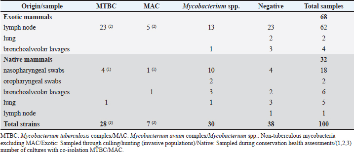
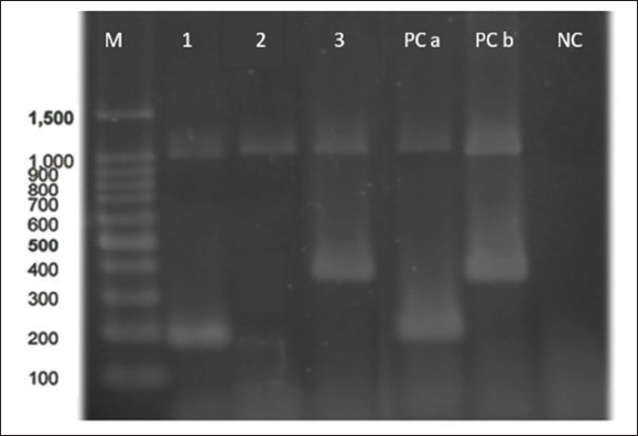
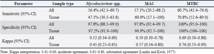
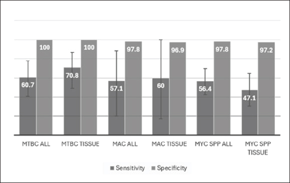
AbstractBackground: Animal tuberculosis caused by Mycobacterium bovis, a member of the Mycobacterium tuberculosis complex (MTBC), remains endemic in Argentina and poses a zoonotic risk at the wildlife–livestock interface. Heterogeneous sampling conditions and limited access to rapid, cost-effective diagnostic tools hinder multi-host wildlife system surveillance. Aim: This study aimed to evaluate the performance of an adapted multiplex polymerase chain reaction (mPCR) method for the post-culture differentiation of MTBC, Mycobacterium avium complex (MAC), and non-tuberculous mycobacteria (NTM) across diverse wildlife sample matrices in Argentina. Methods: We tested 100 cultures (70 tissue-derived, mainly from culled invasive species; 30 derived from minimally invasive samples from living native species). Singleplex PCR (sPCR) was used as a molecular benchmark after culture. Diagnostic sensitivity, specificity (95% confidence interval [CI]), and agreement (Cohen’s κ) were calculated. Results: sPCR confirmed the presence of mycobacteria in 62/100 cultures and identified 65 strains: 28 MTBC, 7 MAC, and 30 NTM; three cultures showed MTBC + MAC co-isolation. MTBC was predominantly recovered from the lymph nodes of exotic mammals (23/28; 82.1%), whereas only four MTBC strains (14.3%) were detected in the minimally invasive samples of native mammals. The adapted mPCR showed high specificity (MTBC, 100%; Mycobacterium genus and MAC, 97.8%). The overall sensitivity was 60.7% for MTBC, 57.1% for MAC, and 56.4% for the Mycobacterium genus, with substantial agreement for MTBC (κ=0.69) and moderate agreement for MAC (κ=0.59) and genus-level (κ=0.52) detection. Performance improved in tissue-derived cultures, with MTBC sensitivity reaching 70.8% (specificity 100%). No cross-reactivity was observed with the tested non-mycobacterial organisms. Conclusion: This mPCR provides a practical and cost-effective approach for the post-culture classification of mycobacteria in wildlife surveillance, especially when tissue sampling is feasible. We recommend expanding the scale of wildlife studies across diverse sample matrices and ecological settings and explicitly incorporating wildlife–livestock–human interface scenarios to characterize the epidemiological role of different wildlife hosts and support integrated, One Health-oriented health surveillance programs. Keywords: Animal tuberculosis; Molecular diagnosis; Surveillance; Wildlife; Zoonoses. IntroductionMycobacteria comprise two principal categories: pathogenic strains and environmental saprophytes. The Mycobacterium tuberculosis complex (MTBC) is the main pathogenic group responsible for tuberculosis in humans and animals (aTB). Nontuberculous mycobacteria (NTM), including most Mycobacterium avium complex (MAC) strains, are primarily environmental organisms. However, specific MAC members exhibit pathogenic behaviors, particularly in veterinary contexts, associated with chronic conditions, such as paratuberculosis in ruminants, resulting in significant economic losses (Carta et al., 2013; Whittington et al., 2019). aTB is a zoonotic disease mainly caused by Mycobacterium bovis within the MTBC. It continues to pose major global health, ecological, and economic challenges (Kanipe and Palmer, 2020). In Latin America, including Argentina, the disease remains endemic in livestock despite longstanding efforts to control it (Res. SENASA 128/12). This epidemiological persistence stems from the remarkable adaptability of the pathogen, demonstrated through its broad host range, capacity for latent infection, and the suboptimal sensitivity of current diagnostic methods (Nuñez-Garcia et al., 2018; Balseiro et al., 2020; Palmer et al., 2020). Historically, local control programs have notably focused on livestock, with limited integration of wildlife surveillance. This gap persists despite increasing evidence of the critical epidemiological role of wildlife in the region (World Organisation for Animal Health, 2024). Wildlife species may act as maintenance or spillover hosts in aTB ecology, with their epidemiological role determined by ecological traits, behavior, and livestock contact (Palmer, 2013; Santos et al., 2022). Transmission dynamics depend on host density, habitat structure, and shared resource use (Triguero-Ocaña et al., 2019). Examples of wildlife species identified as reservoirs of the disease include possum, badger, certain deer species, and wild boar (Gavier-Widén et al., 2009; Fitzgerald and Kaneene, 2013; Nugent et al., 2018; Richomme et al., 2019). The latter two species are invasive exotic species with high ecological adaptability and a broad distribution across the country and are currently under investigation as potential aTB reservoirs in the region (La Sala et al., 2021; Ballari et al., 2024; Barandiaran et al., 2024a). Recent studies in Argentina have confirmed that the wild boar is a significant reservoir of M. bovis, with a prevalence of 11.2%. The spoligotypes are shared with cattle, domestic pigs, and other wildlife, underscoring their role in multihost transmission cycles (Barandiaran et al., 2024a). M. bovis infection in wildlife remains a critical concern not only because of its potential to cause spillback to livestock but also because of its impact on biodiversity. Spillover into native mammalian species can lead to population decline, further threatening their conservation status and disrupting ecosystem stability (Lamattina et al., 2025). The ecological impact of the introduction of M. bovis into wildlife populations has resulted in notable declines of native species, such as the African lion (Panthera leo) in Kruger National Park (Miller et al., 2019). This issue is particularly relevant in Argentina, as more than 22.5% of native mammal species are classified as threatened by national conservation criteria (Ministry of Environment and Sustainable Development, 2019). Furthermore, this threat extends beyond M. bovis. The significant prevalence of NTM (16.36%), including Mycobacterium avium subsp. hominissuis, detected in endangered Argentine wildlife, demonstrates a broader spectrum of shared microbial threats at the wildlife–livestock interface (Barandiaran et al., 2024b). Effective conservation, livestock safety, and public health safeguarding are contingent upon an integrated One Health framework that addresses the convergence of these pathogens in our ecosystems. Effective aTB surveillance in wildlife faces two primary constraints: sample collection limitations and diagnostic methodological challenges. The status of the animal (living or diseased) fundamentally determines sample collection strategies. Ethical considerations mandate minimally invasive techniques during planned captures for free-ranging native species, typically yielding gastric/endotracheal lavages, swabs, or blood samples via chemical restraint (Li et al., 2021; Barandiaran et al., 2024b). Technical limitations further constrain the diagnostic capacity for mycobacterial diseases. Bacterial culture remains the gold standard for detecting infections, and molecular methods offer an essential classification tool (Wahdan et al., 2020). While widely implemented, conventional single-target polymerase chain reactions (PCRs) require substantial time and resources to differentiate mycobacteria relevant to veterinary medicine (Lorente-Leal et al., 2021a,b; Mabe et al., 2024). Although multiple polymerase chain reaction (mPCR) is promising, current validations predominantly use bovine samples from abattoirs, leaving a critical knowledge gap for wildlife applications across diverse field conditions and sample matrices (tissues vs. swabs/lavages). This study aimed to optimize and evaluate an established mPCR post-culture protocol for identifying key veterinary mycobacteria (MTBC, MAC, and NTM) in Argentine wildlife. By improving post-culture differentiation, this approach enhances surveillance in multihost systems and supports zoonosis management. Unlike previous validations, this study focuses on diverse wildlife matrices and integrates ecological and One Health dimensions. Materials and MethodsSample collectionBetween 2018 and 2022, samples were received and processed from free-ranging mammals of different species and geographical locations. This study was conducted at the Laboratory of Tuberculosis Diagnosis, Infectious Diseases Department, Faculty of Veterinary Science, University of Buenos Aires. This study included samples obtained from submandibular lymph nodes (LNs), lungs, bronchoalveolar lavages (BAL), nasopharyngeal swabs (NPS), and oropharyngeal swabs (OPS). Most of the minimally invasive samples (BAL, NPS, and OPS) were collected from living mammals under conservation and translocation programs (Zamboni et al., 2017). Tissue samples (lungs and LNs) were obtained from diseased mammals from National and/or Provincial Parks and Nature Reserves, collected by authorized hunters during invasive species management programs. All samples were stored at –20 °C until processing. All the research was performed under provincially approved hunting license and rewilding projects: https://rewildingargentina.org/iber a-project. Bacteriological cultures and selection criteriaInitially, tissue samples were examined macroscopically for tuberculous-like lesions (TBLL) before culturing. All samples were cultured accordingly. The Petroff decontamination technique was applied before microbiological culture on Löwenstein-Jensen and Stonebrink media (Jorge et al., 2005). Cultures were incubated at 37 °C and monitored weekly for 12 weeks. For this performance evaluation study, 100 bacterial cultures were selected from a biobank obtained between 2018 and 2022. The selection criteria were as follows: 1) growth on Lowenstein-Jensen or Stonebrink medium with colony morphology suggestive of mycobacteria; 2) sufficient biomass for DNA extraction and molecular assays; and 3) representation of the two main sampling types: tissue samples, mainly from culled invasive species, and minimally invasive samples (swabs and lavages), mainly from live native species under conservation programs. This selection process yielded a final set of 70 tissue-derived cultures and 30 minimally invasive sample-derived cultures. DNA extractionMaterials from the 100 selected cultures were suspended in 500 μl of sterile pyrogen-free distilled water in 1.5-ml DNA and RNAase-free microtubes. DNA was extracted by thermal lysis, and the microtubes were subjected to 94 °C for 40 minutes. Finally, the samples were centrifuged at 12,000 rpm for 10 minutes. Molecular identification by singleplex PCR (sPCR)Microtube supernatant (5 μl) was collected for molecular analysis. The sPCR targets used as a benchmark for the post-culture identification of Mycobacterium spp., MTBC, and MAC at the genus level are detailed in the Supplementary Table. These targets were selected based on their broad validity and routine use for the accurate differentiation of mycobacteria in post-culture characterization (Hermans et al., 1990; Telenti et al., 1993; Guerrero et al., 1995). The detailed reaction components and thermal cycling conditions for sPCR are provided in the Supplementary Table. IS6110 amplification was performed using a Touch-down PCR program (Zumárraga et al., 2005) to enhance sensitivity. Horizontal electrophoresis on a 2% agarose gel resolved the amplicons, which we stained with ethidium bromide (0.5 µg/ml) and visualized under UV transillumination. Each study included positive and negative controls. Molecular identification by mPCRThe mPCR method described by Wilton and Cousins (1992) was adapted for the identification of the three groups of mycobacteria under study. Primers originally designed by the authors were used to amplify specific targets: the 16S rRNA gene for the Mycobacterium genus (MYCGEN-f/MYCGEN-r) and MAC (MYCGEN-f/MYCAV-r), and the gene sequence encoding the MPB70 protein for MTBC (TB 1-f/TB1-r). Primer specificity was first assessed in silico using BLAST. Subsequently, optimal annealing temperatures were predicted using the OligoAnalyzer™ online tool and then optimized experimentally using temperature gradient PCR on an IVEMA T21® thermocycler. All primers were purchased from Thermo Fisher Scientific®. Reference strains of M. bovis (AN5) and M. avium (D4) were used as positive controls for MTBC and MAC, respectively. Both reference strains served as positive controls for the Mycobacterium genus. Sterile distilled water was used as a contamination control. Each reaction had a final volume of 25 μl, consisting of 5 μl of template DNA added to 20 μl of a master mix containing: 0.5 units of Taq DNA polymerase (Inbio Highway®, Argentina), 5 μl of 5X reaction buffer, 0.5 μl of 10 mM deoxynucleotide triphosphates (dNTPs), 2 μl of each 10 µM oligonucleotide, and sterile ultrapure distilled water. The key modifications from the original protocol, including the use of commercial master mix components and adjusted reagent concentrations, are detailed in Supplementary Table S1. The amplification program was modified as follows. An initial denaturation at 92°C for 2 minutes, followed by 30 cycles of denaturation at 92°C for 1 minute, annealing at 58°C for 30 seconds, and extension at 72°C for 1 minute, with a final extension at 72°C for 10 minutes. The amplification products were separated by horizontal electrophoresis on a 2% agarose gel (90 V for 50 minutes), stained by immersion in an ethidium bromide solution (0.5 µg/ml), and visualized under UV transillumination. Amplicon sizes were determined using a 100-bp DNA ladder (Inbio Highway®, Argentina). The analytical specificity of the mPCR was experimentally analyzed against template DNA from Corynebacterium, Nocardia, Rhodococcus, Salmonella, and Streptococcus zooepidemicus. Statistical comparison of resultsThe sPCR results were used to evaluate the performance of the adapted mPCR technique. mPCR performance was estimated using Win Episcope 2.0 (online version tool) with a 95% confidence level. The concordance was assessed using Cohen’s kappa coefficient. The degree of agreement was considered according to the empirical scale of Landis and Koch (Landis and Koch, 1977; Abraira, 2000). Given the markedly different origins and characteristics of the analyzed samples (lymph nodes, lung, bronchoalveolar lavages, and swabs), the sample matrix type (especially tissue, which was the most representative) was considered in the statistical comparison between mPCR and sPCR results. Ethical approvalEthical approval and permits for animal experimentation do not apply to this study. ResultsA total of 100 cultures were selected from minimally invasive and tissue matrices, including BAL (n=10), NPS (n=18), OPS (n=2), LNs (n=63), and lung tissue (n=7). TBLL was observed in only 10% of tissues (7/70). Six of these lesions were in the LNs, and one was in the lung tissue. Molecular sPCR resultsCultures were subsequently analyzed by sPCR; 62 samples tested positive for mycobacteria, and 38 samples yielded negative PCR results. Molecular characterization revealed 65 distinct mycobacterial strains. The strain distribution is presented in Table 1. MTBC was confirmed in four of the TBLL cases (three from LNs and one from the lung), whereas no mycobacteria were detected in the remaining three cases. In addition, MTBC was identified in 20 tissue samples without visible lesions. Notably, three cultures from different individuals (two LN samples and one NPS) demonstrated co-isolation of MTBC and MAC strains. MTBC was predominantly identified in exotic LN mammals (33.3%, 21/63). On the contrary, Mycobacterium spp. was more frequently detected in native mammals from NPS (55.6%, 10/18) and BAL (40%, 4/10). MAC strains were identified in 4.8% of LNs (3/63) and 10% of BAL samples (1/10). Among the culture-negative samples (n=38), a significant proportion (20/30, 66.7%) were from living native wildlife specimens. mPCR resultsIn live animals, 53% of cultures were positive (16/30), predominantly identifying Mycobacterium spp. (15 cases) and MAC (1 case). The positivity rate was 47% (33/60) for deceased animals, with a broader distribution that included CMTB (16 cases), Mycobacterium spp. (14 cases), and MAC (5 cases) (Fig. 1). The molecular results of mPCR culture identification (n=100) were compared with those of sPCR (Table 2). Analysis of agreement revealed moderate agreement with Mycobacterium spp. (κ=0.52; 95% CI=0.34–0.69) and MAC (κ=0.59; 95% CI=0.39–0.78), and substantial agreement with MTBC (κ=0.69; 95% CI=0.50–0.88). Furthermore, the mPCR demonstrated consistently high specificity but limited sensitivity compared with the three single PCRs following culture identification across all cultures. Specificity was complete when compared with MTBC (100%; 95% CI=93.3%–100%) and remained nearly as high when compared with Mycobacterium spp. (97.8%; 95% CI=88.2–99.9) and MAC (97.8%; 95% CI=92.4%–99.7%). However, the sensitivity was considerably lower, reaching only 56.4% (31/55; 95% CI=42.3–69.5) when compared with Mycobacterium spp., 57.1% (4/7; 95% CI=20.2–88.2) when compared with MAC, and 60.7% (17/28; 95% CI=40.8–77.8) when compared with MTBC. These limitations were largely attributable to discrepancies between 24 Mycobacterium spp. and 11 MTBC sPCR-positive/mPCR-negative. For tissue-derived cultures, the mPCR sensitivity for MTBC was 70.8% (95% CI=48.9–86.6) with perfect specificity (95% CI=93.3–100). The agreement remained substantial for MTBC (κ=0.76) and moderate for Mycobacterium spp. (κ=0.45) and MAC (κ=0.57). Statistical results are graphical in Figure 2. Finally, the analytical specificity assessment of mPCR demonstrated the absence of cross-reactivity with non-mycobacterial isolates included in this study. Table 1. Distribution of mycobacterial strains identified by sPCR in selected cultures from exotic and native mammals, categorized by matrix type.
Fig. 1. Representative agarose gel electrophoresis of mPCR products. Lanes: M., DNA ladder (100 bp); 1, MTBC wildlife strain; 2, NTM wildlife strain; 3, MAC wildlife strain; 4-5, PCa, MTBC control M. bovis AN5 (372 bp), PCb., MAC control M. avium D4 (180 bp); 6, NC., negative control (sterile water). Both positive control lanes (4 and 5) also show the genus-specific band (1030 bp). Table 2. Diagnostic performance of mPCR: sensitivity, specificity, and kappa value of mPCR compared with those of sPCRs for mycobacterial detection across different wildlife matrices.
DiscussionPerformance of the adapted mPCRInvestigating the presence of pathogenic and nonpathogenic mycobacteria in wildlife that share their environment with livestock from extensive grazing systems, while also interacting with humans, such as veterinarians, hunters, and park rangers, holds significance from the One Health perspective. Implementing mPCR for reliable molecular differentiation of MTBC, MAC, and NTM in wildlife cultures constitutes a relevant methodological advance for aTB research in wildlife. The comparative analysis shows that mPCR has high specificity and moderate sensitivity for detecting mycobacterial strains. However, the interpretation of diagnostic performance should be made cautiously. While the specificity was excellent, the sensitivity was moderate across the targets, indicating that false-negative results remain possible, particularly in matrices with low bacterial loads or heterogeneous mycobacterial biomass. Therefore, mPCR results are best interpreted as confirmatory for MTBC/MAC when positive, but they are not sufficient to rule out infection when negative, especially in minimally invasive sample types. The agreement between mPCR and sPCR was good for MTBC and moderate for Mycobacterium spp. and MAC, which was supported by the statistical results. The detection capacity of MTBC through mPCR significantly increased when the analyzed samples were derived from tissue. This could be related to the type of matrix analyzed, as the tissues consisted of submandibular lymph nodes, which typically harbor a higher bacterial load than other sample types. This results in cultures with greater mycobacterial biomass, which in turn increases the sensitivity of PCR detection, particularly affecting mPCR assays. Conversely, in conservation programs, minimally invasive samples are crucial for health assessments but often contain lower bacterial loads and are more prone to environmental contamination than tissues, which can reduce diagnostic sensitivity (Thomas and Chambers, 2021; Barandiaran et al., 2024b). On the other hand, carcass sampling, predominantly from invasive mammal species culling programs, enables pathological inspection and collection of diagnostically superior samples (e.g., lymph nodes, lungs, liver) (Gürtler et al., 2018). These heterogeneous wildlife matrix conditions directly influence the diagnostic sensitivity of assays and subsequent molecular classification of mycobacteria (Thomas and Chambers, 2021). These results underscore the applicability of this technique for wildlife surveillance contexts where tissue sampling, such as lymph node collection, is readily accessible, as in hunted invasive exotic mammals or road-killed mammals. Overall, these findings highlight the feasibility of implementing surveillance in wildlife populations that are often underrepresented in diagnostic systems. Because clinical interpretation at the individual level is limited in wildlife, this approach’s primary value lies in strengthening population-level monitoring of mycobacterial pathogens at the wildlife–livestock interface.
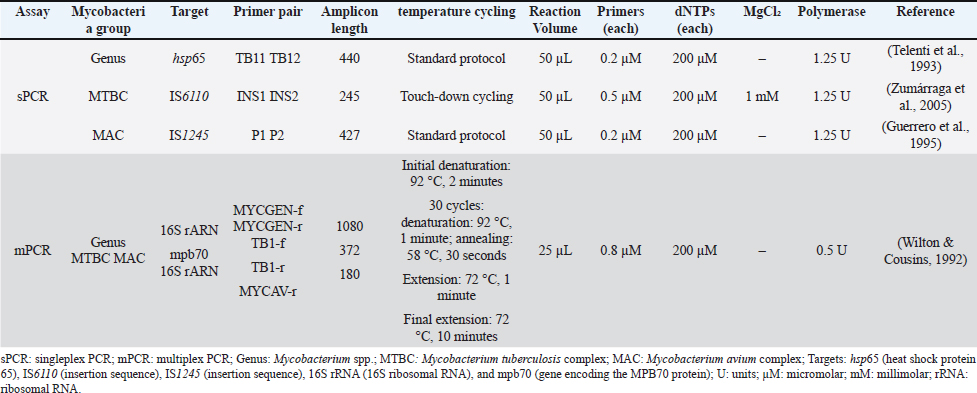
Fig. 2. Comparative identification performance of mPCR versus sPCR assays. It displays the sensitivity (grey) and the specificity (light grey) of the adapted mPCR assay for the detection of MTBC, MAC, and the Mycobacterium genus, for all samples (n=100) and for a tissue sample subset (n=70). Error bars represent 95% confidence intervals. Technical considerations and limitationsThe two selected DNA segments (IS6110, MPB70) for detecting MTBC strains are widely used in both animal and human literature (Wahdan et al., 2020; Lorente-Leal et al., 2021a,b). A recent meta-analysis confirmed that these targets consistently demonstrate high sensitivity and specificity, supporting their robustness for diagnosing aTB (Mabe et al., 2024). Despite this, differences in MTBC detection between mPCR and sPCR could be explained by technical factors such as primer design, chemical reaction stability, variability in target sequence copy number among strains, and primer thermodynamics (Quan et al., 2016). Similarly, the reduced capacity of mPCR to detect and differentiate mycobacterial species could also stem from selection and drift events, commonly observed in mPCR (Bolivar et al., 2014; Mabe et al., 2024). These limitations contributed to the failure of mPCR to detect co-isolations of MAC and MTBC strains in three mixed cultures, which sPCR successfully detected. This finding highlights that multiplex formats may underestimate co-infections and should be interpreted cautiously. Furthermore, the performance of mPCR in Mycobacterium spp. detection may have been influenced by the length of the target sequence (1030 bp), which was the longest among the targets used in this assay. The disparity in target lengths, with MAC and MTBC being significantly shorter (372 and 180 bp, respectively), can lead to uneven amplification efficiency (Kralik and Ricchi, 2017). Moreover, small variations in reaction conditions can have a more significant impact on mPCR compared to sPCR, potentially affecting the reproducibility of results (Markoulatos et al., 2002). These factors highlight the technical challenges associated with mPCR when targeting sequences of varying lengths and underscore the need for careful optimization to ensure consistent and reliable detection. Finally, the interpretation of results should consider the inherent constraints of wildlife surveillance. The limited sample size for certain targets, particularly MAC (n=7), results in wide confidence intervals for sensitivity estimates (20.2%–88.2%), reflecting the practical challenges of obtaining wildlife samples. Furthermore, our sampling strategy intentionally captured two distinct epidemiological contexts: tissue samples from hunted invasive species and minimally invasive samples from native species under conservation. This design provides valuable insights into assay performance across different sample matrices but means prevalence estimates should be contextualized within each host group rather than generalized across wildlife. Epidemiological and ecological implicationssPCR confirmed mycobacteria in 62% of the 100 selected cultures showing signs of bacterial growth. The remaining 38 negative cultures likely reflected contamination or non-mycobacterial overgrowth, possibly due to incomplete decontamination or low mycobacterial loads (Corner et al., 2012). This could be attributed to failures in the decontamination process or low viable mycobacterial load in the original sample (Corner et al., 2012; Lorente-Leal et al., 2021a,b). MTBC strains were predominantly isolated from exotic mammal tissue samples. Of the 28 identified MTBC strains, 23 (82.1%) were isolated from their lymph nodes. On the contrary, only four strains (14.3%) were detected in native mammals. These outcomes underscore the crucial role of exotic species as a potential source of infection in the region. Their high population density and documented interaction with livestock in extensive production systems may contribute to their epidemiological significance (La Sala et al., 2021; Ballari et al., 2024; Barandiaran et al., 2024a). However, given the sampling bias in this study, namely the underrepresentation of minimally invasive samples from native wildlife due to logistical and ethical constraints, additional large-scale research in native species is recommended. This will help clarify their potential role in epidemiology and long-term disease persistence in the region. The detection and characterization of Mycobacterium spp. in wildlife contributes to a deeper understanding of their ecology and transmission dynamics. NTM was detected in 20% (14/70) of tissue samples and in 53.3% (16/30) of minimally invasive samples. This indicates a higher NTM isolation rate in native wildlife. This higher rate may reflect the greater environmental exposure of native hosts, which reside continuously in natural ecosystems as opposed to agricultural settings. Their characteristic behaviors, including foraging, digging, and direct contact with soil and water sources, increase their contact with environmental mycobacteria. Furthermore, MAC was isolated in 7.14% (5/70) of tissue samples and 6.66% (2/30) of minimally invasive samples, representing similar proportions. The detection of MAC has significant implications for the diagnosis of paratuberculosis, particularly in environments where wildlife and livestock share habitats, as interspecies transmission can occur (Varela‐Castro et al., 2022; Barandiaran et al., 2024a). When considering sample type, tissue samples, particularly lymph nodes, showed the highest aTB diagnostic yield. Lymph node tissue yielded the highest diagnostic rates, with 33.8% MTBC positivity (21/62) and 3.2% MTBC–MAC co-infection (2/62), confirming its value as a reference matrix for wildlife tuberculosis surveillance, consistent with previous reports (Réveillaud et al., 2018; Lekko et al., 2021). On the contrary, nasopharyngeal swabs (55.6%) and bronchoalveolar lavages (40%), mainly collected from live individuals of native species, were more frequently associated with MAC and NTM detection. Although such results may reflect environmental contamination during sampling, they might also indicate mucosal colonization by NTM, which is widespread in nature and does not necessarily cause disease in wild animals (Lekko et al., 2020; Barandiaran et al., 2024b). Given that the clinical condition of these animals appeared normal at the time of sampling, whether the detected mycobacteria were responsible for active infection remains unclear. However, sampling bias between native and exotic species, along with differences in the types of matrices analysed, may have influenced this evidence. Therefore, they should be interpreted with caution until further data becomes available to determine whether the observed patterns persist. Public health and integration of One HealthRegarding the observation of TBLL, only 7% (7/70) of the examined tissues showed macroscopic lesions. Among these, the presence of MTBC was confirmed in 57% (4/7) of individuals with TBLL. Notably, MTBC was also isolated from 27% (17/63) of tissue samples in which no macroscopic lesions were observed. This pattern aligns with previous findings that up to 78% of wild animals infected with aTB do not exhibit gross lesions (Réveillaud et al., 2018). Lymph node samples were primarily obtained from apparently healthy exotic mammals, mainly deer and wild boar, that were hunted during culling operations. As meat from these animals is often destined for human consumption, this finding raises concerns from a public health perspective (Ciambrone et al., 2020). Handling and consumption of infected meat, particularly from animals with non-visible lesions, poses a direct risk, compromising both food safety and consumer health (Kmetiuk et al., 2023). Although the proportion of animals with TBLL confirmed as aTB was low, this observation remains relevant to epidemiology. Individuals with visible lesions are more likely to shed M. bovis into the environment, where the pathogen can persist for up to six months under favorable temperature and humidity conditions (Santos et al., 2015; Allen et al., 2021). In this context, indirect interactions (rather than direct contact) are the primary mechanism of interspecies transmission, given the often-asynchronous nature of interactions between livestock and wildlife (Cifuentes, 2018; Ferreira et al., 2024). These outcomes underscore the importance of incorporating ecological interactions and habitat overlaps into aTB control strategies, particularly in areas where domestic and wild species coexist (World Organization for Animal Health, 2024). ConclusionThis study demonstrates that the adapted mPCR assay is a reliable and cost-effective tool for aTB surveillance in Argentine wildlife. The assay achieved perfect specificity (100%) for MTBC and moderate sensitivity (56%–71%) across targets, with optimal performance on tissue-derived cultures. We recommend its adoption for regional surveillance programs targeting invasive species, such as wild boar and axis deer, where lymph node sampling is feasible. Future work should evaluate direct application to tissue samples to enhance field utility. This tool strengthens the diagnostic capacity in resource-limited settings and supports integrated One Health surveillance at the wildlife–livestock interface. AcknowledgmentsL.C.P. is a doctoral fellow, and S.B. is a member of the Scientific Researcher Career (CIC) at CONICET. The authors thank the APN for their support and collaboration throughout this study. We are also grateful to the park rangers and the authorized hunters for their invaluable assistance and dedication during fieldwork and sample collection. We also extend our gratitude to all the members of the Rewilding Argentina Foundation for their cooperation. Conflict of interestThe Authors declare that there is no conflict of interest. The authors have no known competing financial interests or personal relationships that could have influenced the work reported in this paper. The authors have no relevant financial or non-financial interests to disclose. FundingThe authors declare that they received financial support for the research, authorship, and publication of this article. This research was supported by the University of Buenos Aires (UBA) through the UBACyT 2023 Mod I Project (grant number 20020220300094BA). Authors' contributionsLCP was responsible for conceptualization, methodology, investigation, and writing the manuscript. SB provided conceptualization, supervision, funding acquisition, and review and editing. MSPA contributed to formal analysis, review, and editing. MJM conducted the investigation through fieldwork, sample collection, and molecular analysis, managed the resources, and contributed to the review and editing. MW, ATS, JPM, CM, JC, and MM performed the investigation through fieldwork and sample collection, managed the resources, and contributed to the review and editing of the final manuscript. Data availabilityRaw data were generated at the Laboratory of Tuberculosis Diagnosis, Infectious Diseases Department, Faculty of Veterinary Science, University of Buenos Aires, Argentina. Derived data supporting this study’s findings are available from the corresponding author on request. ReferencesAbraira. 2000. El índice kappa. Notas. Estad. 27, 247–249. Allen, A.R., Ford, T. and Skuce, R.A. 2021. Does Mycobacterium tuberculosis var. bovis survival in the environment confound bovine tuberculosis control and eradication? A literature review. Vet. Med. Int. 1, 1–19. Ballari, S.A., La Sala, L.F., Merino, M.L., Carpinetti, B., Winter, M., Gürtler, R.E., Barandarian, S., Cuevas, M.F., Condori, W.E., Tammone, A., Marcos, A. and Barrios-Garcia, M.N. 2024. El jabalí y el cerdo silvestre (Sus scrofa) en la Argentina. Ecol. Austral 34, 401–421. Balseiro, A., Gortázar, C. and Sáez, J.L. 2020. Tuberculosis animal: una aproximación desde la perspectiva de la ciencia y la administración. Minist. Agric. Pesca. Madrid, España. Barandiaran, S., Marfil, M.J., La Sala, L.F., Tammone, A., Condori, W.E., Winter, M., Abate, S., Rosas, A.C., Ponce, L., Carpinetti, B., Serena, M.S., Lozano Calderón, L.C. and Zumárraga, M.J. 2024. Tuberculosis in wild pigs from Argentina. Ecohealth 21, 71–82. Barandiaran, S., Ponce, L., Piras, I., Rosas, A.C., Peña Martinez, J. and Marfil, M.J. 2024. Detection of non-tuberculous mycobacteria in native wildlife species at conservation risk of Argentina. Front. Vet. Sci. 11, 1346514. Bolivar, A.M., Rojas, A. and Garcia-lugo, P. 2014. PCR y PCR-múltiple parámetros críticos y protocolo de estandarización. Av. Biomed. 3, 25–33. Carta, T., Álvarez, J., Pérez De La Lastra, J.M. and Gortázar, C. 2013. Wildlife and paratuberculosis: a review. Res. Vet. Sci. 94, 191–197. Ciambrone, L., Gioffrè, A., Musarella, R., Samele, P., Visaggio, D., Pirolo, M., Clausi, M.T., Di Natale, R., Gherardi, M., Spatari, G., Visca, P. and Casalinuovo, F. 2020. Presence of Mycobacterium bovis in slaughterhouses and risks for workers. Prev. Vet. Med. 181, 105072. Cifuentes, S.P. 2018. Interacción espacio temporal entre jabalí (Sus scrofa) y ganado bovino en el noreste de la Patagonia, M. S. thesis, Universidad Nacional de Río Negro, Río Negro, Argentina. Corner, L.A., Gormley, E. and Pfeiffer, D.U. 2012. Primary isolation of Mycobacterium bovis from bovine tissues: conditions for maximising the number of positive cultures. Vet. Microbiol. 156, 162–171. Ferreira, E.M., Cunha, M.V., Duarte, E.L., Mira, A., Pinto, D., Mendes, I., Pereira, A.C., Pinto, T., Acevedo, P. and Santos, S.M. 2024. Mapping high-risk areas for Mycobacterium tuberculosis complex bacteria transmission: linking host space use and environmental contamination. Sci. Total Environ. 953, 176053. Fitzgerald, S.D. and Kaneene, J.B. 2013. Wildlife reservoirs of bovine tuberculosis worldwide: hosts, pathology, surveillance, and control. Vet. Pathol. 50, 488–499. Gavier-Widén, D., Cooke, M., Gallagher, J., Chambers, M. and Gortázar, C. 2009. A review of infection of wildlife hosts with Mycobacterium bovis and the diagnostic difficulties of the ‘no visible lesion’ presentation. N. Z. Vet. J. 57, 122–131. Guerrero, C., Bernasconi, C., Burki, D., Bodmer, T. and Telenti, A. 1995. A novel insertion element from Mycobacterium avium, IS1245, is a specific target for analysis of strain relatedness. J. Clin. Microbiol. 33, 304–307. Gürtler, R.E., Rodríguez-Planes, L.I., Gil, G., Izquierdo, V.M., Cavicchia, M. and Maranta, A. 2018. Differential long-term impacts of a management control program of axis deer and wild boar in a protected area of north-eastern Argentina. Biol. Invasions 20, 1431–1447. Hermans, P.W., Van Soolingen, D., Dale, J.W., Schuitema, A.R., Mcadam, R.A., Catty, D. and Van Embden, J.D. 1990. Insertion element IS986 from Mycobacterium tuberculosis: a useful tool for diagnosis and epidemiology of tuberculosis. J. Clin. Microbiol. 28, 2051–2058. Jorge, M.C., Alito, A., Bernardelli, A., Canal, A.M., Cataldi, A., Kistermann, J.G., Magnano, G., J.C., Martínez Vivot, M.E., Oriani, D.S., Paolicchi, F.A.P.A.M.R., Schneider, M.I., Torres, M. and Zumárraga, P.M.J. 2005. Manual de diagnóstico de micobacterias de importancia en medicina veterinaria. In Manual de Diagnóstico de Micobacterias de Importancia En Medicina Veterinaria. Diagnóstico CC de. and de la A.A. de Buenos Aires, Argentina: Acosta, Imprenta, pp: 20–46. Kanipe, C. and Palmer, M.V. 2020. Mycobacterium bovis and you: a comprehensive look at the bacteria, its similarities to Mycobacterium tuberculosis, and its relationship with human disease. Tuberculosis 125, 102006. Kmetiuk, L.B., Biondo, L.M., Pedrosa, F., Favero, G.M. and Biondo, A.W. 2023. One Health at gunpoint: impact of wild boars as exotic species in Brazil - a review. One Health 17, 100577. Kralik, P. and Ricchi, M. 2017. A basic guide to real time PCR in microbial diagnostics: definitions, parameters, and everything. Front. Microbiol. 8, 1–9. La Sala, L.F., Burgos, J.M., Scorolli, A.L., VanderWaal, K. and Zalba, S.M. 2021. Trojan hosts: the menace of invasive vertebrates as vectors of pathogens in the Southern Cone of South America. Biol. Invasions 23, 2063–2076. Lamattina, D., Martinez, M.F., Couto, E.M., Scarry, C., Tujague, M.P., Arrabal, J.P., Di Nucci, D.L., Lestani, E.A., Bombelli, D., López, M.A., Sasoni, N., Piloni, R., Kim, A., Zenobi, C., Marfil, M.J., Trigo, R., Pérez, N.E., Cáceres, M.G. and Salomón, O.D. 2025. Detection of Mycobacterium bovis in free-ranging Sapajus nigritus, Argentina. Zoonoses Public Health 72, 95–99. Landis, J.R. and Koch, G.G. 1977. The measurement of observer agreement for categorical data. Biometrics 33, 159–174. Lekko, Y., Che-Amat, A., Ooi, P., Omar, S., Ramanoon, S., Mazlan, M., Jesse, F., Jasni, S. and Ariff Abdul-razak, M. 2021. Mycobacterium tuberculosis and avium complex investigation among Malaysian free-ranging wild boar and wild macaques at wildlife-livestock-human interface. Animals 11, 3252. Lekko, Y.M., Ooi, P.T., Omar, S., Mazlan, M., Ramanoon, S.Z., Jasni, S., Jesse, F.F.A. and Che-Amat, A. 2020. Mycobacterium tuberculosis complex in wildlife: review of current applications of antemortem and postmortem diagnosis. Vet. World 13, 1822–1836. Li, H., Chen, Y., Machalaba, C.C., Tang, H., Chmura, A.A., Fielder, M.D. and Daszak, P. 2021. Wild animal and zoonotic disease risk management and regulation in China: examining gaps and One Health opportunities in scope, mandates, and monitoring systems. One. Health. 13, 100301. Lorente-Leal, V., Farrell, D., Romero, B., Álvarez, J., De Juan, L. and Gordon, S.V. 2021. Performance and agreement between WGS variant calling pipelines used for bovine tuberculosis control: toward international standardization. Front. Vet. Sci. 8, 1–12. Lorente-Leal, V., Liandris, E., Pacciarini, M., Botelho, A., Kenny, K., Loyo, B., Fernández, R., Bezos, J., Domínguez, L., De Juan, L. and Romero, B. 2021. Direct PCR on tissue samples to detect Mycobacterium tuberculosis complex: an alternative to the bacteriological culture. J. Clin. Microbiol. 59, 1–14. Mabe, L., Muthevhuli, M., Thekisoe, O. and Suleman, E. 2024. Accuracy of molecular diagnostic assays for detection of Mycobacterium bovis: a systematic review and meta-analysis. Prev. Vet. Med. 226, 106190. Markoulatos, P., Siafakas, N. and Moncany, M. 2002. Multiplex polymerase chain reaction: a practical approach. J. Clin. Lab. Anal. 16, 47–51. Miller, M.A., Buss, P., Sylvester, T.T., Lyashchenko, K.P., Deklerk-Lorist, L.M., Bengis, R., Hofmeyr, M., Hofmeyr, J., Mathebula, N., Hausler, G., Helden, P.V., Stout, E., Parsons, S.D.C. and Olea-Popelka, F. 2019. Mycobacterium bovis in free-ranging lions (Panthera leo): evaluation of serological and tuberculin skin tests for detection of infection and disease. J. Zoo. Wildl. Med. 50(7), 7–15. Ministry of Environment and Sustainable Development. 2019. Lista de especies exóticas invasoras, potencialmente invasoras y criptogénicas. Anexo 1. Argentina. Nugent, G., Gormley, A.M., Anderson, D.P. and Crews, K. 2018. Roll-back eradication of bovine tuberculosis (TB) from wildlife in New Zealand: concepts, evolving approaches, and progress. Front. Vet. Sci. 5, 1–11. Nuñez-Garcia, J., Downs, S.H., Parry, J.E., Abernethy, D.A., Broughan, J.M., Cameron, A.R., Cook, A.J., De La Rua-domenech, R., Goodchild, A.V., Gunn, J., More, S.J., Rhodes, S., Rolfe, S., Sharp, M., Upton, P.A., Vordermeier, H.M., Watson, E., Welsh, M., Whelan, A.O., Woolliams, J.A., Clifton-Hadley, R.S. and Greiner, M. 2018. Meta-analyses of the sensitivity and specificity of ante-mortem and post-mortem diagnostic tests for bovine tuberculosis in the UK and Ireland. Prev. Vet. Med. 153, 94–107. Palmer, M.V. 2013. Mycobacterium bovis: characteristics of wildlife reservoir hosts. Transbound. Emerg. Dis. 60, 1–13. Palmer, M.V., Thacker, T.C., Rabideau, M.M., Jones, G.J., Kanipe, C., Vordermeier, H.M. and Ray Waters, W. 2020. Biomarkers of cell-mediated immunity to bovine tuberculosis. Vet. Immunol. Immunopathol. 220, 109988. Quan, Z., Haiming, T., Xiaoyao, C., Weifeng, Y., Hong, J. and Hongfei, Z. 2016. Development of one-tube multiplex polymerase chain reaction (PCR) for detecting Mycobacterium bovis. J. Vet. Med. Sci. 78, 1873–1876. Réveillaud., Desvaux, S., Boschiroli, M., -L.., Hars, J., Faure., Fediaevsky, A., Cavalerie, L., Chevalier, F., Jabert, P., Poliak, S., Tourette, I., Hendrikx, P. and Richomme, C. 2018. Infection of wildlife by Mycobacterium bovis in France assessment through a national surveillance system. Front. Vet. Sci. 5, 1–16. Richomme, C., Courcoul, A., Moyen, J.L.., Reveillaud., Maestrini, O., De Cruz, K., Drapeau, A. and Boschiroli, M.L. 2019. Tuberculosis in the wild boar: frequentist and Bayesian estimations of diagnostic test parameters when Mycobacterium bovis is present in wild boars but at low prevalence. PLos One 14, 222661. Santos, N., Almeida, V., Gortázar, C. and Correia-Neves, M. 2015. Patterns of Mycobacterium tuberculosis-complex excretion and characterization of super-shedders in naturally infected wild boar and red deer. Vet. Res. 46, 1–10. Santos, N., Colino, E.F., Arnal, M.C., De Luco, D.F., Sevilla, I., Garrido, J.M., Fonseca, E., Valente, A.M., Balseiro, A., Queirós, J., Almeida, V., Vicente, J., Gortázar, C. and Alves, P.C. 2022. Complementary roles of wild boar and red deer to animal tuberculosis maintenance in multi-host communities. Epidemics 41, 100633. Telenti, A., Marchesi, F., Balz, M., Bally, F., Böttger, E.C. and Bodmer, T. 1993. Rapid identification of mycobacteria to the species level by polymerase chain reaction and restriction enzyme analysis. J. Clin. Microbiol. 31, 175–178. Thomas, R. and Chambers, M. 2021. Review of methods used for diagnosing tuberculosis in captive and free-ranging non-bovid species (2012–2020). Pathogens 10, 584. Triguero-Ocaña, R., Barasona, J.A., Carro, F., Soriguer, R.C., Vicente, J. and Acevedo, P. 2019. Spatio-temporal trends in the frequency of interspecific interactions between domestic and wild ungulates from Mediterranean Spain. PLos One. 14, 211216. Varela-Castro, L., Barral, M., Arnal, M.C., Fernández De Luco, D., Gortázar, C., Garrido, J.M. and Sevilla, I.A. 2022. Beyond tuberculosis: diversity and implications of non-tuberculous mycobacteria at the wildlife–livestock interface. Transbound. Emerg. Dis. 69, e2978–e2993. Wahdan, A., Riad, E.M. and Enany, S. 2020. Genetic differentiation of Mycobacterium bovis and Mycobacterium tuberculosis isolated from cattle and human sources in Egypt (Suez Canal area). Comparative Immunol. Microbiol. Infect. Dis. 73, 101553. Whittington, R., Donat, K., Weber, M.F., Kelton, D., Nielsen, S.S., Eisenberg, S., Arrigoni, N., Juste, R., Sáez, J.L., Dhand, N., Santi, A., Michel, A., Barkema, H., Kralik, P., Kostoulas, P., Citer, L., Griffin, F., Barwell, R., Moreira, M.A.S., Slana, I., Koehler, H., Singh, S.V., Yoo, H.S., Chávez-Gris, G., Goodridge, A., Ocepek, M., Garrido, J., Stevenson, K., Collins, M., Alonso, B., Cirone, K., Paolicchi, F., Gavey, L., Rahman, M.T., De Marchin, E., Van Praet, W., Bauman, C., Fecteau, G., Mckenna, S., Salgado, M., Fernández-Silva, J., Dziedzinska, R., Echeverría, G., Seppänen, J., Thibault, V., Fridriksdottir, V., Derakhshandeh, A., Haghkhah, M., Ruocco, L., Kawaji, S., Momotani, E., Heuer, C., Norton, S., Cadmus, S., Agdestein, A., Kampen, A., Szteyn, J., Frössling, J., Schwan, E., Caldow, G., Strain, S., Carter, M., Wells, S., Munyeme, M., Wolf, R., Gurung, R., Verdugo, C., Fourichon, C., Yamamoto, T., Thapaliya, S., Di Labio, E., Ekgatat, M., Gil, A., Alesandre, A.N., Piaggio, J., Suanes, A. and De Waard, J.H. 2019. Control of paratuberculosis: who, why and how. A review of 48 countries. BMC Vet. Res. 15, 198. Wilton, S. and Cousins, D. 1992. Detection and identification of multiple mycobacterial pathogens by DNA amplification in a single tube. Genome Res. 1, 269–273. World Organisation for Animal Health. 2024. Guidelines for the control of Mycobacterium tuberculosis complex in livestock. Paris, France. Zamboni, T., Di Martino, S. and Jiménez-Pérez, I. 2017. A review of a multispecies reintroduction to restore a large ecosystem: the Iberá rewilding program (Argentina). Perspect. Ecol. Conservation. 15, 248–256. Zumárraga, M.J., Meikle, V., Bernardelli, A., Abdala, A., Tarabla, H., Romano, M.I. and Cataldi, A. 2005. Use of touch-down polymerase chain reaction to enhance the sensitivity of Mycobacterium bovis detection. J. Vet. Diagn. Invest. 17, 232–238. Supplementary Table
Supplementary Table. Detailed PCR reaction mix composition for singleplex PCR (sPCR) and multiplex PCR (mPCR) assays used for the identification of Mycobacterium spp., Mycobacterium tuberculosis complex (MTBC), and Mycobacterium avium complex (MAC) in wildlife samples. sPCR: singleplex PCR; mPCR: multiplex PCR; Genus: Mycobacterium spp.; MTBC: Mycobacterium tuberculosis complex; MAC: Mycobacterium avium complex; Targets: hsp65 (heat shock protein 65), IS6110 (insertion sequence), IS1245 (insertion sequence), 16S rRNA (16S ribosomal RNA), and mpb70 (gene encoding the MPB70 protein); U: units; µM: micromolar; mM: millimolar; rRNA: ribosomal RNA. | ||
| How to Cite this Article |
| Pubmed Style Ponce LC, Capra J, Marfil MJ, Winter M, Santos AT, Martínez JP, Moyano C, Mesplet M, Aguirreburualde MSP, Barandiaran S. Advances in animal tuberculosis surveillance in Argentine wildlife using multiplex PCR. Open Vet. J.. 2026; 16(8): 5434-5445. doi:10.5455/OVJ.2026.v16.i8.36 Web Style Ponce LC, Capra J, Marfil MJ, Winter M, Santos AT, Martínez JP, Moyano C, Mesplet M, Aguirreburualde MSP, Barandiaran S. Advances in animal tuberculosis surveillance in Argentine wildlife using multiplex PCR. https://www.openveterinaryjournal.com/?mno=294362 [Access: August 08, 2026]. doi:10.5455/OVJ.2026.v16.i8.36 AMA (American Medical Association) Style Ponce LC, Capra J, Marfil MJ, Winter M, Santos AT, Martínez JP, Moyano C, Mesplet M, Aguirreburualde MSP, Barandiaran S. Advances in animal tuberculosis surveillance in Argentine wildlife using multiplex PCR. Open Vet. J.. 2026; 16(8): 5434-5445. doi:10.5455/OVJ.2026.v16.i8.36 Vancouver/ICMJE Style Ponce LC, Capra J, Marfil MJ, Winter M, Santos AT, Martínez JP, Moyano C, Mesplet M, Aguirreburualde MSP, Barandiaran S. Advances in animal tuberculosis surveillance in Argentine wildlife using multiplex PCR. Open Vet. J.. (2026), [cited August 08, 2026]; 16(8): 5434-5445. doi:10.5455/OVJ.2026.v16.i8.36 Harvard Style Ponce, L. C., Capra, . J., Marfil, . M. J., Winter, . M., Santos, . A. T., Martínez, . J. P., Moyano, . C., Mesplet, . M., Aguirreburualde, . M. S. P. & Barandiaran, . S. (2026) Advances in animal tuberculosis surveillance in Argentine wildlife using multiplex PCR. Open Vet. J., 16 (8), 5434-5445. doi:10.5455/OVJ.2026.v16.i8.36 Turabian Style Ponce, Loreana Carla, Johanna Capra, María Jimena Marfil, Marina Winter, Agostina Tammone Santos, Jorge Peña Martínez, Cecilia Moyano, María Mesplet, María Sol Perez Aguirreburualde, and Soledad Barandiaran. 2026. Advances in animal tuberculosis surveillance in Argentine wildlife using multiplex PCR. Open Veterinary Journal, 16 (8), 5434-5445. doi:10.5455/OVJ.2026.v16.i8.36 Chicago Style Ponce, Loreana Carla, Johanna Capra, María Jimena Marfil, Marina Winter, Agostina Tammone Santos, Jorge Peña Martínez, Cecilia Moyano, María Mesplet, María Sol Perez Aguirreburualde, and Soledad Barandiaran. "Advances in animal tuberculosis surveillance in Argentine wildlife using multiplex PCR." Open Veterinary Journal 16 (2026), 5434-5445. doi:10.5455/OVJ.2026.v16.i8.36 MLA (The Modern Language Association) Style Ponce, Loreana Carla, Johanna Capra, María Jimena Marfil, Marina Winter, Agostina Tammone Santos, Jorge Peña Martínez, Cecilia Moyano, María Mesplet, María Sol Perez Aguirreburualde, and Soledad Barandiaran. "Advances in animal tuberculosis surveillance in Argentine wildlife using multiplex PCR." Open Veterinary Journal 16.8 (2026), 5434-5445. Print. doi:10.5455/OVJ.2026.v16.i8.36 APA (American Psychological Association) Style Ponce, L. C., Capra, . J., Marfil, . M. J., Winter, . M., Santos, . A. T., Martínez, . J. P., Moyano, . C., Mesplet, . M., Aguirreburualde, . M. S. P. & Barandiaran, . S. (2026) Advances in animal tuberculosis surveillance in Argentine wildlife using multiplex PCR. Open Veterinary Journal, 16 (8), 5434-5445. doi:10.5455/OVJ.2026.v16.i8.36 |


