





| Research Article | ||
Open Vet. J.. 2026; 16(8): 5446-5458
Open Veterinary Journal, (2026), Vol. 16(8): 5446–5458 Research Article Protective effects of soybean extract on menopause-induced inflammation and apoptosis in ratsAsih Anggraeni1,2*, Soetrisno Soetrisno1,2, Ida Nurwati1,3, Uki Retno Budihastuti1,2, Eti Poncorini Pamungkasari1,4, Brian Wasita1,5 and Paramasari Dirgahayu1,61Doctoral Program of Medical Sciences, Faculty of Medicine, Universitas Sebelas Maret, Surakarta, Indonesia 2Department of Obstetrics and Gynecology, Faculty of Medicine, Universitas Sebelas Maret, Surakarta, Indonesia 3Department of Biochemistry, Faculty of Medicine, Universitas Sebelas Maret, Surakarta, Indonesia 4Department of Public Health, Faculty of Medicine, Universitas Sebelas Maret, Surakarta, Indonesia 5Department of Anatomical Pathology, Faculty of Medicine, Universitas Sebelas Maret, Surakarta, Indonesia 6Department of Parasitology & Micology, Faculty of Medicine, Universitas Sebelas Maret, Surakarta, Indonesia *Corresponding Author: Asih Anggraeni. Doctoral Program of Medical Sciences and Department of Obstetrics and Gynecology, Faculty of Medicine, Universitas Sebelas Maret, Surakarta, Indonesia. Email: asihanggraeni [at] staff.uns.ac.id Submitted: 30/09/2025 Revised: 22/04/2026 Accepted: 04/05/2026 Published: 08/08/2026 © 2025 Open Veterinary Journal
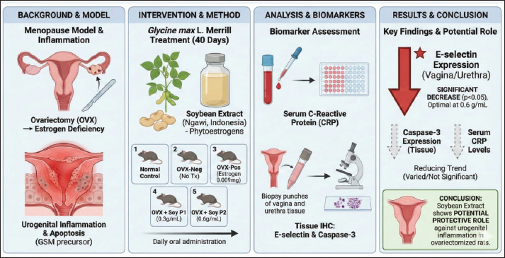
AbstractBackground: Menopause-induced estrogen deficiency triggers inflammatory responses and promotes apoptosis in urogenital tissues, contributing to genitourinary syndrome of menopause. Soybean extract contains phytoestrogenic isoflavones. Aim: To investigate the effects of soybean extract against menopause-induced inflammation and apoptosis in ovariectomized rats. Methods: Forty female Rattus norvegicus, Wistar strain were randomly allocated into five experimental groups (n=8 per group): a normal control group (KN), an ovariectomized negative control group (KNeg), an ovariectomized positive control group receiving estrogen therapy (0.009 mg/day; KPos), and two ovariectomized treatment groups administered soybean extract at doses of 300 mg/ml/day (P1) and 600 mg/ml/day (P2) for 40 days following ovariectomy. Serum C-reactive protein (CRP) concentrations were quantified using an enzyme-linked immunosorbent assay, whereas E-selectin and caspase-3 expression in vaginal and urethral tissues were assessed by immunohistochemistry (IHC). Results: Soybean extract treatment induced lower E-selectin expression in both vaginal and urethral tissues (p < 0.05), with the highest dose (600 mg/ml/day) having the most pronounced effect. IHC analysis demonstrated that E-selectin expression was markedly lower in the P2 group (29.17% ± 16.86% in vaginal tissue and 13.33% ± 11.69% in urethral tissue) than in the negative control group (KNeg: 60.00% ± 14.14% in vaginal tissue and 35.00% ± 5.48% in urethral tissue). Similarly, caspase-3 expression was lower in the treatment groups (P2: 25.00% ± 10.95% vaginal, 18.33% ± 7.53% urethral) than in the negative control (KNeg: 33.33% ± 15.06% vaginal, 20.83% ± 9.17% urethral), although the difference was statistically significant. Serum CRP levels were also lower in the soybean extract groups (P2: 2.02 ± 0.43 ng/ml) than in the negative control (KNeg: 2.78 ± 0.80 ng/ml), but the difference was not statistically significant. Conclusion: Glycine max L. Merrill extract significantly decreased the expression of E-selectin in ovariectomized rats, showing a potential protective role. The effects on caspase-3 and CRP were observed as a reducing trend; however, they were not statistically significant in all comparisons. Keywords: CRP, Caspase-3, E-selectin, Soybean extract, Urogenital atrophy. IntroductionMenopause is defined as the permanent cessation of menstruation due to a reduction or complete loss of ovarian activity, leading to a marked decline in estrogen levels (Davis et al., 2023; Peacock et al., 2023). Menopause manifests with various discomforts, including night sweats, sleep disturbances, and vaginal and urethral atrophy, of which vaginal dryness, as well as atrophy of the vagina and urethra, is commonly reported among women (Nappi et al., 2019; Bleibel et al., 2022). Approximately 50% of postmenopausal women experience vaginal and urethral atrophy, a condition also referred to as genitourinary syndrome of menopause (GSM), characterized by symptoms such as dyspareunia, urinary disturbances, burning sensations, and vaginal pruritus (Gandhi et al., 2016; Carlson and Nguyen, 2024). Many cells undergo atrophy during menopause, leading to decreased cellular integrity and function (Osorio et al., 2016). Mitochondrial dysfunction in postmenopausal women increases reactive oxygen species (ROS) production, resulting in oxidative stress (Osorio et al., 2016). These changes contribute to vaginitis, characterized by vaginal wall thinning and vaginal-urethral atrophy (Micks et al., 2023). This process begins with an inflammatory response triggered by ROS, which promotes type I collagen degradation and epithelial apoptosis through the caspase pathway (Chen et al., 2015; Rafiq et al., 2025). Elevated cleaved caspase-3 levels reflect active apoptotic processes (Zhang et al., 2017). ROS induces an oxidative stress environment in the setting of inflammation (Guerra-Castellano et al., 2018). Tumor necrosis factor induces rapid transcription of genes regulating inflammation, cell survival, proliferation, and differentiation (Kalliolias and Ivashkiv, 2016). Tumor necrosis factor alpha (TNF-α), in addition to activating Nicotinamide Adenine Dinucleotide Phosphate (NADPH) oxidase, can also induce E-selectin expression on inflamed endothelium (Zhang et al., 2024). The extravasation of circulating monocytes and dendritic cells extravasate into inflamed tissues, contributing to immunopathological responses and cell death” (Silva et al., 2018). Cell surfaces express estrogen receptors that interact with estrogens, including phytoestrogens, particularly soy-derived compounds. These interactions activate intracellular signaling pathways characterized by increased intracellular calcium levels. Elevated calcium levels lead to the formation of a calcium–calcineurin complex, which inactivates inhibitor kappa beta (IκB). This process enables the activation and nuclear translocation of nuclear factor kappa beta (NF-κB). Subsequently, NF-κB subsequently initiates the transcription of genes involved in cell cycle regulation (Osorio et al., 2016; Lecomte et al., 2017). The NF-κβ is maintained in an inactive state in the cytoplasm through its association with the inhibitory protein Iκβ (Guo et al., 2024). Interleukin-6 (IL-6) expression may also be stimulated by NF-κβ, subsequently promoting hepatocytes to synthesize C-reactive protein (CRP) as part of the acute-phase response (Ngwa et al., 2022). CRP-influenced endothelial dysfunction is linked to atherothrombosis, abnormal lipid regulation, and impaired vascular regeneration (Sproston and Ashworth, 2018). These effects involve interactions with cytokines and hormones such as angiotensin II, aldosterone, endothelin-1, platelet-derived growth factor, transforming growth factor-beta, and TNF-α, all of which can activate NADPH oxidase and further enhance ROS production (Tarafdar and Pula, 2018). Herbal extracts provide multiple classes of bioactive molecules, such as flavonoids, polyphenols, terpenoids, alkaloids, carotenoids, and other phytochemicals, that can scavenge ROS and reduce oxidative stress (Ramana et al., 2018). These compounds help restore and support endogenous antioxidant systems (superoxide dismutase, glutathione, and catalase), thereby limiting cellular damage (Mo et al., 2018; Salehi et al., 2018). Many herbal compounds can suppress pro-inflammatory cytokines and reduce lipid peroxidation (Imran et al., 2019). Bioactive substances, such as carotenoids and polyphenols, have been shown to prevent atherosclerosis, reduce cancer risk, and contribute to cellular “rejuvenation” (Testai and Calderone, 2017; Abd-Rabou et al., 2023a; Kishta et al., 2025). These effects are attributed to combined antioxidant, anti-inflammatory, and signaling-modulatory actions on key pathways involved in vascular health, cell proliferation, and apoptosis (Abd-Rabou et al., 2023b). Phytoestrogens, which are plant-derived compounds with estrogen-like properties, have gained increasing attention as alternative therapeutic agents (Desmawati and Sulastri, 2019). The two major groups of phytoestrogens are isoflavones and lignans, with soybeans serving as a rich source of isoflavones (Kim, 2021). When cells are exposed to phytoestrogens, particularly those derived from soy, these compounds are expected to bind to their corresponding receptors on the cell surface (Lecomte et al., 2017). Phytoestrogens also exert antioxidant effects by scavenging free radicals, thereby mitigating oxidative stress (Hariri et al., 2021). Despite the availability of effective therapies for genitourinary atrophy, hormone replacement therapy remains costly and associated with potential adverse effects, including increased risks of breast and ovarian cancer, which limits its acceptance and long-term use (Harrison and Shanahan, 2023). Natural and plant-derived compounds have gained increasing scientific attention for their therapeutic potential, particularly in conditions involving hormonal decline and chronic inflammation (Shoaib et al., 2023). Many phytochemicals possess antioxidant, anti-inflammatory, and estrogen-modulating properties that may help restore tissue homeostasis and counteract menopause-related pathophysiology (Desmawati and Sulastri, 2019). Previous studies have demonstrated that herbal bioactive molecules can attenuate oxidative damage, regulate inflammatory mediators, and improve endocrine function, supporting their use as safer alternatives to hormone-based therapies (Panche et al., 2016; Ahmad et al., 2023; Zoheir et al., 2025). In this context, soybean isoflavones, a class of phytoestrogens with selective estrogen receptor modulator (SERM)-like behavior, represent a promising intervention to mitigate estrogen deficiency-associated inflammation and apoptosis. Therefore, this study aimed to investigate the effects of soybean extract (Glycine max L. Merrill), a more affordable phytoestrogen with SERM-like properties, on menopause-induced inflammation and apoptosis in ovariectomized rats by assessing the expression of CRP, E-selectin, and caspase-3 (Thongon et al., 2017). The overall research framework is shown in Figure 1.
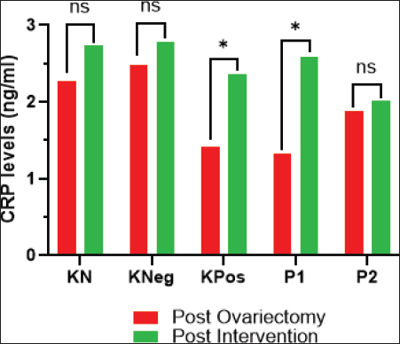
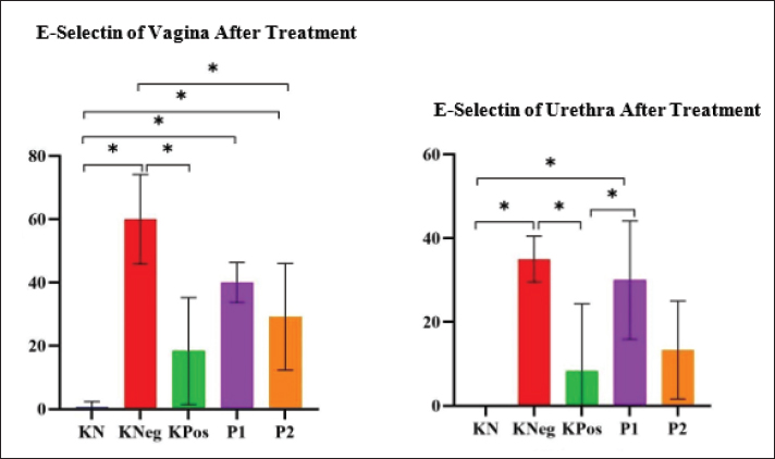
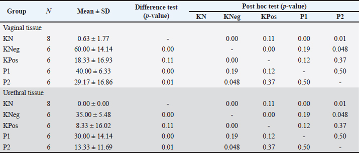
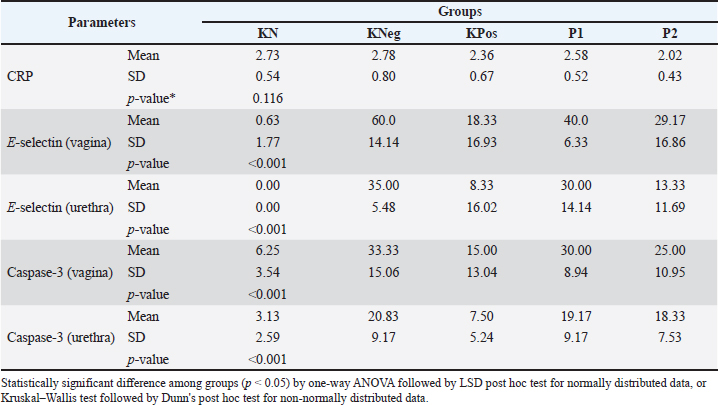
Fig. 1. The overall research framework. Materials and MethodsThis experimental study employed a controlled laboratory design incorporating post-ovariectomy pretest–posttest and post-test only measurements and was conducted at the Faculty of Medicine, Universitas Sebelas Maret, from September 2024 to August 2025. Forty female Wistar rats (Rattus norvegicus) aged 4 months and weighing 200–300 g were included in the study. Animals were acclimatized for 1 week using standard laboratory conditions with a 12-hour light and dark cycle, ambient temperature maintained at 22℃ ± 2°C, and free access to standard pellet feed (BR-2) and reverse osmosis water. Animals were randomly allocated into five groups with eight rats per group: a normal control group without ovariectomy (KN), a negative control group with ovariectomy and no treatment (KNeg), a positive control group with ovariectomy receiving estrogen therapy at a dose of 0.009 mg per day (KPos), and two treatment groups with ovariectomy receiving soybean extract at doses of 300 mg/ml per day (P1) and 600 mg/ml per day (P2). Bilateral ovariectomy using a double dorsolateral incision technique. Ketamine hydrochloride (Ketamil®) administered intraperitoneally at a dose of 40 mg/kg body weight. The ovaries were identified using the periovarian fat pad, ligated, and surgically removed. Postoperative care consisted of topical analgesic and antibiotic application to the incision site for 3 consecutive days, as previously described (Anggraeni et al., 2025). Serum CRP measurementBlood samples of approximately 1.5 ml were collected from the medial canthus, specifically the sinus orbitalis, utilizing Ketamine hydrochloride (Ketamil®) at 21 days after ovariectomy to obtain baseline post-ovariectomy serum CRP levels. Additional blood samples were collected for post-intervention analysis after 40 days of treatment. Blood sampling prior to ovariectomy was performed exclusively for hormonal assessment and did not include CRP measurements. Serum CRP concentrations were measured using an enzyme-linked immunosorbent assay (ELISA) with a rat CRP quantitative kit (rat CRP; PTX1 ELISA Kit). The CRP rabbit monoclonal antibody (mAb, No. A19003) was used in this study. Treatment preparationOne estrogen tablet (Progynova®, 2 mg) was crushed using a mortar and dissolved in 20 ml of distilled water to obtain a 0.1 mg/ml stock solution. A volume of 0.09 ml of the estrogen solution was administered daily to each rat and mixed with 2 ml of 1% Na-CMC based on the target dose of 0.009 mg estrogen per rat per day prior to oral gavage. The estrogen dose was set at 0.009 mg/day per rat, and the administered volume complied with the recommended limits for oral gavage in rats, not exceeding 2 ml per 100 g body weight, with a maximum of 5 ml per administration (Effendi et al., 2015; Negara et al., 2023). We prepared the soybean extract from soybean kernels using ethanol maceration. We obtained soybeans (100 g) from Ngawi, East Java, Indonesia, and extracted them with 500 ml of ethanol by kinetic maceration using a rotary shaker at 180 rpm for 4 hours. The extraction was repeated twice, and the combined extracts were concentrated using a rotary evaporator to obtain a dry extract (Sartini et al., 2014). Treatment administrationTreatment was initiated on day 21 following ovariectomy and continued for 40 consecutive days. During the treatment period, we administered soybean extract sourced from Ngawi, Indonesia, together with estrogen orally by gavage. Soy extract was prepared by diluting the designated amount with 1 ml of distilled water to achieve doses of 300 and 600 mg/ml/day (Akyun et al., 2019). We prepared the estrogen by dissolving powdered Progynova tablets in 1% Na-CMC and administered it at a dose of 0.009 mg/day (Effendi et al., 2015). Immunohistochemical analysisVaginal and urethral tissues were collected for histopathological and immunohistochemical examination at the end of the treatment period. Tissue samples were fixed in formalin, processed using standard histological techniques, embedded in paraffin, and sectioned at a thickness of 4 to 5 µm. Immunohistochemical staining was performed following deparaffinization by incubating tissue sections overnight at 4°C with primary antibodies against E-selectin (SELE Rabbit pAb, No. A2191) and Caspase-3 (Rabbit pAb, No. A11319). To ensure the reliability of immunohistochemistry (IHC) quantification, E-selectin and Caspase-3 expression were analyzed using the percentage of positive cells method within five randomly selected high-power fields (400× magnification) per specimen. To eliminate potential bias, the scoring process was conducted double-blinded by two independent pathologists. Cell positivity was strictly defined as distinct brown-red staining in the cytoplasm or cell membrane. Any discrepancies between the two pathologists were resolved through consensus or consultation with a third expert pathologist. This stringent double-blind scoring process confirmed the consistency of the near-zero percentage values observed in the normal control (KN) group for both markers, thereby validating the high specificity and sensitivity of the IHC protocol in detecting minimal basal expression. Statistical analysisStatistical analyses were performed using the Statistical Package for the Social Sciences version 25.0 (IBM Corp., Armonk, NY). Data distribution was assessed using the Shapiro–Wilk test, and Levene’s test was used to evaluate the homogeneity of variances. Variables were considered normally distributed when p > 0.05. For normally distributed data, group comparisons were conducted using one-way analysis of variance, followed by the least significant difference post hoc test when the overall significance and homogeneity assumptions were met. For non-normally distributed data, the Kruskal–Wallis test was applied, followed by Dunn’s post hoc test for pairwise comparisons. A significance level of α=0.05 was used for all statistical tests, and p-values <0.05 were considered statistically significant (Maxwell and Delaney, 2004). Ethical approvalThe study protocol was approved by the Ethics Committee of the Faculty of Medicine, Universitas Sebelas Maret (No. 207/UN27.06.11/KEP/EC/2024). ResultsLevels of CRPA statistically significant difference in serum CRP levels was observed among groups at baseline (post-ovariectomy) (p=0.037). However, post-hoc least significant difference analysis showed no significant difference between the ovariectomized control group (KNeg) and the normal control group (KN) (p > 0.05), suggesting that the overall difference likely reflected intergroup variability rather than a meaningful baseline imbalance showed lower CRP levels than the untreated ovariectomized control group (KNeg: 2.78 ± 0.80 ng/ml). No statistically significant differences in CRP levels were found among groups after 40 days of treatment (p=0.166) (Fig. 2). When compared descriptively, both soybean extract groups (P1 and P2) demonstrated lower mean CRP levels than the untreated ovariectomized group (KNeg: 2.78 ± 0.80 ng/ml), with values of 2.21 ± 0.52 ng/ml (P1) and 2.02 ± 0.43 ng/ml (P2). The estrogen-treated group (KPos) also showed a lower mean CRP level (2.36 ± 0.67 ng/ml) than the KNeg group. CRP levels increased after the intervention period across all groups. Within-group comparisons, however, the magnitude of the increase was smaller in the treatment groups, particularly in P2. Despite these observations, the differences were not statistically significant, and no definitive treatment effect on CRP levels was established. Vaginal tissueA significant difference in E-selectin expression was observed among the groups (p < 0.001) (Table 1, Fig. 3). The untreated ovariectomized group (KNeg) showed the highest expression (60.00% ± 14.14%), whereas the normal control group (KN) showed minimal expression (0.63% ± 1.77%). Compared with the untreated ovariectomized group (KNeg), the estrogen therapy (KPos: 18.33% ± 16.93%) and soybean extract groups demonstrated significantly lower E-selectin expression. A statistically significant reduction was observed in the KPos group (p < 0.001) and in the high-dose soybean extract group (P2: 29.17 ± 16.86%, p < 0.05). The P1 group also showed lower mean expression than the KNeg group, although the difference was less pronounced. These findings indicate that estrogen therapy and soybean extract, particularly at higher doses, were associated with reduced endothelial inflammatory marker expression in vaginal tissue (Fig. 4). Uretheral tissueA significant overall difference in the expression of E-selectin was also observed (p < 0.001). The KNeg group had higher expression (35.00% ± 5.48%) than the normal control (KN: 0.00% ± 0.00%). Estrogen therapy significantly reduced E-selectin expression compared with KNeg (KPos: 8.33% ± 16.02%, p=0.01). The high-dose soybean extract group (P2: 13.33% ± 11.69%) also showed lower mean expression compared to KNeg; however, this difference was not statistically significant (p=0.06). The P1 group showed intermediate values between KNeg and P2.
Fig. 2. Comparison of mean CRP levels among experimental and control groups across study phases (post-ovariectomy and post-intervention) of menopausal rats (Rattus norvegicus, Wistar strain) (* for significant p-value <0.05, ns, nonsignificant).
Fig. 3. Bar chart comparing the expression of E-selectin in the vagina and urethra of menopausal model rats (Rattus norvegicus, Wistar strain) after treatment across different experimental groups. (* for significant p -value <0.05, ns, non-significant. Table 1. Analysis of E-selectin expression.
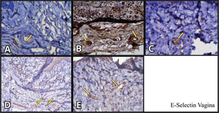
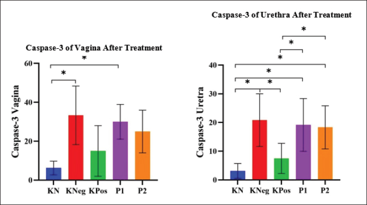
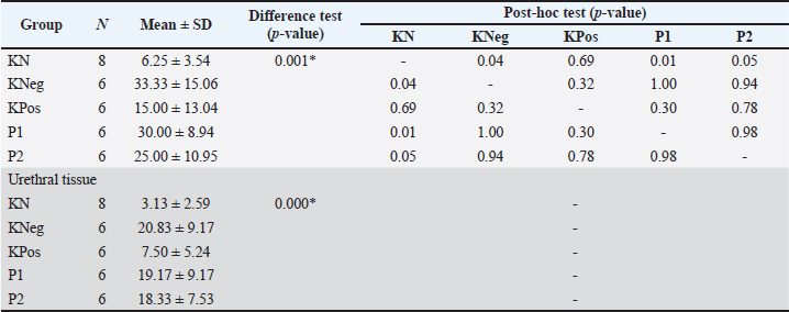
Fig. 4. E-selectin expression in vaginal tissue immunohistochemical staining was predominantly identified in the cytoplasm of vascular endothelial cells, indicated by brown staining (arrows). In the normal control group (A), minimal E-selectin expression was observed, indicating very weak staining in the endothelial cells. Strong brown cytoplasmic staining was observed in the negative control group (B), indicating increased E-selectin expression associated with inflammatory conditions. The positive control group (C) showed moderate E-selectin expression in the ECs. In treatment group 1 (D), E-selectin expression appeared to be reduced compared with the negative control group, with weaker brown staining in endothelial cells. E-selectin expression was further decreased in treatment group 2 (E), showing faint cytoplasmic staining, showing a greater reduction in the expression of endothelial inflammatory markers. All images were captured at a magnification of 400×. Thus, while reductions in E-selectin expression were observed in the treatment groups, statistically significant effects in urethral tissue were primarily observed in the estrogen-treated group (Fig. 5). Significant differences in caspase-3 expression were observed across all treatment groups in vaginal tissue (p=0.001) (Table 2, Fig. 6). The ovariectomized control group exhibited the highest Caspase-3 expression (KNeg: 33.33% ± 15.06%), indicating high apoptotic activity. Post-hoc analysis showed that estrogen therapy (KPos: 15.00% ± 13.04%) and both soybean extract treatments (P1 and P2) resulted in lower Caspase-3 expression than KNeg, although the statistical significance varied. The P2 group (25.00% ± 10.95%) showed lower expression compared with KNeg, although the difference was not statistically significant (p=0.94). Mean Caspase-3 expression compared to the normal control group (KN: 6.25% ± 3.54%), P1 group still showed significantly higher expression than KN but lower to KNeg (P1: 30.00% ± 8.94%; p=0.01), while the KPos and P2 groups showed values closer to normal while the KPos and P2 groups showed values closer to the normal control group (KN), with no statistically significant differences (p=0.321 and p=0.936, respectively).
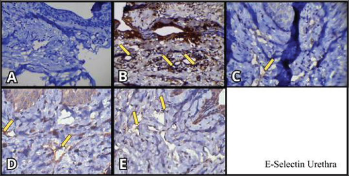
Fig. 5. Similar patterns were observed in urethral tissue immunohistochemical staining, where E-selectin expression was predominantly identified in the cytoplasm of vascular endothelial cells (arrows). E-selectin expression was absent or minimal in the normal control group (A), showing no significant brown staining. Strong brown cytoplasmic staining was observed in endothelial cells in the ovariectomized negative control group (B), indicating markedly increased E-selectin expression. E-selectin expression was reduced in the positive control group receiving estrogen therapy (C), with weaker brown staining compared to the negative control group. In treatment group 1 (D), moderate E-selectin expression was observed, showing reduced staining intensity compared with that in the negative control group. In treatment group 2 (E), E-selectin expression was lower than that in the negative control group and comparable to that in the estrogen therapy group, indicating a reduction in the expression of endothelial inflammatory markers following treatment. All images were captured at a magnification of 400×.
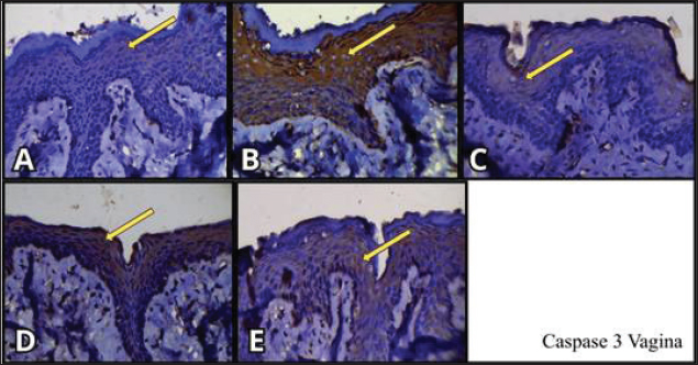
Fig. 6. Bar chart comparing vaginal and urethral Caspase-3 expression in menopausal model rats (Rattus norvegicus, Wistar strain) after treatment across different experimental groups. (* for significant p-value <0.05, ns for non-significant). We evaluated caspase-3 expression in vaginal tissue using IHC, which demonstrated cytoplasmic staining in endothelial cells of blood vessels (arrows) (Fig. 7). Panel A represents the normal control group, Panel B represents the negative control, and Panel C represents the positive control. Panels D and E illustrate caspase-3 expression in groups 1 and 2, respectively. All images were captured at 400× magnification. Table 2. Analysis of caspase-3 expression (IHC).
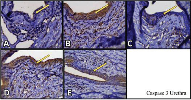
IHC was used to assess caspase-3 expression in urethral tissue, demonstrating cytoplasmic staining in blood vessel endothelial cells (arrows) (Fig. 8). Panel A shows the normal control group, Panel B shows the negative control, and Panel C shows the positive control. Panels D and E depict caspase-3 expression in groups 1 and 2, respectively. All images were obtained at 400× magnification. In urethral tissue, significant between-group differences in Caspase-3 expression were observed (p < 0.001). The ovariectomized control group KNeg exhibited higher expression (20.83% ± 9.17%) than the normal control group KN (3.13% ± 2.59%). Estrogen therapy significantly lowers Caspase-3 expression (KPos: 7.50% ± 5.24%, p=0.02 vs. KNeg), indicating that treatments with soybean extract from Ngawi Indonesia showed varying degrees of reduction. However, compared to the normal control group (KN), both soybean extract from Ngawi Indonesia treatment groups (P1: 19.17% ± 9.17%; P2: 18.33% ± 7.53%) still maintained significantly higher caspase-3 expression (p=0.00 for both), indicating incomplete restoration of normal apoptotic levels. The overall post-treatment comparison of serum CRP levels, E-selectin and Caspase – 3 expression across all experimental groups is summarized in Table 3. DiscussionCRP and E-selectin as inflammatory biomarkersEstrogen deficiency following menopause promotes oxidative stress and activates inflammatory pathways, leading to increased expression of systemic and local inflammatory markers (Desmawati and Sulastri, 2019; Hariri et al., 2021). In this study, the administration of soybean extract was associated with lower mean CRP levels compared with the untreated ovariectomized group; however, these differences were not statistically significant. Therefore, the present findings cannot confirm a definitive effect of soybean extract on systemic CRP levels. The potential mechanism by which soybean extract may influence CRP levels is related to its isoflavone content, particularly genistein and daidzein, which exhibit phytoestrogenic and antioxidant properties. Isoflavones can bind to estrogen receptors and partially mimic estrogen activity, thereby modulating intracellular signaling pathways. In estrogen-deficient conditions, increased ROS activate redox-sensitive transcription factors such as NF-κB, which subsequently induces the production of pro-inflammatory cytokines, including IL-6. Since IL-6 is a major stimulator of hepatic CRP synthesis, inhibition of NF-κB activation may reduce CRP production indirectly (Ngwa et al., 2022). However, given that the CRP reduction in this study was not statistically significant, the proposed mechanism remains speculative and requires further confirmation.
Fig. 7. Caspase-3 expression in vaginal tissue immunohistochemical staining was observed as brown cytoplasmic staining in epithelial and endothelial cells, as indicated by the arrows. Caspase-3 expression was minimal in the normal control group (A), showing weak staining in the epithelial layer. Strong brown cytoplasmic staining was observed in the negative control group (B), indicating increased caspase-3 expression associated with increased apoptotic activity. Caspase-3 expression was reduced in the positive control group (C) compared to the negative control group, with weaker staining intensity. In treatment group 1 (D), caspase-3 expression appeared to be lower than that in the negative control group, showing mild cytoplasmic staining. In treatment group 2 (E), caspase-3 expression was further reduced and appeared comparable to the positive control group, indicating decreased apoptotic marker expression following treatment. All images were captured at a magnification of 400×.
Fig. 8. Caspase-3 expression in urethral tissue immunohistochemical staining was observed as brown cytoplasmic staining in epithelial and endothelial cells, as indicated by the arrows. Caspase-3 expression was low in the normal control group (A), showing minimal cytoplasmic staining. Strong brown cytoplasmic staining was observed in the negative control group (B), indicating increased caspase-3 expression and higher apoptotic activity. Caspase-3 expression was reduced in the positive control group receiving estrogen therapy (C) compared to the negative control group, with weaker staining intensity. Caspase-3 expression remained relatively high with moderate staining intensity in treatment group 1 (D), indicating partial reduction compared to the negative control group. In treatment group 2 (E), caspase-3 expression showed a slight reduction but remained higher than that in the normal control group, suggesting that the treatment reduced apoptotic activity but did not fully restore caspase-3 expression to normal levels. All images were obtained at 400× magnification. Table 3. Summary of post treatment CRP, E-selectin, and Caspase-3 expression across experimental groups.
In contrast to CRP, a significant reduction in E-selectin expression was observed in vaginal tissue following soybean extract administration, particularly at higher doses. E-selectin is an endothelial adhesion molecule that plays a critical role in leukocyte recruitment during inflammation (Silva et al., 2018). The reduction in E-selectin expression shows that the soybean extract may attenuate local endothelial activation. This effect is likely mediated through the modulation of NF-κB signaling, which regulates the transcription of adhesion molecules, including E-selectin, as well as through the antioxidant activity of isoflavones that reduce ROS-mediated endothelial activation (Lecomte et al., 2017; Kim, 2021). Caspase-3 expression was higher in ovariectomized rats than in normal controls, reflecting increased apoptotic activity associated with estrogen deficiency. Soybean extract administration was associated with lower mean caspase-3 expression compared to untreated ovariectomized rats; however, these reductions were not statistically significant, and expression levels remained higher than in the normal control group. This indicates that while soybean extract may influence apoptotic pathways, it did not fully restore apoptosis to physiological levels in this study. The potential mechanism underlying caspase-3 modulation involves isoflavones’ antioxidant properties, which may reduce mitochondrial oxidative stress and stabilize cellular homeostasis. Reduced oxidative stress can limit the activation of the intrinsic apoptotic pathway, thereby decreasing the activation of caspase-3 (Guerra-Castellano et al., 2018; Ghafoor et al., 2024). In addition, phytoestrogenic activity may partially compensate for estrogen deficiency, contributing to cell survival signaling (Thangavel et al., 2019). Nevertheless, given the lack of consistent statistical significance, these mechanisms should be cautiously interpreted. In comparison with previous studies, phytoestrogens modulate inflammatory and apoptotic pathways in estrogen-deficient models (Lecomte et al., 2017; Thangavel et al., 2019; Kim, 2021). However, the findings of this study should not be interpreted as superior to those of previous studies. Instead, they provide additional supporting evidence that soybean-derived isoflavones may influence local inflammatory markers, particularly E-selectin, in urogenital tissues. Differences in results across studies may be attributed to variations in dosage, treatment duration, experimental models, and outcome measures. Overall, the present findings show that soybean extract may exert a modulatory effect on local inflammatory processes, particularly endothelial activation, while its effects on systemic inflammation and apoptosis remain inconclusive. To better elucidate these mechanisms, further studies with larger sample sizes, improved statistical approaches, and additional biomarkers are required. ConclusionGlycine max L. Merrill extract from Ngawi, Indonesia, demonstrated a significant reduction in E-selectin expression in this menopausal rat model showing a potential modulatory effect on local inflammatory processes. However, its effects on systemic inflammation (CRP) and apoptosis (caspase-3) were not consistently significant. These findings indicate preliminary biological activity rather than definitive therapeutic efficacy. Further studies, including well-designed clinical trials, are required to confirm its potential role in the management of GSM. AcknowledgmentsThe authors would like to express their sincere gratitude to the Animal Testing Laboratory, Anatomical Pathology Laboratory, and Biomedical Laboratory of the Faculty of Medicine, Sebelas Maret University, as well as the Department of Pharmacy, Faculty of Mathematics and Natural Sciences, Sebelas Maret University, for providing research facilities and technical support. The authors would like to thank Hanifah Karim, MD, and 'Aininna 'Izzah Zafira for their invaluable assistance as research assistants and for their contributions to manuscript editing. Conflict of interestThe authors declare that they have no known competing financial or non-financial interests that could have influenced the work reported in this paper. FundingThis research received no specific grant from any funding agency in the public, commercial, or not-for-profit sectors. The authors received no financial support for the research, authorship, and publication of this article. Authors' contributionsA.A: Conceptualization, methodology, administrative support, data curation, and formal analysis. S: Administrative support, data curation, and data collection. I.N. and U.R.B.: Data curation and collection. P.D., E.P.P., and B.W.: Formal analysis and data interpretation. All authors contributed to manuscript writing, critically reviewed the manuscript for important intellectual content, approved the final version of the manuscript, and agreed to be accountable for all aspects of the work. Data availabilityThe datasets generated and analyzed during this study are not publicly available due to institutional restrictions. The datasets are available from the corresponding author upon reasonable request. ReferencesAbd-Rabou, A., Melegy, W., Kishta, M. and El-Ganzuri, M. 2023a. Nano-encapsulation and apoptotic impact of green tea polyphenol and epigallocatechin-3-gallate on breast cancer cells in vitro. Egypt. J. Chem. 116, e220; doi:10.21608ejchem.2023.224011.8285 Abd-Rabou, A.A., Mwaheb, M.A., Sayed, O.N., Mohamed, S.H. and Kishta, M.S. 2023b. 5-fluorouracil synergized with raloxifene and cytosine β-D-arabinofuranoside to combat colorectal cancers in vitro via controlling lipolysis. J. Pharmacol. Toxicol. 12(1), 14–23; doi:10.3923jpt.2017.14.23 Ahmad, R.A., Al-Hamdany, A.N., Al-Dahmoshi, H.O., Ghasemzadeh-Moghaddam, H., Salleh, M. and Al-Dahmoshi, H.O. 2023. Herbal medicine A promising therapeutic approach in modern healthcare. Curr. Bioactive. Sci. 3(1), 14–21. Akyun, I.K., Fajariyah, S. and Mahriani, M. 2019. Efek ekstrak etanol kedelai hitam (Glycine soja) terhadap ketebalan dermis mencit (Mus musculus L) pasca unilateral ovariektomi. J. Biol. Udayana 23(2), 80–87. Anggraeni, A., Soetrisno, S., Nurwati, I., Budihastuti, U.R., Pamungkasari, E.P., Wasita, B. and Dirgahayu, P. 2025. Experimental model of menopausal genitourinary syndrome Ovariectomy and vaginectomy protocols in rats. Open Vet. J. 15(8), 3624–3630; doi: 10.5455OVJ.2025.v15.i8.25 Bleibel, B. and Nguyen, H. 2022 Vaginal Atrophy. In: StatPearls [Internet]. Treasure Island, FL: StatPearls Publishing. Available via https://www.ncbi.nlm.nih.gov/books/NBK559297/ Carlson, K. and Nguyen, H. 2024. Genitourinary syndrome of menopause. Treasure Island, FL: StatPearls Publishing. Available via httpswww.ncbi.nlm.nih.govbooksNBK559297 Chen, Z., Zhang, Z., Zhang, H. and Xie, B. 2015. ‘Analysis of the oxidative stress status in nonspecific vaginitis and its role in vaginal epithelial cell apoptosis. BioMed. Res. Int. 2015, g1–g7; doi:10.11552015795656 Davis, S.R., Pinkerton, J., Santoro, N. and Simoncini, T. 2023. Menopause—biology, consequences, supportive care, and therapeutic options. Cell 186(19), 4038–4058. Desmawati, D. and Sulastri, D. 2019. ‘Phytoestrogens and their health effect. Macedonian J. Med. Sci. 7(3), 495–499; doi:10.3889oamjms.2019.086 Effendi, E.M., Hera, H.M. and Listya, M.J. 2015. Aktifitas Estrogenik Ekstrak Etanol 70% Herba Kemangi (Ocimum americanum L.) pada Tikus Putih Betina (Rattus norvegicus) Premenopause. Fitofarmaka Jurnal. Ilmiah. Farmasi. 5(1), 190; doi:10.33751jf.v5i1.190 Gandhi, J., Chen, A., Dagur, G., Suh, Y., Smith, N., Cali, B. and Khan, S.A. 2016. Genitourinary syndrome of menopause: an overview of clinical manifestations, pathophysiology, etiology, evaluation, and management. Am. J. Obstetrics Gynecol. 215(6), 704–711; doi:10.1016j.ajog.2016.07.045 Ghafoor, K., Park, S.B., Lee, D.U., Kim, S.H., Kim, M.O. and Jeong, J.B. 2024. Poncirin attenuates microplastic-induced hepatotoxicity by modulating oxidative stress, inflammation, and apoptosis in rats. J. King Saud Univ–Sci. 2024, 103475; doi: 10.1016j.jksus.2024.103475 Guerra-Castellano, A., Díaz-Quintana, A., Pérez-Mejías, G., Elena-Real, C.A., González-Arzola, K., García-Mauriño, S.M., De La Rosa, M.A. and Díaz-Moreno, I. 2018. Oxidative stress is tightly regulated by cytochrome c phosphorylation and respirasome factors in mitochondria. Proc. Natl. Acad. Sci. USA. 115, 7955–7960; doi:10.1073pnas.1806833115 Guo, Q., Jin, Y., Chen, X., Ye, X., Shen, X., Lin, M., Zeng, C., Zhou, T. and Zhang, J. 2024. NF-κB in biology and targeted therapy: new insights and translational implications. Signal Transduct. Target. Ther. 9(1), 53; doi: 10.1038/s41392-024-01757-9 Hariri, M., Ghasemi, A., Baradaran, H.R., Mollanoroozy, E. and Gholami, A. 2021. Beneficial effect of soy isoflavones and soy isoflavones plus soy protein on serum concentration of C-reactive protein among postmenopausal women an updated systematic review and meta-analysis of randomized controlled trials. Complement. Ther. Med. 59, 102715. Harrison, G.H. and Shanahan, M.M. 2023. Hormone replacement therapy. In: StatPearls [Internet]. Treasure Island, FL: StatPearls Publishing. Available via https://www.ncbi.nlm.nih.gov/books/NBK493191/ Imran, M., Rauf, A., Tareq, A.I., Nadeem, M., Shariati, M.A., Imtiaz, A.K., Imran, A., Orhan, I.E. and Rizwan, M. 2019. Luteolin, a flavonoid, as an anticancer agent: a review. Biomed. Pharmacother. 112, 108612; doi: 10.1016/j.biopha.2019.108612 Kalliolias, G.D. and Ivashkiv, L.B. 2016. TNF biology, pathogenic mechanisms and emerging therapeutic strategies. Nat. Rev. Rheumatol. 12(1), 49–62; doi: 10.1038/nrrheum.2015.169 Kim, I.S. 2021. Current perspectives on the beneficial effects of soybean isoflavones and their metabolites for humans. Antioxidants 10(7), 1064. Kim, S.H., Dhungana, S.K., Kim, I.D., Adhikari, A. and Kim, J.H. 2024. Effect of illite treatment on quality characteristics and antioxidant activity of broccoli (Brassica oleracea L. var. italica) sprouts. Molecules 29(18), 4347. Kishta, M.S., Abd-Rabou, A.A., Sarkissian, G.K., Elwakil, A.I., Elsabry, D.M., Zagzoug, Y.M., Hussein, S.R. and Abdallah, A.N. 2025. Exosome mediated delivery of epigallocatechin 3 gallate as a novel approach to alleviate psoriasis symptoms through cytokine and apoptotic pathway modulation. Sci. Rep. 15(1), 30013. Lecomte, S., Demay, F., Ferrière, F. and Pakdel, F. 2017. Phytochemicals targeting estrogen receptors beneficial rather than adverse effects. Int. J. Mol. Sci. 18(7), 1381. Maxwell, S.E. and Delaney, H.D. 2004. Designing experiment and analyzing data A model comparison perspective, 2nd ed. New York, London, UK: Psychology Press, Taylor & Francis Group, pp: 208–226. Micks, E., Reed, S. and Mitchell, C. 2023. The postmenopausal vaginal microbiome and genitourinary syndrome of menopause. Clin. Obstet. Gynecol. 67(1), 79–88; doi: 10.1097/grf.0000000000000832 Mo, W., Wu, X., Jia, G., Zhao, H., Chen, X., Tang, J., Wu, C., Cai, J., Tian, G., Wang, J. and Liu, G. 2018. Roles of dietary supplementation with arginine or N-carbamylglutamate in modulating the inflammation, antioxidant property, and mRNA expression of antioxidant-relative signaling molecules in the spleen of rats under oxidative stress. Anim. Nutr. 4(3), 322–328; doi:10.1016j.aninu.2018.02.003 Nappi, R.E., Seracchioli, R., Salvatore, S., Cagnacci, A., Di Paolantonio, T. and Busacca, M. 2019. Impact of vulvovaginal atrophy of menopause prevalence and symptoms in Italian women according to the EVES study. Gynecol. Endocrinol. 35, 453–459; doi:10.108009513590.2018.1563883 Negara, K.S.P., Suyatmi, Susilawati, T.N., Prawestri, W., Setyawan, S. and Meididewi, D. 2023. Prosedur Operasional Baku Laboratorium Hewan Coba FK UNS. Surakarta, Indonesia: Universitas Sebelas Maret Press, pp. 42–48. Ngwa, D.N., Pathak, A. and Agrawal, A. 2022. IL-6 regulates induction of C-reactive protein gene expression by activating STAT3 isoforms. Mol. Immunol. 146, 50–56; doi:10.1016j.molimm.2022.04.003 Osorio, F.G., Soria-Valles, C., Santiago-Fernández, O., Freije J, M.P. and López-Otín, C. 2016. NF-κB signaling as a driver of aging. Int. Rev. Cell Mol. Biol. 2016, 133–174; doi:10.1016bs.ircmb.2016.04.003 Panche, A.N., Diwan, A.D. and Chandra, S.R. 2016. Flavonoids: an overview. J. Nutr. Sci. 5, 47; doi: 10.1017/jns.2016.41 Peacock, K., Carlson, K. and Ketvertis, K.M. 2023. Menopause. In: StatPearls [Internet]. Treasure Island, FL: StatPearls Publishing. Available via https://www.ncbi.nlm.nih.gov/books/NBK507826/ Rabou, A., Kamal, M., Alharbi, H.Y., Aljohani, M.S., El Atawy, M.A. and Kishta, M.S. 2025. Modulation of PI3KAKT signaling and DFT modeling via selected pharmaceutical compounds attenuates carrageenan-induced inflammation and oxidative stress in rats. Naunyn. Schmiedeberg. Arch. Pharmacol. 398(6), 7583–7603; doi:10.1007s00210-024-03689-1 Rafiq, M., Tariq, M., Hayat, S., Al-Harbi, M., Al-Ghamdi, A.A. and Al-Johani, S. 2025. Interplay between oxidative stress and inflammation mechanistic insights and toxicological implications. Toxicol. Chem. Eng. 2025, 102988; doi:10.1016j.tice.2025.102988 Ramana, K.V., Reddy, A.B.M., Majeti, N.V.R.K. and Singhal, S.S. 2018. Therapeutic potential of natural antioxidants. Oxid. Med. Cell. Longev. 2018, 1–3; doi: 10.1155/2018/9471051 Salehi, B., Mishra, A., Nigam, M., Sener, B., Kilic, M., Sharifi-Rad, M., Fokou, P., Martins, N. and Sharifi-Rad, J. 2018. Resveratrol: a double-edged sword in health benefits. Biomedicines 6(3), 91; doi: 10.3390/biomedicines6030091 Sartini, M., Djide, S., Permana, D. and Ismail. 2014. Ekstraksi Isoflavon Kedelai dan Penentuan Kadarnya Secara Ultra Fast Liquid Chromatography (UFLC). Jurnal Sainsmat 3(2), 130–134. Silva, M., Videira, P.A. and Sackstein, R. 2018. E-selectin ligands in the human mononuclear phagocyte system implications for infection, inflammation, and immunotherapy. Front. Immunol. 8(1), 1878; doi:10.3389fimmu.2017.01878 Sproston, N.R. and Ashworth, J.J. 2018. Role of C-reactive protein at sites of inflammation and infection. Front. Immunol. 9, 754; doi: 10.3389/fimmu.2018.00754 Shoaib, S., Ansari, M.A., Ghazwani, M., Hani, U., Jamous, Y.F., Alali, Z., Wahab, S., Ahmad, W., Weir, S.A., Alomary, M.N., Yusuf, N. and Islam, N. 2023. Prospective epigenetic actions of organo-sulfur compounds against cancer: perspectives and molecular mechanisms. Cancers 15(3), 697; doi: 10.3390/cancers15030697 Testai, L. and Calderone, V. 2017. Nutraceutical value of citrus flavanones and their implications in cardiovascular disease. Nutrients 9(5), 502; doi: 10.3390/nu9050502 Tarafdar, A. and Pula, G. 2018. The role of NADPH oxidases and oxidative stress in neurodegenerative disorders. J. Mol. Sci. 19(12), 3824. Thangavel, P., Puga-Olguín, A., Rodríguez-Landa, J.F. and Zepeda, R.C. 2019. Genistein as potential therapeutic candidate for menopausal symptoms and other related diseases. Molecules 24(21), 3892. Thongon, N., Boonmuen, N., Suksen, K., Wichit, P., Chairoungdua, A., Tuchinda, P., Suksamrarn, A., Winuthayanon, W. and Piyachaturawat, P. 2017. Selective estrogen receptor modulator (SERM)-like activities of diarylheptanoid a phytoestrogen from Curcuma comosa, in breast cancer cells, pre-osteoblast cells, and rat uterine tissues. J. Agricult. Food. Chem. 65(17), 3490–3496. Zoheir, K.M., Ali, N.I., Ashour, A.E., Kishta, M.S., Othman, S.I., Rudayni, H.A., Rashad, A.A. and Allam, A.A. 2025. Lipoic acid improves wound healing through its immunomodulatory and anti-inflammatory effects in a diabetic mouse model. J. Diabetes Metabolic Disord. 24, 56. Zhang, Y., Chen, X., Gueydan, C. and Han, J. 2017. Plasma membrane changes during programmed cell deaths. Cell Res. 28(1), 9–21; doi: 10.1038/cr.2017.133 Zhang, J., Huang, S., Zhu, Z., Gatt, A. and Liu, J. 2024. E-selectin in vascular pathophysiology. Front. Immunol. 15, 1401399; doi: 10.3389/fimmu.2024.1401399 | ||
| How to Cite this Article |
| Pubmed Style Anggraeni A, Soetrisno S, Nurwati I, Budihastuti UR, Pamungkasari EP, Wasita B, Dirgahayu P. Protective effects of soybean extract on menopause-induced inflammation and apoptosis in rats. Open Vet. J.. 2026; 16(8): 5446-5458. doi:10.5455/OVJ.2026.v16.i8.37 Web Style Anggraeni A, Soetrisno S, Nurwati I, Budihastuti UR, Pamungkasari EP, Wasita B, Dirgahayu P. Protective effects of soybean extract on menopause-induced inflammation and apoptosis in rats. https://www.openveterinaryjournal.com/?mno=287470 [Access: August 08, 2026]. doi:10.5455/OVJ.2026.v16.i8.37 AMA (American Medical Association) Style Anggraeni A, Soetrisno S, Nurwati I, Budihastuti UR, Pamungkasari EP, Wasita B, Dirgahayu P. Protective effects of soybean extract on menopause-induced inflammation and apoptosis in rats. Open Vet. J.. 2026; 16(8): 5446-5458. doi:10.5455/OVJ.2026.v16.i8.37 Vancouver/ICMJE Style Anggraeni A, Soetrisno S, Nurwati I, Budihastuti UR, Pamungkasari EP, Wasita B, Dirgahayu P. Protective effects of soybean extract on menopause-induced inflammation and apoptosis in rats. Open Vet. J.. (2026), [cited August 08, 2026]; 16(8): 5446-5458. doi:10.5455/OVJ.2026.v16.i8.37 Harvard Style Anggraeni, A., Soetrisno, . S., Nurwati, . I., Budihastuti, . U. R., Pamungkasari, . E. P., Wasita, . B. & Dirgahayu, . P. (2026) Protective effects of soybean extract on menopause-induced inflammation and apoptosis in rats. Open Vet. J., 16 (8), 5446-5458. doi:10.5455/OVJ.2026.v16.i8.37 Turabian Style Anggraeni, Asih, Soetrisno Soetrisno, Ida Nurwati, Uki Retno Budihastuti, Eti Poncorini Pamungkasari, Brian Wasita, and Paramasari Dirgahayu. 2026. Protective effects of soybean extract on menopause-induced inflammation and apoptosis in rats. Open Veterinary Journal, 16 (8), 5446-5458. doi:10.5455/OVJ.2026.v16.i8.37 Chicago Style Anggraeni, Asih, Soetrisno Soetrisno, Ida Nurwati, Uki Retno Budihastuti, Eti Poncorini Pamungkasari, Brian Wasita, and Paramasari Dirgahayu. "Protective effects of soybean extract on menopause-induced inflammation and apoptosis in rats." Open Veterinary Journal 16 (2026), 5446-5458. doi:10.5455/OVJ.2026.v16.i8.37 MLA (The Modern Language Association) Style Anggraeni, Asih, Soetrisno Soetrisno, Ida Nurwati, Uki Retno Budihastuti, Eti Poncorini Pamungkasari, Brian Wasita, and Paramasari Dirgahayu. "Protective effects of soybean extract on menopause-induced inflammation and apoptosis in rats." Open Veterinary Journal 16.8 (2026), 5446-5458. Print. doi:10.5455/OVJ.2026.v16.i8.37 APA (American Psychological Association) Style Anggraeni, A., Soetrisno, . S., Nurwati, . I., Budihastuti, . U. R., Pamungkasari, . E. P., Wasita, . B. & Dirgahayu, . P. (2026) Protective effects of soybean extract on menopause-induced inflammation and apoptosis in rats. Open Veterinary Journal, 16 (8), 5446-5458. doi:10.5455/OVJ.2026.v16.i8.37 |


