





| Research Article | ||
Open Vet. J.. 2026; 16(5): 3178-3191 Open Veterinary Journal, (2026), Vol. 16(5): 3178-3191 Research Article Protective effect of Commiphora gileadensis against cadmium toxicity in male ratsAbdullah S.M. Aljohani1, Ahmed A. Aliiri1, Amal S. Soliman2*, Khaled H. Musa3, Mohamed Alreshoodi4 and Ibrahim M. El-Ashmawy1,51Department of Medical Biosciences, College of Veterinary Medicine, Qassim University, Buraidah, Saudi Arabia 2Department of Basic Sciences, High Institute of Engineering and Technology, Ministry of Higher Education, Alexandria, Egypt 3Department of Food Science and Human Nutrition, College of Agricultural and Nutrition, Qassim University, Buraidah, Saudi Arabia 4Salam Veterinary Group, Buraidah, Saudi Arabia 5Department of Pharmacology, Faculty of Veterinary Medicine, Alexandria University, Alexandria, Egypt *Corresponding Author: Amal S. Soliman. Department of Basic Sciences, High Institute of Engineering and Technology, Ministry of Higher Education, Alexandria, Egypt. Email: soliman.amal [at] yahoo Submitted: 27/09/2025 Revised: 11/03/2026 Accepted: 27/03/2026 Published: 31/05/2026 © 2025 Open Veterinary Journal
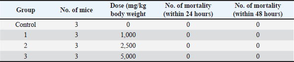
ABSTRACTBackground: Male reproductive organs are highly susceptible to oxidative damage. Cadmium (Cd) is classified as one of the 10 most toxic environmental heavy metals that cause testicular harm. Exposure to Cd alters the reproductive system in males. Free radical generation by Cd by testicular lipid peroxidation. Natural antioxidants may alleviate the hazard effects of Cd. Commiphora gileadensis (CG) sap, wood, bark, leaves, and seeds have notable therapeutic features. CG sap is a candidate to mitigate Cd toxicity. Aim: This study aimed to evaluate the antioxidant potential of oleo-resin exudate (sap) from Commiphora gileadensis excised bark in vitro and its prevention of reproductive alterations in male rats exposed to Cd chloride exposure. Methods: The antioxidant potentials of CG sap in vitro were estimated based on the total phenolic and flavonoid contents, 2,2-diphenyl-1-picrylhydrazyl radical-scavenging activity, and 2,2’-azinobis-3-ethylbenzothiazoline-6-sulfonic acid free radical scavenging activity. The reducing activities were also performed using copper (cupric reducing antioxidant capacity) and ferric (ferric reducing antioxidant power). Explored how CG sap influences Cd toxicity in the reproductive systems of male rats. Seven groups (six rats each) were created from 42 healthy male adult albino rats. Group 1 (C) received distilled water as a control. Group 2 (Cd): 50 mg Cd chloride/l drinking water (equal to 5 mg/kg/day). Group 3 was administered 100 mg/kg of CG sap. Group 4 was administered 400 mg CG sap/kg. Group 5 was administered 100 mg CG sap/kg plus 50 mg Cd chloride/l. Group 6 was administered 400 mg CG sap/kg b. wt. plus 50 mg Cd chloride/l. Group 7 (V) was administered 1% Tween 80, vehicle (1 ml/rat). Results: Rats suffering from Cd poisoning exhibited significant decreases in the weights of their testis, epididymis, and sex glands, as well as in sperm count, sperm motility, and overall biochemical antioxidant capacity. Co-administration of CG sap in a dose-dependent manner significantly reduced the size of the testis, epididymis, and sex accessory glands of rats intoxicated with Cd. Furthermore, this treatment reduced the catalase levels and total antioxidant capacity induced by Cd toxicity. Conclusion: It can be concluded that CG sap may be utilized as a natural candidate for protection against environmental Cd toxicity. Keywords: Antioxidants, Cadmium, CG, Male rats, Reproductive organs. IntroductionCadmium (Cd) is one of the most persistent environmental pollutants, occurring in the Earth’s crust and entering human habitats through numerous industrial and lifestyle-related activities. As a result, large populations across the globe are affected by long-term, low-grade exposure (Bhardwaj et al., 2021; Zhou et al., 2022). Its continuous circulation in agricultural, industrial, and domestic settings make avoidance extremely difficult (Bhardwaj et al., 2021; Zhang et al., 2022). Tobacco smoke is considered one of the most concentrated non-occupational sources of Cd, and agricultural application of phosphate fertilizers contributes significantly to soil contamination and subsequent food supply (Habib et al., 2019; Chen et al., 2022). Even minimal Cd intake can be harmful because the element is slowly eliminated from the body, remaining in tissues for 20–40 years and gradually accumulating in organs such as the kidneys, skeletal system, and reproductive tract (Zhao et al., 2017; Venditti et al., 2021). Once absorbed, Cd promotes the formation of reactive oxygen species (ROS), provoking oxidative disturbances, inflammatory cascades, cellular dysfunction, and gene regulation abnormalities that manifest as various health complications in both humans and animals (Badr et al., 2019; Yang et al., 2019). Testicular toxicity is one of the most characteristic outcomes of Cd intoxication, with male reproductive impairment attracting considerable attention (Chemek et al., 2018; Yang et al., 2019; Kumar and Sharma, 2019). Higher Cd levels have been documented in smokers, corresponding with increased oxidative imbalance and weakened antioxidant systems in semen, especially among infertile males (Kumar and Sharma, 2019; Chen et al., 2022). Cd disrupts the blood–testis barrier architecture, triggering germ cell death, structural degeneration, Sertoli and Leydig cell function deterioration, hormonal irregularities, erectile issues, abnormal spermatogenesis, poor semen parameters, and ultimately reduced fertility (Kumar and Sharma, 2019; Basal et al., 2023). Experimental findings also demonstrate that Cd can interfere with the hypothalamic–pituitary–gonadal axis sperm development and injure sperm nuclear DNA (Kumar and Sharma, 2019; Mitra et al., 2020). Natural antioxidant defenses are indispensable for maintaining the functional stability of male reproductive tissues (He et al., 2020; Moradi et al., 2020; Kalehoei et al., 2023). Because Cd-induced oxidative stress is regarded as the principal mechanism underlying tissue injury, organs with high lipid content and limited antioxidant reserves (such as the testes) are particularly susceptible (Habib et al., 2019; Kumar and Sharma, 2019). Consequently, many studies have been dedicated to pinpointing the precise molecular pathways affected by Cd and identifying therapeutic agents capable of counteracting its harmful effects (Habib et al., 2019; Moradi, 2021). Medicinal plants have historically been central to healthcare systems worldwide and continue to provide essential bioactive compounds for the pharmaceutical sector (İPEK et al., 2024; Doğan et al., 2025; Unzile et al., 2025). According to the World Health Organization, approximately 80% of the global population still relies on herbal medicines or plant-derived constituents for basic medical needs (Varijakzhan et al., 2020). Plant-based remedies are viewed as valuable alternatives to chemically synthesized drugs because they often exhibit strong therapeutic potential with relatively fewer adverse effects (Rosic et al., 2024; Alasgarova et al., 2025; Evcil et al., 2025). Therefore, considerable research has focused on exploring phytochemicals with antioxidant capacities capable of mitigating the toxicity induced by heavy metals, including lead, Cd, and mercury, in living organisms (Ashry et al., 2010; Oda and El-Ashmawy, 2012). More recent investigations have particularly emphasized the capacity of plant-based antioxidants to protect cells and tissues from Cd-induced oxidative injury in both experimental animals and human studies (Aldaddou et al., 2022 and 2023; Zhu et al., 2025). Consequently, the antioxidant defense capacity must be improved to counter endogenous and exogenous oxidative stress reagents. This can be accomplished using synthetic or natural antioxidant supplements (Ahmed et al., 2019). Commiphora gileadensis L., a member of the Burseraceae family, is a small dioecious shrub that grows naturally in the Red Sea, Mediterranean Basin, and areas bordering Saudi Arabia (Shalabi and Otaif, 2022). Standing 2–4 m tall, it is characterized by a dark green, stout trunk, thin, peeling bark, and compound leaves arranged in alternating fascicles on short lateral branches (Eslamieh 2011; Yaniv and Dudai, 2014). Commiphora gileadensis has held profound cultural, religious, and medicinal value for millennia, traditionally known as the balm of Makkah, Mecca myrrh, opobalsam, or the biblical balm of Gilead (Al-Herthi et al., 2020; Alhazmi et al., 2022). Its sap, bark, wood, leaves, flowers, and seeds have been integral to both the Ayurvedic and Arabian folk medicine systems. Historical uses include the treatment of inflammatory conditions, gastrointestinal disorders (e.g., constipation and abdominal pain), joint ailments, headaches, coronary artery disease, gynecological imbalances, and obesity (Alqethami et al., 2017). The plant’s resin was particularly valued as a natural analgesic and a potent wound-healing agent, attributed to its beneficial effects on skin integrity and repair (Tajbakhsh et al., 2021; Althurwi et al., 2022; Alhazmi et al., 2022). Modern scientific inquiry has increasingly validated these traditional applications. Extracts of C. gileadensis have demonstrated a wide spectrum of pharmacological activities, including antioxidant, antimicrobial, antiviral, antidiabetic, anticoagulant, hepatoprotective (notably against confluent hepatic necrosis), and cytotoxic effects (Al-Mahbashi et al., 2015; Al-Hazmi et al., 2020; Al-Hazmi et al., 2022; Farid et al., 2022). In vitro studies have reported significant inhibition of prostate, liver, and other cancer cell lines, underscoring its potential as an anticancer agent (Shen et al., 2012; Wineman et al., 2015; Bouslama et al., 2019; Al-Zahrani et al., 2022). Most previous studies have focused on various parts of the plant, such as leaves, stem, seeds, and rare on its sap, and our study is a pioneer. The therapeutic efficacy of plants is largely linked to their rich phytochemical profile. Essential oils and extracts contain bioactive terpenoids such as β-pinene, β-caryophyllene, terpinene-4-ol, δ-cadinene, commigileadin, and canophyllal, as well as phenolic compounds and flavonoids—including quercetin and mearnsetin (Dudai et al., 2017; Abdallah et al., 2022; Mohamed et al., 2025). Additionally, recent analytical work on the Commiphora gileadensis (CG) leaf extract has further identified prenylated flavonoids, coumarin, chalcone, and even an alkaloid in the butanol fraction of stem bark extracts, highlighting the chemical complexity underlying its bioactivity (Bin Mokaizh et al., 2024; Ahmed et al., 2024). Despite these advances, critical knowledge gaps remain. Notably, there is a scarcity of research exploring its effects on reproductive health, particularly concerning Cd-induced male infertility in preclinical models such as rats. Given the well-established role of oxidative stress in Cd-induced reproductive toxicity and the antioxidant potential of C. gileadensis, this study aimed to evaluate whether CG sap administration could alleviate Cd-induced reproductive organ damage in male rats. Materials and MethodsAnimalsBased on the method of Aldaddou et al. (2022). Forty-two adult male albino rats, each weighing between 200 and 220 g, were procured from the College of Pharmacy at Qassim University in the Kingdom of Saudi Arabia. The animals were maintained in cages and provided with laboratory animal feed pellets and water for 2 weeks before the start of the experiment. This period served for adaptation, ensuring their healthy condition, and eliminating any animals exhibiting signs of emaciation or having no diseases. A suitable number of adult mice should be used to determine the Lethal Dose50 (LD50)of C. gileadensis sap. Collection of C. gileadensisCommiphora gileadensis was attained in Spring 2022 from a high-altitude location known as the Alaab Valley, located in the western part of Saudi Arabia. Preparation of C. gileadensis sapThe growing tips of C. gileadensis branches were lopped, leaving a 5-mm distance from the tips. Subsequently, the exuding sap was promptly collected following the incision and was stored at a temperature of −20°C until ready for examination (Iluz et al., 2010). In vitro antioxidant activity assessmentOne gram of plant sap was combined with 10 ml of ethanol to evaluate the antioxidant activity. The mixture was agitated using a magnetic stirrer (IKA, Staufen, Germany) operating at 1,000 revolutions per minute. Determination of the total phenolic contentTotal phenolic content (TPC) was assessed using the Folin-Ciocalteu method (Slinkard and Singleton, 1977), with gallic acid as the standard reference. In this procedure, 100 μl of either the standard or a blank plant sap sample was oxidized using 500 μl of a diluted Folin-Ciocalteu reagent. After a 5-minute reaction time, the mixture was neutralized by adding 1 ml of a sodium carbonate solution (7.5%, w/v) and then incubated for 120 minutes before measuring the absorbance at 765 nm. The results are expressed as gallic acid equivalents (mg GAE/g). Determination of total flavonoid contentThe TFC was evaluated using the aluminum chloride colorimetric technique, as previously described (Chang et al., 2002). The absorbance was recorded at 405 nm. A calibration curve was created with quercetin as the standard, and the TFC was represented in terms of mg quercetin equivalents/g (mg QE/g). Determination of antioxidant activity using cupric reducing antioxidant capacity (CUPRAC)The CUPRAC assay was conducted according to the methodology established by Apak et al. (1981)and subsequently modified by Annapurna et al. (2021). Plant sap at different concentrations was combined with 1 ml of a 0.01 M Cu (II) chloride solution. Subsequently, 1 ml of 7.5 × 10³ M neocuproine solution (NC) and 1 ml of an ammonium acetate buffer at pH 7.0 was added. The mixture was then incubated in the dark at room temperature for 30 minutes. The absorbance of the resulting color was measured at 440 nm using a reference blank following incubation. Trolox served as the standard antioxidant. The experiment was performed in triplicate, and the results were expressed as Trolox equivalents per gram of dry weight. Measurement of antioxidant activity via ferric reducing antioxidant power (FRAP)The FRAP assay method was performed in accordance with the methodology outlined by Musaa et al. (2015)to evaluate antioxidant activity using Trolox as the reference standard. To prepare the FRAP reagent, a mixture was created using 300 mM acetate buffer, 10 mM TPTZ (2,4,6-tri(2-pyridyl)-s-triazine) dissolved in a 40 mM HCl solution, and 20 mM FeCl3·6H2O, adhering to a volumetric ratio of 10:1:1. In the assay process, 1,900 µl of the FRAP reagent was combined with 100 µl of the sample, standard, or blank solution and then incubated for 30 minutes before measuring the absorbance at 595 nm. The results are expressed as Trolox equivalents per gram of dry weight. Determination of antioxidant activity using the 2,2-di-phenyl-1-picrylhydrazyl (DPPH) radical-scavenging methodThe DPPH assay method described by Al-Altaie and Addai (2021)was used to evaluate radical-scavenging activity, using Trolox as the reference standard. During the experiment, 3 ml of DPPH solution at a concentration of 40 mg/l was mixed with 100 μl of the sample, the standard, or a blank control. The mixture was then incubated for 30 minutes before measuring the absorbance at 517 nm. The results are expressed as Trolox equivalents per gram of dry weight. Determination of antioxidant activity using the 2,20-azinobis (3-ethylbenzothiazoline-6-sulfonic acid) (ABTS) free radical-scavenging methodThe Trolox equivalent antioxidant capacity or ABTS assay was adapted from the procedure outlined by Ali et al. (2020a,b). This technique uses Trolox as a reference standard. To produce ABTS radical cations, 7 mM ABTS was oxidized with 2.45 mM potassium persulfate, and the resulting solution was left in the dark at room temperature for 12 hours. The ABTS solution was then diluted with distilled water to achieve an absorbance of 1.000 ± 0.02 at 734 nm. A 1-ml portion of the ABTS cation solution was combined with 100 μl of the sample extract, standard, or blank, and the absorbance reduction at 734 nm was recorded after 5 minutes. The results are expressed as Trolox equivalents per gram of dry weight. Median LD50 of C. gileadensis sapThe study involved determining the LD50 that resulted in the death of 50% of the subjects, which comprised 12 male Swiss albino mice weighing between 20 and 25 g each. To ensure fairness, the mice were evenly divided into four groups, each consisting of three animals. All mice were provided with food and water and allowed a 7-day acclimation period in the laboratory environment before the experiment began. Following an overnight fast, the control group received oral administration of 1% Tween 80 solution. Meanwhile, the treatment groups were orally administered C. gileadensis sap, which had been dissolved in a 1% Tween 80 solution, at doses of 1.0, 2.5, and 5.0 g/kg of b. wt. (Benzie and Strain, 1996). The animals were closely monitored for signs of mortality, changes in general behavior, or other physiological activities. This observation period was extended for the initial 4 hours post-sap administration and then continued at 24-hour intervals for the subsequent 48 hours. GroupingRats were randomly allocated into seven equal groups (n=6) and administered subsequent treatments. Group 1 (C) was administered distilled water. Group 2 (Cd) was administered 50 mg Cd chloride/l drinking water (equivalent to 5 mg/kg body weight/day). Group 3 (CG 100) was administered 100 mg CG sap/kg. Group 4 (CG 400) administered 400 mg CG sap/kg. Group 5 (CG100 + Cd) was administered 100 mg CG sap/kg plus 50 mg Cd chloride/l. Group 6 (CG400+Cd) was administered 400 mg CG sap/kg b. wt. plus 50 mg Cd chloride/l. Group 7 (V) was administered 1% Tween 80 as a vehicle (1 ml/rat). Rats received the plant sap orally by gavage after dissolving in 1% Tween 80, Cd chloride was given at the drinking water (50 mg/l) (Musaa et al., 2015) for 55 days. Sample collection and processingAfter the end of drug administration (at day 55 from the beginning), the following processes were performed: blood sampling and serum preparation for biochemical analysis, reproductive organ index (ROI) weights, epididymal semen collection for sperm motility and count, and histopathological findings in the testis. Blood samplesBlood samples were collected in test tubes from six rats per experimental group after euthanasia. Serum preparationBlood was centrifuged at 15,000 rpm for 15 minutes. To separate serum and store at −18oC until was used for biochemical analysis. Biochemical analysisLipid peroxidation was calculated by measuring malondialdehyde (MDA) levels following the procedure described by Mahfouz et al. (1986). The total antioxidant capacity (TAC) of serum samples was assessed using the method described by Benzie and Strain (1996)and catalase (CAT) activity was measured using the method described by Sinha (1972). ROI weightsThe animals were sacrificed, and the reproductive organs (testes, epididymis, and accessory sex organs) were dissected, examined grossly, and weighed using the following equation: Organ weight/body weight × 100 Preparation of serumBlood samples were allowed to clot; they were centrifuged for approximately 15 minutes at a speed of 3,000 revolutions per minute. This serum was subsequently stored in a −20°C until needed for biochemical analysis (Aldaddou et al., 2022). Semen investigationSperm motilityA clean dry slide was placed on a heated microscope stage and allowed to warm at 38°C. Using a small pipette, a drop of semen from the vas deferens was mixed with a drop of 0.9% saline on the slide. The proportion of sperm exhibiting progressive motility was evaluated and documented following the procedure described by Aldaddou et al. (2022). Sperm countA hemocytometer and pipette of red blood corpuscles estimation were used to count epididymal sperm. One epididymis was minced in a 5-ml solution of 5% NaHCO3, and the rats from each group were used. The filtrate was withdrawn up to a volume of 0.5, and the pipette was filled with 5% sodium bicarbonate solution to mark 101. The contents of the pipette were mixed vigorously. A few drops of fluid were blown out, and a small amount of the diluted sperm suspension was placed at the edge of the cover slide. The semen was drawn under a cover slip, and the spermatozoa were counted using a high-power objective lens following the procedure outlined by Aldaddou et al. (2022). Histopathological examinationUpon concluding the drug treatment phase, the rats from each group were euthanized. Following this, thorough macroscopic assessment was performed as described by Aldaddou et al. (2022). Then, testicular samples were carefully removed and stored in a 10% buffered formalin solution. These tissues were dehydrated (in ethanol), cleared (in xylene), and infiltrated by using paraffin wax (melting point 60°C) in an oven at temperature 62°C–65°C. Blocks were prepared for sectioning by embedding the tissues in paraffin wax at 60°C in metal molds. Then, using (Leica) rotary microtome, the tissues were sectioned at 5 µ and stained with the routine stain hematoxylin and eosin (H&E). A Distrene 80, dibutyl phthalate, xylene was placed on the section and then covered with a cover slip. Sections of the tissues were examined using Leica light microscope (Japan) to assess the changes in testicular morphology, using the method outlined by El-Ashmawy and Youssef (1999). Statistical analysisThe results are presented as the mean ± standard error of mean. To evaluate the effect of the seven treatment groups on various biochemical data, a one-way analysis of variance was achieved, followed by Tukey’s multiple tests as a post hoc analysis. A p-value of 0.05 was considered statistically significant. All analyses were performed using the Statistical Package for the Social Sciences (IBM SPSS Statistics, version 26). Ethical approvalThe current work was approved by the Animal Ethics Committee of Qassim University (No. 10024902). All experimental procedures were conducted in accordance with the institutional guidelines for animal welfare and care. ResultsMedian LD50 of C. gileadensis sapNone of the tested doses of CG, even at levels as high as 5,000 mg/kg of body weight, resulted in any fatalities within the various groups (Table 1). Table 1. The number of mortalities of different groups of mice with different doses.
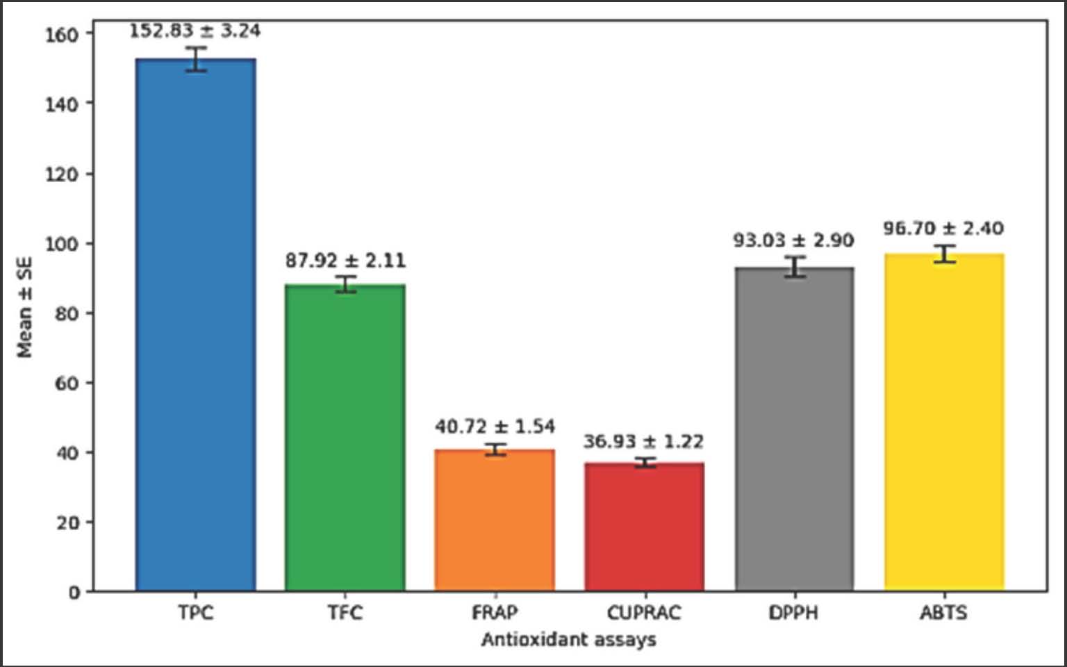
In vitro assessment of antioxidant propertiesAs illustrated in Figure 1, the in vitro assessment of antioxidant activity revealed that the sap of C. gileadensis contained a high content of TPC (152.83 ± 3.24 mg GAE/g), TFC (87.92 ± 2.11 QE/ g), high percentages of ABTS (96.70 ± 2.40 μ mole TE/g) and DPPH (93.03 ± 2.90 μ mole TE/g) radical-scavenging activities, and a good metal chelating capacity by assaying FRAP ( 40.72 ± 1.54 μ mole TE/g) and CUPRAC (36.93 ± 1.54 μ mole TE/g) percentages values.
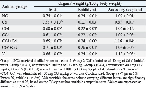
Fig. 1. In vitro assessment of antioxidant activity TPC, CUPRAC, TFC, FRAP, DPPH radical-scavenging activity, and ABTS radical cation assay. # values are expressed as mean ± S.E. Effect of CG sap and Cd chloride on the weights of reproductive organsThe results revealed that the administration of C. gileadensis sap insignificantly (p ≥ 0.05) affected the testis, epididymis, and accessory sex glands compared with the values of the control groups (Table 2). The findings revealed a significant decrease (p < 0.05) in the weights of the testis, epididymis, and accessory sex glands in the Cd chloride-treated group compared with all other experimental groups. Additionally, the simultaneous administration of C. gileadensis sap demonstrated a significant dose-dependent protection, effectively mitigating the adverse effects of Cd chloride on the weights of the testis, epididymis, and accessory sex glands (Table 2). Table 2. Effect of CG sap and Cd chloride on organ weights in rats.
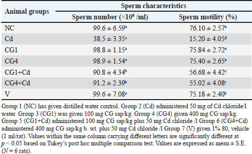
Sperm characteristicsThe findings indicated that administering C. gileadensis sap had no effect on sperm count and motility compared with the control groups (Table 3). The results indicated a notable (p < 0.05) reduction in both sperm count and motility in the Cd chloride-treated group compared with all other groups in the study. Co-administration of C. gileadensis sap plus Cd chloride significantly (p < 0.05) improved the effects of Cd chloride on sperm count and motility; however, these values remained significantly lower than the control values (Table 3). Table 3. Effect of CG sap and Cd chloride on sperm characteristics in rats.
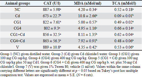
Effect of CG sap and Cd chloride on serum antioxidant concentrationsThe administration of C. gileadensis sap insignificantly (p ≥ 0.05) affected the serum levels of CAT, total antioxidants, and MDA compared with the values of the control groups (Table 4). Meanwhile, the present findings demonstrated a significant (p < 0.05) rise in serum MDA values and reduction in the concentrations of CAT and total antioxidants in the Cd chloride-exposed group compared with all other groups. Conversely, the simultaneous administration of C. gileadensis sap in conjunction with Cd chloride led to a noteworthy elevation in the serum levels of both CAT and total antioxidants, with these measurements exhibiting no statistically significant difference from those recorded in the control groups (Table 4). The biochemical restoration of antioxidant markers was consistent with the histopathological findings, which revealed preserved seminiferous tubule structure in rats treated with CG. Table 4. Effect of CG sap and Cd chloride on serum CAT MDA and TAC activities in rats.
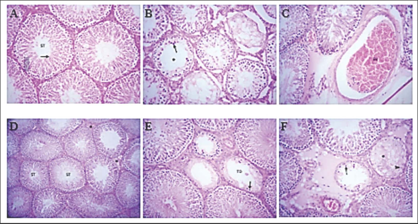
Histopathological examinationHistopathological examination of the testis was extended to evaluate and observe the effect of plant sap in curing and preventing Cd toxicity. Initially, the negative control group (untreated) exhibited a normal histological appearance of seminiferous tubules, active spermatogenesis, and the presence of spermatids within the lumen (Fig. 2A). In contrast, Cd chloride exposure induced marked seminiferous tubule degeneration, germinal epithelium vacuolization, and spermatogenic cell loss (Fig. 2B). Treatment with 400 mg/kg of CG sap markedly preserved the testicular architecture, demonstrating almost normal spermatogenesis alongside mild interstitial edema (Fig. 2C). In contrast, the 100 mg/kg dose provided only partial protection (Fig. 2E).
Fig. 2. Photomicrograph of the effects of plant sap on testicular tissue morphology upon Cd exposure. (A) control group; (B) Cd group; (C) plant sap group; (D) Cd + plant sap group; (E) Cd + plant sap group; (F) Cd + plant sap group (H&E stain; magnification: A, B, and C are 100× and E and F are 400×). A: Testis of control rats showing normal histological appearance of seminiferous tubules (ST), spermatogenesis, and spermatids (arrow) in the lumen. H&E stain × 200. B: Testis of rats administered with 50 mg Cd chloride showing degenerated tubules lined by one or two layers of vacuolated germinal epithelium (arrow) with reduced spermatogenesis and absence of spermatozoa in the lumen (asterisk). H&E stain × 200. C: Testis of rats administered 400 mg plant sap/kg body weight showing mild interstitial edema (asterisk). H&E stain × 200. D: Testis of rats administered 400 mg plant sap/kg body weight and 50 mg Cd chloride showing normal histological appearance of ST and mild interstitial edema (asterisk). H&E stain × 100. E: Testis of rats administered 100 mg plant sap/kg body weight and 50 mg Cd chloride showing testicular degeneration (TD) in a few ST, lined by one or two layers of vacuolated germinal epithelium (arrow) with reduced spermatogenesis. H&E stain × 400. F: Testis of rats administered 400 mg plant sap/kg body weight and 50 mg Cd chloride showing TD of individual ST, lined by a single layer of Sertoli cells (arrowhead) and containing fibrillar eosinophilic material (asterisk) in the lumen or lined by two layers of vacuolated germinal epithelium (arrow). H&E stain × 400. DiscussionThis study is the first to use C. gileadensis sap to alleviate the oxidative damage induced by Cd poisoning in male reproductive organs. Plants possess various bioactive compounds with significant antioxidant properties. Research on the antioxidant activity of diverse plant species may enhance our understanding of their potential as sources of novel antioxidant compounds (But et al., 2024; Mittal et al., 2025; Zhu et al., 2025). Plants inhabit environments with extreme conditions and develop a range of adaptive strategies. Among these strategies is the prevention of oxidative stress, which involves maintaining ROS levels at non-hazardous levels. This highlights the need to examine the antioxidant properties of key plant species, such as C. gileadensis. The defensive mechanisms against oxidative stress are attributed to the normal antioxidants found in herbaceous plants and spices. These aromatic ingredients harbor polyphenols, flavonoids, and phenolic compounds, which act as free radical scavengers (Zhao et al., 2025; Salem et al., 2025). Antioxidant properties of CG sapCommiphora gileadensis sap exhibited strong antioxidant activity, as evidenced by its DPPH, ABTS, FRAP, and CUPRAC values. Previous findings on parts of the CG other than its sap, such as leaves and bark, have been confirmed. Ahmed et al. (2023)reported the DPPH value of CG to be 75.27%, which is lower than the value reported in the present study. However, Ahmed et al. (2023)and Al Al-Mulaiky and Al-Farga (2020)stated higher values and stated the ABTS value of CG to be 98, which is higher than the value obtained in this study. In the same direction, TPC values were assessed by Ahmed et al. (2023) and Safhi et al. (2022)and were 101.47 and 92.54 mg GAE/g, respectively. Additionally, Safhi et al. (2022)reported the TFC value of CG to be 77.13 mg QE/g, which is lower than the value reported in this study. Furthermore, the highest antioxidant enzyme activity content was present with values of 16.87, 60.87, 35.76, and 27.98 U/mg for superoxide dismutase, peroxidase, CAT, and ascorbate peroxidase. Interestingly, Bin Mokaizh et al. (2024) used ultrasonic-assisted extraction in CG leaf extract to improve the yield and recovery of phenolic compounds. TPC and TFC were 96.55 and 31.66, respectively, and GC-MS analysis detected 25 phytochemicals not previously identified. Shadid et al. (2023)reported that CG steam-distilled essential oil exhibited higher antioxidant activity than ascorbic acid. The major constituents in the essential oil were β-myrcene, nonane, verticiol, β-phellandrene, β-cadinene, terpinen-4-ol, β-eudesmol, α-pinene, cis-β-copaene and verticillol, which might be responsible for the antioxidant activity. However, no reports exist in the literature on the FRAP and CUPRAC values. The differences between the results may be due to the geographical locations of the samples, different portions used, and extraction conditions, such as the methodology and diluents used in the extraction (Chaves et al., 2020; Safhi et al., 2022). Commiphora gileadensis sap has a wide safety margin as confirmed in this study as LD50 was above 5-g/kg b. wt. These results are aligned with those of Al-Mahbashi et al. (2015) who reported that C. gileadensis sap is safe up to 5 g/kg b. wt. Cd-induced reproductive toxicityThe testicles are the most vulnerable organs to Cd poisoning, with strong evidence linking Cd to impaired function, even at levels too low to detect (Siu et al., 2009; Blanco et al., 2010). Cd intoxication induces a dose-dependent decrease in testicular weight and function (El-Ashmawy and Youssef, 1999; Abarikwu et al., 2013; Erboga et al., 2016). When Cd enters the body, it enters the bloodstream and accumulates in the kidneys (Satarug and S, 2018), liver, and gut (Tinkov et al., 2018a). It is removed gradually through the kidneys, urine, saliva, and milk. Cd exposure can cause various negative outcomes, such as testicular damage, liver and kidney issues, lung swelling, bone softening, and harm to the adrenal and blood-forming systems (Tinkov et al., 2018b). Animals exposed to Cd showed a marked decrease in the expression of nuclear factor (erythroid-derived 2)-like-2 factor (Nrf2) in the testicular tissue. Nrf2 provides cellular protection against ROS via different mechanisms (Ma, 2013). In the present study, Cd chloride administration significantly decreased the weights, sperm count, and motility of the testis, epididymis, and accessory sex glands. These results align with previous research conducted by Adamkovicova et al. (2016); Erboga et al. (2016); Abarikwu et al. (2013) and El-Ashmawy and Youssef (1999)who reported that Cd chloride had detrimental effects on reproductive organ weights, sperm count, and motility. One of the main mechanisms by which Cd is toxic is through oxidative stress. Cd interferes with sulfhydryl groups, affecting the redox state of cells and the activity of proteins, including mitogen-activated protein kinases. Subsequently, ROS levels increase (Zhu et al., 2011). MDA, which results from lipid peroxidation, was elevated in Cd-exposed testes, while CAT and TAC levels were decreased. These results are consistent with those of Genchi et al. (2020); Koyu et al. (2006)and Carageorgiou et al. (2025). The testis is susceptible to oxidative damage due to the abundance of polyunsaturated fatty acids present in the sperm’s plasma membrane (Zhu et al., 2011). The decrease in reproductive organ weights might be due to lipid peroxidation, antioxidant suppression, and apoptosis, as previously recorded with Cd administration (El-Refaiy and Eissa, 2013). In terms of histopathology, the outcomes of this study support those of previous studies, which showed that Cd damages the testis (Babaknejad et al., 2018; Shojaeepour et al., 2021). Earlier animal studies have shown that Cd decreases testicular weight, sperm count, and testosterone levels (Ren et al., 2012). In addition, Cd diminishes sperm count by disrupting the cell cycle, DNA repair mechanisms, and cell proliferation processes. These adverse changes could occur after a single dose of Cd (Akinloye et al., 2006). Histopathological examination of the testis revealed tissue necrosis, seminiferous tubule degeneration, and spermatogenic damage. Biochemical and histological evidence of the protective effectFlora is used to cure several illnesses because of its antioxidant content, which is a characteristic of traditional medicine (Mahmoudi et al., 2017). Antioxidants combat oxidation by attaching to free radicals, chelating catalytic metals, reducing Cd uptake (Winiarska-Mieczan and A, 2015; Winiarska-Mieczan and A, 2018), and scavenging oxygen (Akunna et al., 2017). Our results established that C. gileadensis sap contains high levels of total phenolic and flavonoid content. In addition to higher ABTS radical-scavenging activity and the best DPPH scavenging activity, CUPRAC and FRAP assays showed good metal chelating capacity. Coadministration of 400 mg/kg CG sap restored serum CAT to 32.1 ± 2.7 U/ml, comparable to controls (35.2 ± 2.4 U/ml), and decreased MDA levels by 28% compared with Cd-only rats. Therefore, it could restore fertility and normal sperm production and improve sperm count and motility. Proposed mechanisms (chelation and ROS scavenging)This study confirms the positive effects of C. gileadensis on Cd-induced infertility and sperm quality parameters. This may be due to the following: C. gileadensis sap chelates Cd owing to its high content of polyphenolic and flavonoid compounds (TFC and TPC), good metal chelating capacity (FRAP and CUPRAC), and potential radical-scavenging antioxidant activity (ABTS and DPPH). Because the combination of several antioxidant assays allows a more complete evaluation of a sample’s antioxidant properties, these assays provide complementary information about the interaction between radicals and samples (López-Alarcón and Denicola, 2013). The identified antioxidant mechanisms provide important suggestions for the protective effects of CG sap. Antioxidant activity is closely correlated with phenolic and flavonoid content (Ma and Huang, 2014; Çolak et al., 2020; Işık, 2020; Butkeviciute et al., 2021). Limitations and suggestions for future workAlthough the present study demonstrates the protective effect of CG sap in rats, further studies are needed to identify the active compounds, elucidate the molecular mechanisms, identify several pathways, such as the Nfr2 pathway, gene expression and assess potential applications in humans and other mammals. ConclusionThis study is the first to use C. gileadensis sap to investigate Cd-induced reproductive toxicity. Administering Cd chloride induced adverse effects on the weights of male reproductive organs, semen characteristics, and decreased antioxidative status. Co-administration of CG sap with Cd significantly mitigated the hazardous effects by reducing antioxidant status, metal chelation, and radical scavenging. The sap was found to be safe, as there were no signs of toxicity in the study of acute toxicity. However, a noteworthy constraint of the current study is the absence of hormonal assay or clarification of some pathways employed to impede the molecular mechanisms implicated in Cd-induced testicular toxicity, thereby verifying the protective efficacy of CG sap. Additionally, a phytochemical investigation is proposed to isolate the active fraction and pure compound. AcknowledgmentsThe researchers would like to acknowledge the Deanship of Graduate Studies and Scientific Research at Qassim University for financial support (Qu-APC-2024-9/1). Conflict of interestThe authors declare no conflict of interest. FundingNo specific grant was provided for this study. Authors’ contributionAll authors were actively involved in research design, execution, data analysis, data interpretation, and manuscript writing. All authors have approved the final version for publication. Data availabilityAll study data are included in this manuscript. Any other related data can be obtained by sending it to the corresponding author. ReferencesAbarikwu, S.O., Iserhienrhien, B.O. and Badejo, T.A. 2013. Rutin-and selenium-attenuated cadmium-induced testicular pathophysiology in rats. Hum. Exp. Toxicol. 32, 395–406. Abdallah, H.M., Mohamed, G.A., Ibrahim, S.R.M., Koshak, A.E., Alnashri, I., Alghamdi, A., Aljohani, A. and Khairy, A. 2022. Commigileadin A: a new triterpenoid from C. gileadensis aerial parts. Pharm. Mag. 18, 256. Adamkovicova, M., Toman, R., Martiniakova, M., Omelka, R., Babosova, R., Krajcovicova, V., Grosskopf, B. and Massanyi, P. 2016. Sperm motility and morphology changes in rats exposed to Cd and diazion. Reprod. Biol. Endoc. 14(42), 2–4. Ahmed, A.F., Shi, M., Liu, C. and Kang, W. 2019. Comparative analysis of antioxidant activities of essential oils and fennel (Foeniculum vulgare Mill.) extracts from Egypt and China. Food. Hum. Wellness. 8, 67–72. Ahmed, H., Rashed, M.M., Almoiliqy, M., Abdalla, M., Bashari, M., Zaky, M.Y., Hailin, Z., Naji, T.A., Eibaid, A. and Wang, J. 2023. Antioxidant activity and total phenolic compounds of C. gileadensis extracts obtained by ultrasonic-assisted extraction, with antiaging and cytotoxicity monitoring activities. Food. Sci. Nutr. 11(6), 3506–3515. Ahmed, H., Zaky, M.Y., Rashed, M., Almoiliqy, M., Al-Dalali, S., Eldin, Z.E., Bashari, M., Cheikhyoussef, A., Alsalamah, S.A., Alghonaim, I.M., Alhudhaibi, A.M., Wang, J. and Jiang, L.P. 2024. UPLC-qTOF-MS phytochemical profile f Commiphora gileadensis leaf extract via integrated ultrasonic-microwave-assisted technique and synthesis of silver nanoparticles for enhanced antibacterial properties. Ultrason. Sonochem. 107(6), 106923. Akinloye, O., Arowojolu, A.O., Shittu, O.B. and Anetor, J.I. 2006. Cadmium toxicity: a possible cause of male infertility in rats. Reprod. Biol. 6, 17–30. Akunna, G., Obikili, E., Anyawu, G. and Esom, E. 2017. Evidence for spermatozoa toxicity and the oxidative damage of cadmium exposure in rats. J. Pharmacol. Toxicol. 12, 50–56. Al-Altaie, D.M. and Addai, Z.R. 2021. Determination of antioxidant compounds, antibacterial activity and broccoli mineral content. Indian J. Forensic. Med. Toxicol. 15, 123–130. Alasgarova, N., Baran, A., Yıldıztekin, M., Ganbarov, D., Babayeva, S., Güneş, Z. and Evcil, M. 2025. Synthesis of plant-derived selenium nanoparticles from Lankaran-Astara tea (Camellia sinensis L.) plant and evaluation of their activities on different enzymes. Adv. Biol. Earth. Sci. 10(2), 262–269. Aldaddou, W.A., Aljohani, A.S., Ahmed, I.A., Al-Wabel, N.A. and El- Ashmawy, I.M. 2022. Ameliorative effect of methanolic extract of Tribulus terrestris L. on nicotine and lead-induced sperm quality degeneration in male rats. J. Ethnopharmacol. 295, 115337. Aldaddou, W.A., Aljohani, A.S.M., Adewale Ahmed, I., Al-Wabel, N.A. and El-Ashmawy, I.M. 2023. Methanolic extract of Salvia officinalis L. reduces lead and nicotine-induced degeneration of sperm quality in male rats. Chem. Biodiversity. 20(7), e202300115. Al-Hazmi, A., Aldairi, A.F., Alghamdi, A., Alomery, A., Mujalli, A., Obaid, A.A., Farrash, W.F., Allahyani, M., Halawani, I., Aljuaid, A., Alharbi, S.A., Almehmadi, M., Alharbi, M.S., Khan, A.A., Jastaniah, M.A. and Alghamdi, A. 2022. Antibacterial effects of Commiphora gileadensis methanolic extract on wound healing. Molecules 27, 3320. Al-Hazmi, A.S., Albeshi, B.M., Alsofyani, E.M., Alherthi, M.N., Aljuaid, M.M., Alfifi, O.A., Alshaer, R.S., Alsaadi, R.S., Alomery, A.M. and Almehmadi, M.M. 2020. Research article in vitro and in vivo antibacterial effect of Commiphora gileadensis methanolic extract against methicillin-resistant Staphylococcus aureus (MRSA) and Pseudomonas aeruginosa. Pak. J. Biol. Sci. 23, 1676–1680. Al-Herthi, M., Aljuaid, M., Alfifi, O., Alshaer, R., Al Saadi, R., Al Mehmadi, M., Eid, E. and Hawash, Y. 2020. In vitro and in vivo antibacterial effect of methanolic extract of Commiphora gileadensis against methicillin-resistant Staphylococcus aureus (MRSA) and Pseudomonas aeruginosa. Pak. J. Biol. Sci. 23, 1676–1680. Ali, A., Derar, D.R., Abdel-El-Moniem, E.M. and Al Mundarij, T.I. 2020a. Cadmium in seminal plasma of fertile and infertile male dromedary patients. Biol. Trace Element. Res. 193, 162–165. Ali, H.A., Mohamed, S.H., Alharbi, H.F. and Algheshairy, R.M. 2020b. Synergism between probiotics and herbs for the management of type 2 diabetes in rats. Intern. J. Pharm. Pharm. Sci. 12(5), 17–25. Al-Mahbashi, H.M., El-Shaibany, A. and Saad, F.A. 2015. Evaluation of acute toxicity and antimicrobial effects of Bisham bark extract (Commiphora gileadensis) L. J. Chem. Pharm. Res. 7, 810–814. Al-Mulaiky, Y.Q. and Al-Farga, A. 2020. Evaluation of the antioxidant enzyme content, phenolic content and antibacterial activity of C. gileadensis grown in Saudi Arabia. Main Group Chem. 19, 329–343. Alqethami, A., Hawkins, J.A. and Teixidor-Toneu, I. 2017. Medicinal plants used by women in Mecca: urban, Muslim, and gendered knowledge. J. Ethnobiol. Ethnomed. 13(1), 1–24. Althurwi, H.N., Salkini, M.A.A., Soliman, G.A., Ansari, M.N., Ibnouf, E.O. and Abdel-Kader, M.S. 2022. Wound healing potential of Commiphora gileadensis stems essential oil and chloroform Extract. Separations 9, 254. Al-Zahrani, S.A., Bhat, R.S., Al-Onazi, M.A., Alwhibi, M.S., Soliman, D.A., Aljebrin, N.A., Al-Suhaibani, L.S. and Al Daihan, S. 2022. Anticancer potential of biogenic silver nanoparticles using Commiphora gileadensis stem extract against human colon cancer. Green. Process. Synth. 11, 435–444. Annapurna, A.S., Abhirami, D. and Umesh, T.G. 2021. A comparative study of phytochemicals and bioactivities of Curcuma amygdala leaf extracts Ada and Curcuma karnatakakensis. South. Afri. J. Botany. 142, 441–450. Apak, R., Güçlü, K., Özyürek, M. and Karademir, S.E. 1981. Novel total antioxidant capacity index for dietary polyphenols and vitamins C and E using their cupric ion reducing capability in the presence of neocuproine: CUPRAC method. J. Agric. Food. Chem. 52, 7970–7981. Ashry, K.M., El-Sayed, Y.S., Khamiss, R.M. and El-Ashmawy, I.M. 2010. Oxidative stress and immunotoxin effects of lead and their reduction with myrrh (Commiphora molmol) emulsion. Food Chem. Toxicol. 48(1), 236–241. Babaknejad, N., Bahrami, S., Moshtaghie, A.A., Nayeri, H., Rajabi, P. and Iranpour, F.G. 2018. Cadmium testicular toxicity in male Wistar rats: protective roles of zinc and magnesium. Biol. Trace. Element. Res. 185, 106–115. Badr, G.M., Elsawy, H. and Sedky, A. 2019. Protective effects of quercetin supplementation short-term toxicity of cadmium-induced hematological impairment, hypothyroidism and testicular disease in albino rats. Environ. Sci. Pollut. Res. 26(8), 8202–8211. Basal, W.T., Issa, A.M., Abdelalem, O. and Omar, A.R. 2023. S. officinalis restores semen quality and testicular functionality in cadmium-intoxicated male rats. Sci. Rep. 13(1), 20808. Benzie, I.F.F. and Strain, J.J. 1996. The ferric reducing ability of plasma (FRAP) as a measure of ‘antioxidant power’: the FRAP assay. Anal. Biochem. 239, 70–76. Bhardwaj, J.K., Paliwal, A. and Saraf, P. 2021. Effects of heavy metals on the reproduction rate owing to Infertility. J. Biochem. Mole. Toxicol. 35(8), e22823. Bin Mokaizh, A.A., Nour, A.H. and Kerboua, K. 2024. Ultrasonic-assisted extraction to enhance the recovery of bioactive phenolic compounds from Commiphora gileadensis leaves. J. Ultsonch. 105, 106852. Blanco, A., Moyano, R., Molina López, A.M., Blanco, C., Flores-Acuña, R., García-Flores, J.R., Espada, M. and Monterde, J.G. 2010. Preneoplastic and neoplastic changes in the Leydig cells population in mice exposed to low doses of cadmium. Toxicol. Indust. Health. 26, 451–457. Bouslama, L., Kouidhi, B., Alqurashi, Y.M., Chaieb, K. and Papetti, A. 2019. Virucidal effect of guggulsterone isolation from Commiphora gileadensis. Planta Med. 85, 1225–1232. But, A.E., Pop, R.M., Binsfeld, G.F., Ranga, F., Orăsan, M.S., Cecan, A.D., Morar, I.I., Chera, E.I., Bonci, T.I., Usatiuc, L.O., Țicolea, M., Cătoi, F.A., Pârvu, A.E. and Ghergie, M.C.D. 2024. The phytochemical composition and antioxidant activity of Matricaria recutita blossoms and Zingiber officinale Rhizome ethanol extracts. Nutrients 7(1), doi: 10.3390/nu17010005 Butkeviciute, A., Petrikaite, V., Jurgaityte, V., Liaudanskas, M. and Janulis, V. 2021. Antioxidant, anti-inflammatory, and cytotoxic activity of extracts from some commercial apple cultivars in two colon and glioblastoma human cell lines. Antioxidants 10, 1098. Carageorgiou, H., Tzotzes, V., Sideris, A., Zarros, A. and Tsakiris, S. 2025. Cadmium effects on brain acetylcholinesterase activity and antioxidant status of adult rats: modulation by zinc, calcium and L-cysteine co-administration. Basic Clin. Pharmacol. Toxicol. 97, 320–324. Chang, C.-C., Yang, M.-H., Wen, H.-M. and Chern, J.C. 2002. Estimation of total flavonoid content in propolis by two complementary colorimetric methods. J. Food Drug Anal. 10(3), 3. Chaves, N., Santiago, A. and Alías, J.C. 2020. Quantification of the antioxidant activity of plant extracts: analysis of sensitivity and hierarchization based on the method used. Antioxidants 9(1), 76; doi:10.1016/j.antioxidant.2020.09.016 Chemek, M., Venditti, M., Boughamoura, S., Mimouna, S.B., Messaoudi, I. and Minucci, S. 2018. Involvement of testicular DAAM1 expression in zinc protection against cadmium induced male rat reproductive toxicity. J. Cell. Physiol. 233(1), 630–640. Chen, Z., Zuo, Z. and Chen, K. 2022. Activated Nrf-2 pathway by vitamin E to attenuate testicular injuries of withs with sub chronic cadmium exposure. Biol. Trace. Elem. Res. 200(4), 1722–1735. Çolak, S., Çömlekcioğlu, N. and Aygan, A. 2020. Antioxidant investigation of antioxidant and antimicrobial activities of plant extracts of Urtica dioica L. Eurasian. J. Bio. Chem. Sci. 3, 206–212. Doğan, S., Ahmadian, A.M.E., Baran, M.F., Mohamed, A.J., Baran, A., Eftekhari, A., Aktepe, N. and Aktaş, H. 2025. Anticancer, antioxidant, and antimicrobial activities of E. angustifolia L leaf extract. Drug Design Develop. Therapy 19, 7719–7734. Dudai, N., Shachter, A., Satyal, P. and Setzer, W. 2017. Chemical composition and monoterpenoid enantiomeric distribution of African Bdellium (Commiphora gileadensis) essential oils. Medicines 4, 66. El-Ashmawy, I.M. and Youssef, S.A. 1999. The antagonistic effect of chlorpromazine on cadmium toxicity. Toxicol. Appl. Pharmacol. 161, 34–39. El-Refaiy, A.I. and Eissa, F.I. 2013. Histopathology and cytotoxicity as biomarkers treated rats with cadmium and some therapeutic agents. Saudi. J. Biol. Sci. 20, 265–280. Erboga, M., Kanter, M., Aktas, C., Bozdemir Donmez, Y., Fidanol Erboga, Z., Aktas, E. and Gurel, A. 2016. Antiapoptotic and antioxidant effects of caffeic acid phenethyl ester on cadmium-induced testicular toxicity in rats. Biol. Trace Elem. Res. 171, 176–184. Eslamieh, J. 2011. Commiphora gileadensis. Cactus Succul. J. 83, 206–210. Evcil, M., Kurt, B., Baran, A., Mouhoub, A. and Karakaplan, M. 2025. Development, characterization and application of chitosan-based formulation incorporationorating Crataegus orientalis extract for food consumption action. Adv. Biol. Earth Sci. 10(2), 208–225. Farid, M.M., Aboul Naser, A.F., Salem, M.M., Ahmed, Y.R., Emam, M. and Hamed, M.A. 2022. Chemical compositions of Commiphora opobalsamum stem bark in the treatment of liver complications in rats with streptozotocin-induced diabetes: the role of oxidative stress and DNA damage. Biomarkers 27, 671–683. Genchi, G., Sinicropi, M.S., Lauria, G., Carocci, A. and Catalano, A. 2020. The effects of cadmium toxicity. Intern. J. Environ. Res. Public Health 17(11), 3782. Habib, R., Wahdan, S.A., Gad, A.M. and Azab, S.S. 2019. Infliximab abrogates cadmium-induced testicular damage and spermiotoxicity via enhancement of steroidogenesis and suppression of inflammation and apoptosis mediators. Ecotoxicol. Environ. Saf. 182, 109398. He, Y., Zou, L. and Luo, W. 2020. Heavy metal exposure, oxidative stress and semen quality: exploring associations and mediation effects in reproductive-aged men. Hemosphere 244, 125498. Iluz, D., Hoffman, M., Gilboa-Garber, N. and Amar, Z. 2010. Medicinal properties of Commiphora gileadensis. Afr. J. Pharm. Pharmacol. 4, 516–520. İPEK, P., Baran, A., Cebe, D.B., Ahmadian, E., Eftekhari, A. and Baran, M.F. 2024. Antioxidant properties of allium turcicum Özhatay & cowley plant extract, its effects on the proliferation and migration of cancer cells. Front. Pharmacol. 15(2024), 1438634. Işık, M. 2020. Anticholinergic, antioxidant activity and LC–MS/MS analysis of ethanol extract from Salvia officinalis L. Int. J. Life. Sci. Biotech. 3, 51–61. Kalehoei, E., Moradi, M., Azadbakht, M., Zhaleh, H., Abadi, S.A.L., Mahdiuni, H. and Gharzi, A. 2023. Therapeutic effects of L-Arginine, L-Carnitine and mesenchymal stem cell-conditioned medium on endometriosis-induced oocyte poor quality in an experimental mouse model. J. Obstet. Gynecol. Res. 49(4), 1180–1188. Koyu, A., Gokcimen, A., Ozguner, F., Bayram, D.S. and Kocak, A. 2006. Evaluation of the effects of cadmium on rat’s liver. Mol. Cell. Biochem. 284, 81–85. Kumar, S. and Sharma, A. 2019. Cadmium toxicity: effects on human reproduction and fertility. Rev. Environ. Health. 34(4), 327–338. López-Alarcón, C. and Denicola, A. 2013. Evaluating the antioxidant capacity of natural products: a review of chemical and cellular-based assays. Anal. Chim. Acta. 763, 1–10. Ma, Y. and Huang, H. 2014. Characterization and comparison of phenols, flavonoids, and isoflavones in soymilk and their correlations with antioxidant activity. Int. J. Food Sci. Technol. 49, 2290–2298. Ma, Q. 2013. The role of nrf2 in oxidative stress and toxicity. Annu. Rev. Pharmacol. Toxicol. 53, 401–426. Mahfouz, M.O., Hariprasad, C.H., Shaffie, I.A. and Sadasivudu, B. 1986. Serum malondialdehyde levels in patients with myocardial infarction and chronic renal failure. IRCS. Med. Sci. 14, 1110–1111. Mahmoudi, R., Honarmand, Z., Karbalay-Doust, S., Jafari-Barmak, M., Nikseresht, M. and Noorafshan, A. 2017. Using curcumin to prevent structural impairments of testicles in rats induced by sodium metabisulfite. EXCLI. J. 16, 583. Mitra, S., Varghese, A.C., Mandal, S., Bhattacharyya, S., Nandi, P., Rahman, S.M., Kar, K.K., Saha, R., Roychoudhury, S. and Murmu, N. 2020. Lead and cadmium exposure induces male reproductive dysfunction by modulating the expression profiles of apoptotic and survival signal proteins in tea-garden workers. Reprod. Toxicol. 98, 134–148. Mittal, G., Dhali A., Prasad R., Nurani, K.M. and Găman, M.A. 2025. Plant extracts with antioxidant and hepatoprotective benefits for liver health: a bibliometric analysis of drug delivery systems. World. J. Gastroenterol. 31(18), 105836. Mohamed, T.A., Zayed, A., AlSherif, E.A., Farag, M.A. and Abdallah, H.M. 2025. NMR-based metabolomics for unraveling oleo-gum resin metabolome of rare species: insights into taxonomic variation and tree sex differences. J. Nat. Med. 79(4), 807–820; doi:10.1016/j.jnm.2025 Moradi, M., Karimi, I., Ahmadi, S. and Mohammed, L.J. 2020. Necessity of antioxidant inclusion in caprine and ovine semen extenders: a systematic review complemented with computational insight. Reprod. Domestic. Animals. 55(9), 1027–1043. Moradi, M. 2021. Melatonin attenuates testicular dysfunction and sperm damage induced by bleomycin, etoposide, and cisplatin (BEP)-based chemotherapy: an experimental study. Fertil. Steril. 116(3), 341. Musaa, K.H., Abdullaha, A. and Subramaniamb, V. 2015. Flavonoid profile and antioxidant activity of the pink guava. Skin 172(36b), 308–357. Oda, S.S. and El-Ashmawy, I.M. 2012. Adverse effects of boldenone undecylenate, an anabolic steroid, on the reproductive functions of male rabbits. Intern. J. Exper. Pathol. 93(3), 172–178. Ren, X.M., Wang, G.G., Xu, D.Q., Luo, K., Liu, Y.X., Zhong, Y.H. and Cai, Y.Q. 2012. The protection of selenium on cadmium-induced inhibition of spermatogenesis via activating testosterone synthesis in mice. Food. Chem. Toxicol. 50, 3521–3529. Rosic, G., Selakovic, D. and Omarova, S. 2024. Cancer signaling, cell/gene therapy, diagnosis and role of nanobiomaterials. Adv. Biol. Earth Sci. 9(Special Issue), 11–34. Safhi, F.A., Alshamrani, S.M., Jalal, A.S., El-Moneim, D.A., Alyamani, A.A. and Ibrahim, A.A. 2022. Genetic characterization of some Saudi Arabia’s accessions from Commiphora gileadensis using physio-biochemical parameters, molecular markers, DNA barcoding analysis and relative gene expression. Genes 13, 2099. Salem, M.A., Khalil, H.M.A., Manaa, E.G., Bass, A.K.A., Osama, N., Samaka, R.M., Ibrahim, M.T. and Hamdan, D.I. 2025. Antioxidant potential of selected apiaceae plant extracts: a study focused on the chemical composition and neuroprotective effect of Coriandrum sativum L. extract against lead (Pb)-induced neurotoxicity in rats. Biol. Trace. Elem. Res. 03(12), 6093–6114. Satarug, S. 2018. Dietary cadmium intake and its effects on kidney function. Toxics 6, 15. Shadid, K.A., Shakya, A.K., Naik, R.R., Al-Qaisi, T.S., Oriquat, G.A., Atoom, A.M. and Farah, H.S. 2023. Exploring the chemical constituents, antioxidant, xanthine oxidase and COX inhibitory activity of Commiphora gileadensis commonly grown wild in Saudi Arabia. Molecules 28(5), 2321; doi:10.3390/molecules28052321 Shalabi, L.F. and Otaif, F.S. 2022. Commiphora Jacq (Burseraceae) in Saudi Arabia: botanical, phytochemical, and ethnobotanical notes. Ecologies 3, 38–57. Shen, Y., You, Y. and Zhu, K. 2023. The traditional Chinese medicine Qiangjing tablet prevents blood-T182, barrier injury induced by CdCl2 through the PI3K/Akt/Rictor signaling pathway. Environ. Toxicol. 38(3), 591–603. Shen, T., Li, G.H., Wang, X.-N. and Lou, H.-X. 2012. Genus commiphora: a traditional uses, phytochemistry, and pharmacology. J. Ethnopharmacol. 142, 319–330. Shojaeepour, S., Dabiri, S., Dabiri, B., Imani, M., Fekri Soofi Abadi, M. and Hashemi, F. 2021. Histopathological findings of testicular tissue following cadmium toxicity in rats. Iranian J. Pathol. 16(4), 348. Sinha, A.K. 1972. Colorimetric assay of catalase. Anal. Biochem. 47, 389–394; doi:10.1016/0003-2697(72)90132-7 Siu, E.R., Mruk, D.D., Porto, C.S. and Cheng, C.Y. 2009. Cadmium-induced testicular injury. Toxicol. Appl. Pharmacol. 238, 240–249. Slinkard, K. and Singleton, V.L. 1977. Total phenol analysis: automation and comparison with manual methods. Am. J. Enol. Viticulture 28, 49–55. Tajbakhsh, E., Khamesipour, A., Hosseini, S.R., Kosari, N., Shantiae, S. and Khamesipour, F. 2021. The effects of medicinal herbs and marine natural products on wound healing of cutaneous leishmaniasis: a systematic review. Microbial. Pathogenesis. 161, 105235. Tinkov, A.A., Filippini, T., Ajsuvakova, O.P., Skalnaya, M.G., Aaseth, J., Bjørklund, G., Gatiatulina, E.R., Popova, E.V., Nemereshina, O.N. and Huang, P.-T. 2018. Cadmium and atherosclerosis: a review of toxicological mechanisms and a meta-analysis of epidemiological data and studies. Environ. Res. 162(1), 240–260. Tinkov, A.A., Gritsenko, V.A., Skalnaya, M.G., Cherkasov, S.V., Aaseth, J. and Skalny, A.V. 2018. Gut as a target for Cd toxicity. Environ. Pollut. 235, 429–434. Unzile, Y., Ince-Erguc, E., Ozturk, I., Okudan, E.S. and Kirci, D. 2025. Evaluation of cytotoxic and antimicrobial activities of methanolic extract from Cystoseira foeniculacea and Sargassum vulgare. Adv. Biol. Earth Sci. 10(2), 347–358. Varijakzhan, D., Chong, C., -M.., Abushelaibi, A., Lai, K.-S. and Lim, S.-H.E. 2020. Middle Eastern plant extracts: an alternative to modern medicines. Molecules 25(5), 1126. Venditti, M., Ben, M., Romano, I., Messaoudi, R., Reiter, J. and Minucci, S. 2021. Evidence of melatonin ameliorative effects on the blood-testis barrier and alterations in sperm quality induced by cadmium in the rat testis. Ecotoxicol. Environ. Saf. 226, 112878. Wineman, E., Douglas, I., Wineman, V., Sharova, K., Jaspars, M., Meshner, S., Bentwich, Z., Cohen, G. and Shtevi, A. 2015. Commiphora gileadensis Sap extract induces cell cycle-dependent death in immortalized keratinocytes and human dermoid carcinoma cells. J. Herb. Med. 5, 199–206. Winiarska-Mieczan, A. 2015. The potential protective effect of green, black, red and white tea infusions against the adverse effects of cadmium and lead during chronic exposure - a rat model study. Regulatory Toxicol. Pharmacol. 73, 521–529. Winiarska-Mieczan, A. 2018. Tea’s protective effect against lead and cadmium-induced oxidative stress—a review. Biometals 31, 909–926. Yang, S.H., He, J.B., Yu, L.H., Li, L., Long, M., Liu, M.D. and Li, P. 2019. Protective Role of curcumin in cadmium-induced testicular injury in mice by attenuating oxidative stress via Nrf2/ARE pathway. Environ. Sci. Pollut. Res. 26(33), 34575–34583. Yaniv, Z. and Dudai, N. 2014. Medicinal and aromatic plants of the Middle- East; Springer: berlin/Heidelberg. Berlin, Germany: Springer, vol. 2, pp: 67–150. Zhang, Q., Xu, W., Kong, Z., Wu, Y. and Liu, Y. 2023. cadmium exposure-induced rat testicular dysfunction and its mechanism of chronic stress. Food. Chem. Toxicol. 182, 114181. Zhao, L., Yue Z Wang., Qin, G., Ma, J., Tang, H. and D Yin. 2025. Smilax glabra roxb. alleviates cisplatin-induced acute kidney injury in mice by activating the Nrf2/HO-1 signaling pathway. Phytomedicine 139, 156550. Zhao, L.L., Ru, Y.F., Liu, M., Tang, J.N., Zheng, J.F., Wu, B., Gu, Y.H. and Shi, H.J. 2017. Reproductive effects of cadmium on sperm function and early embryonic development in vitro. PLoS One 12(11), 186727226. Zhou, J., Zhang, Y. and Zeng, L. 2022. Paternal exposure to cadmium affects testosterone synthesis by reducing the testicular cholesterol pool in offspring-induced cells mice. Ecotoxicol. Environ. Saf. 242, 113947. Zhu, H., Li, K., Liang, J., Zhang, J. and Wu, Q. 2011. Changes in the levels of DNA methylation in the testis and liver of SD rats neonatally exposed to 5-aza-2′-deoxycytidine and cadmium. J. Appl. Toxicol. 31, 484–495. Zhu, Y., Tian, M., Lu, S., Qin, Y., Zhao, T., Shi, H., Li, Z. and Qin, D. 2025. The antioxidant role of aromatic plant extracts in managing neurodegenerative diseases: a comprehensive review. Brain Res. Bull. 222, 111253. | ||
| How to Cite this Article |
| Pubmed Style Aljohani AS, Aliiri AA, Soliman AS, Musa KH, Alreshoodi M, El-ashmawy IM. Protective effect of Commiphora gileadensis against cadmium toxicity in male rats. Open Vet. J.. 2026; 16(5): 3178-3191. doi:10.5455/OVJ.2026.v16.i5.58 Web Style Aljohani AS, Aliiri AA, Soliman AS, Musa KH, Alreshoodi M, El-ashmawy IM. Protective effect of Commiphora gileadensis against cadmium toxicity in male rats. https://www.openveterinaryjournal.com/?mno=286824 [Access: June 26, 2026]. doi:10.5455/OVJ.2026.v16.i5.58 AMA (American Medical Association) Style Aljohani AS, Aliiri AA, Soliman AS, Musa KH, Alreshoodi M, El-ashmawy IM. Protective effect of Commiphora gileadensis against cadmium toxicity in male rats. Open Vet. J.. 2026; 16(5): 3178-3191. doi:10.5455/OVJ.2026.v16.i5.58 Vancouver/ICMJE Style Aljohani AS, Aliiri AA, Soliman AS, Musa KH, Alreshoodi M, El-ashmawy IM. Protective effect of Commiphora gileadensis against cadmium toxicity in male rats. Open Vet. J.. (2026), [cited June 26, 2026]; 16(5): 3178-3191. doi:10.5455/OVJ.2026.v16.i5.58 Harvard Style Aljohani, A. S., Aliiri, . A. A., Soliman, . A. S., Musa, . K. H., Alreshoodi, . M. & El-ashmawy, . I. M. (2026) Protective effect of Commiphora gileadensis against cadmium toxicity in male rats. Open Vet. J., 16 (5), 3178-3191. doi:10.5455/OVJ.2026.v16.i5.58 Turabian Style Aljohani, Abdullah S.m., Ahmed A. Aliiri, Amal S. Soliman, Khaled H. Musa, Mohamed Alreshoodi, and Ibrahim M. El-ashmawy. 2026. Protective effect of Commiphora gileadensis against cadmium toxicity in male rats. Open Veterinary Journal, 16 (5), 3178-3191. doi:10.5455/OVJ.2026.v16.i5.58 Chicago Style Aljohani, Abdullah S.m., Ahmed A. Aliiri, Amal S. Soliman, Khaled H. Musa, Mohamed Alreshoodi, and Ibrahim M. El-ashmawy. "Protective effect of Commiphora gileadensis against cadmium toxicity in male rats." Open Veterinary Journal 16 (2026), 3178-3191. doi:10.5455/OVJ.2026.v16.i5.58 MLA (The Modern Language Association) Style Aljohani, Abdullah S.m., Ahmed A. Aliiri, Amal S. Soliman, Khaled H. Musa, Mohamed Alreshoodi, and Ibrahim M. El-ashmawy. "Protective effect of Commiphora gileadensis against cadmium toxicity in male rats." Open Veterinary Journal 16.5 (2026), 3178-3191. Print. doi:10.5455/OVJ.2026.v16.i5.58 APA (American Psychological Association) Style Aljohani, A. S., Aliiri, . A. A., Soliman, . A. S., Musa, . K. H., Alreshoodi, . M. & El-ashmawy, . I. M. (2026) Protective effect of Commiphora gileadensis against cadmium toxicity in male rats. Open Veterinary Journal, 16 (5), 3178-3191. doi:10.5455/OVJ.2026.v16.i5.58 |


