





| Research Article | ||
Open Vet. J.. 2026; 16(5): 3155-3165 Open Veterinary Journal, (2026), Vol. 16(5): 3155-3165 Research Article Neutrophil-to-Lymphocyte Ratio as a Prognostic Biomarker in Experimental Tongue Squamous Cell Carcinoma Induced by 4-Nitroquinoline-1-Oxide—A Pilot StudyAkeem Moradehun Bamiro1,2, Zul Izhar Mohd Ismail1,3, Sharifah Emilia Tuan Sharif3,4, Tengku Ahmad Damitri Al-Astani Tengku Din3,5, Nasibah Mohamad3,6, Luqman Afiq Mohamad Ishak3,7, Samir Acherar8, Philippe Arnoux8, Céline Frochot8 and Norhafiza Mat Lazim3,7*1Department of Anatomy, School of Medical Sciences, Universiti Sains Malaysia Health Campus, Kelantan, Malaysia 2Department of Anatomy, Lagos State University College of Medicine, Lagos, Nigeria 3Hospital Pakar Universiti Sains Malaysia, Kelantan, Malaysia 4Department of Pathology, School of Medical Sciences, Universiti Sains Malaysia Health Campus, Kelantan, Malaysia 5Department of Chemical Pathology, School of Medical Sciences, Universiti Sains Malaysia Health Campus, Kelantan, Malaysia 6Department of Radiology, School of Medical Sciences, Universiti Sains Malaysia Health Campus, Kelantan, Malaysia 7Department of Otorhinolaryngology-Head & Neck Surgery, School of Medical Sciences, Universiti Sains Malaysia Health Campus, Kelantan, Malaysia 8Reactions and Chemical Engineering Laboratory, Université de Lorraine, LRGP-CNRS, Nancy, France *Corresponding Author: Norhafiza Mat Lazim. Department of Otorhinolaryngology-Head & Neck Surgery, School of Medical Sciences, Universiti Sains Malaysia Health Campus, Kelantan, Malaysia. Email: norhafiza [at] usm.my; norhafizamatlazim [at] gmail.com Submitted: 25/09/2025 Revised: 03/03/2026 Accepted: 15/03/2026 Published: 31/05/2026 © 2025 Open Veterinary Journal
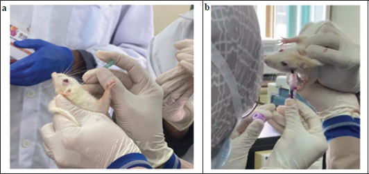
ABSTRACTBackground: Oral squamous cell carcinoma of the tongue is the most common type of head and neck malignancy and is associated with poor survival. Although inflammatory blood indices, such as the neutrophil-to-lymphocyte ratio (NLR), platelet-to-lymphocyte ratio (PLR), and lymphocyte-to-monocyte ratio (LMR), have prognostic value in human cancers, their role in preclinical oral cancer models is poorly defined. Aim: To evaluate the prognostic performance of post-induction NLR, PLR, and LMR for short-term survival in rats with 4-nitroquinoline-1-oxide (4NQO)-induced tongue squamous cell carcinoma. Methods: Female Sprague Dawley rats were exposed to 50 ppm 4NQO in drinking water for 20 weeks. Complete blood counts were obtained at baseline and after tumor induction to compute systemic inflammatory indices. 18 rats were included in the survival study, and the overall survival was defined as the time from the post-induction blood assessment (start of the 42-day observation period) to death/euthanasia, with animals alive at day 42 right-censored. Receiver operating characteristic (ROC) analysis with Youden’s index was used to select the most sensitive model and derive an optimal cutoff for 42-day mortality. Kaplan–Meier curves with log-rank testing were used to compare survival between the biomarker strata. Results: The ROC analysis demonstrated good discrimination for NLR [area under the curve (AUC) 0.901], while PLR (AUC 0.358) and LMR (AUC 0.265) showed poor discrimination. The optimal NLR cutoff was 0.5463 (sensitivity, 0.889; 1-specificity, 0.111; Youden’s index, 0.778). Kaplan–Meier analysis showed markedly poorer survival in rats with NLR > 0.55, with 8/9 deaths versus 1/9 deaths in the ≤ 0.55 group (log-rank p < 0.001). Conclusion: Post-induction NLR derived from routine complete blood counts modestly discriminated 42-day survival in a 4NQO-induced rat model of tongue squamous cell carcinoma, supporting its use as a potential prognostic biomarker in resource-limited preclinical settings and as a basis for larger, controlled studies. Keywords: 4-Nitroquinoline-1-oxide, Blood cell count, Clinical decision rule, Mouth neoplasm, Squamous cell carcinoma. IntroductionOral cancer is the most common type of head and neck cancer (Sexton et al., 2024), accounting for 177,000 deaths annually (Ashwini et al., 2024). Tongue cancer is the most common type of oral cancer (Migueláñez-Medrán et al., 2019). Smoking and alcohol consumption are the major risk factors of this disease (Migueláñez-Medrán et al., 2019; Ashwini et al., 2024). Despite advanced treatment modalities, the overall survival (OS) of this disease remained unchanged, underscoring the need for improved prognostic markers and treatment strategies (Ashwini et al., 2024). Prolonged exposure to carcinogens causes chronic inflammation, and the persistent inflammatory state within the tissue acts as a key hallmark that promotes cancer development and progression (Tripathi et al., 2025). Inflammatory immune cells, such as neutrophils and monocytes, secrete pro-survival and pro-migratory factors needed for unfettered tumor growth and metastasis. Monocytes differentiate into tumor-associated macrophages, which release cytokines and chemokines. These factors promote tumor initiation, angiogenesis, invasion, and metastasis while simultaneously suppressing T cell-mediated anti-tumor immunity. Consequently, an inflammation-driven, immunosuppressive environment is created, which is associated with more aggressive disease and poorer outcomes (Ravindranathan et al., 2021; Tripathi et al., 2025). In line with this, inflammatory indicators in the bloodstream have emerged as promising biomarkers for gauging cancer severity and prognosis (Zhou et al., 2014). Recently, neutrophil-to-lymphocyte ratio (NLR), platelet-to-lymphocyte ratio (PLR), and lymphocyte-to-monocyte ratio (LMR), which are ratios derived from routine complete blood counts (CBC), have gained attention as easily accessible measures of systemic inflammation and immunity in patients with chronic disease and cancer. These CBC-derived indices reflect the balance between pro-tumor inflammatory cells (neutrophils, monocytes, and platelets) and anti-tumor immune cells (lymphocytes) (Hu et al., 2022). A growing body of evidence links these biomarkers to cancer outcomes. For instance, Pingdong et al. (2022) reported that patients with laryngeal cancer with higher NLR and PLR and lower LMR showed a higher 5-year mortality than those with lower NLR and PLR and higher LMR. The roles of these biomarkers have been reported in esophageal cancers (Feng et al., 2014), colorectal cancer (Han et al., 2018), and advanced gastric cancer (Song et al., 2017). The 4-nitroquinoline-1-oxide (4NQO) model of oral carcinogenesis provides a valuable and controlled experimental platform for investigating these inflammation-driven biomarkers. Chronic administration of 4NQO (a water-soluble chemical carcinogen) induces tongue squamous cell carcinoma (TSCC) in rodents through a multi-step process of initiation, promotion, and progression, closely mimicking the developmental process in humans. This model reliably produces dysplasia and invasive carcinoma in the tongue and oral cavity, providing opportunities to track systemic inflammatory changes during tumor development (Ribeiro and Salvadori, 2007; Zigmundo et al., 2022). In this pilot component of a larger photodynamic therapy (PDT) study, we induced tongue squamous cell carcinoma in rats with 4NQO for over 20 weeks and then observed them for a further 6 weeks before initiating treatment. During this pre-treatment period, we examined whether the post-induction values of NLR, LMR, and PLR could predict the model’s short-term survival. As the study was not specifically designed for survival endpoints, these findings should be regarded as exploratory and hypothesis-generating. Although NLR, PLR, and LMR are increasingly reported as prognostic markers in clinical head and neck cancer cohorts, their role in preclinical oral cancer models is poorly defined. These models remain important because they allow these inflammatory indices to be examined under controlled conditions, with defined carcinogen exposure and longitudinal sampling, and with less influence from clinical confounders, such as comorbidities, concurrent infections, medications, or heterogeneous treatment histories. Materials and MethodsAnimals and housingTwenty-three inbred female Sprague Dawley rats (8 weeks old, 190–260 g) were procured from The Animal Research and Services Center, Universiti Sains Malaysia Health Campus. The use of this age and sex of rats has been reported in a study by Lin et al. (2022). After a week of acclimatization, three rats were housed per cage under controlled conditions of a 12-hour light–dark cycle, temperature of 22°C ± 2°C, and humidity of 50% ± 10%. Food was provided throughout the study, while water was available ad libitum during acclimation and was resumed only after the cessation of carcinogen exposure at week 20. Experimental designThis study was initially designed to evaluate the therapeutic effects of PDT on 4NQO-induced TSCC. All animals were first subjected to cancer induction and subsequently randomized into positive control and treatment groups before PDT intervention. During the planned pretreatment observational period of 42 days, spontaneous mortality was observed. This unanticipated outcome provided an opportunity to explore the prognostic relevance of post-induction hematological biomarkers on short-term survival. Survival analyses based on post-induction biomarkers were reported as an observational component of the study. At this stage, no formal untreated control group was incorporated because the primary intervention phase had not yet started (Khozeimeh et al., 2019). 4NQO protocolTongue carcinogenesis was induced using 4-nitroquinoline-1-oxide (4NQO; Sigma-Aldrich, ≥98% purity, Lot WXBF0119V). A 500-ppm stock solution was prepared by dissolving 500 mg of 4NQO in 50 ml of acetone and diluting it with 950 ml of tap water. This solution was vortexed and stored in an aluminum foil-wrapped bottle. A 50 ppm working solution was freshly prepared from the stock solution and administered twice weekly in light-protected bottles for 20 weeks (Ribeiro & Salvadori, 2007). 4NQO solution consumption was recorded weekly per cage. Blood sampling and analysisBlood samples (0.5 ml) were collected from conscious rats via the submandibular vein at baseline and after 20 weeks of 4NQO administration. Before sampling, the hair around the venipuncture site was shaved to improve vessel visualization. The animal was gently restrained by holding the skin over the back and neck while stabilizing the tail to ensure venous congestion and minimize head and body movement. Venipuncture was performed using a 21-gauge sterile needle inserted slightly posterior to the mandibular joint at the submandibular vein’s anatomical location (Fig. 1). Once the required blood volume was obtained, the restraint was released to allow the blood flow to cease. The puncture site was then gently compressed with a sterile cotton swab for approximately 30 seconds to achieve adequate hemostasis before the animal was returned to its cage (Ott Joslin, 2009; Port Louis et al., 2023). Samples were collected into ethylenediaminetetraacetic acid-coated tubes and gently inverted several times to prevent clotting. CBC was performed using a Sysmex multispecies automated hematology analyzer. The NLR, PLR, and LMR were computed from the absolute values.
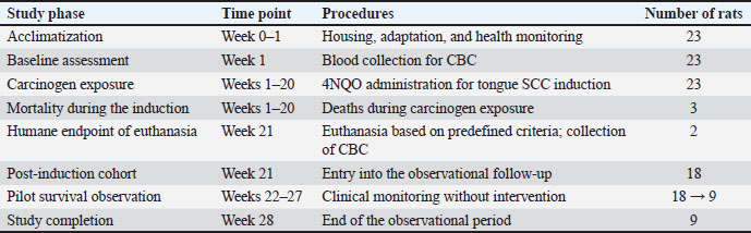
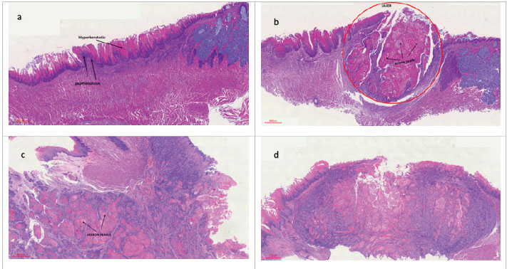
Fig. 1. Submandibular venous blood collection in Sprague–Dawley rats (a) Identification and puncture of the submandibular vein using a 21-gauge needle inserted slightly posterior to the mandibular joint in a gently restrained rat. The hair around the sampling site was shaved to facilitate venipuncture visualization. (b) Blood collection from the submandibular vein while the rat is manually restrained to maintain venous congestion and minimize movement. Endpoints and follow-upThe rats were weighed weekly and monitored for physical and behavioral abnormalities, including significant weight loss, ulceration, reduced food intake, lethargy, abnormal posture, and labored breathing. Humane endpoints included >10% body weight loss in 1 week, >20% progressive weight loss, tumor size of >2 cm, ulceration or infection, or inability to ambulate (Olfert et al., 1998). Rats that met these criteria were euthanized by intraperitoneal administration of a ketamine–xylazine anesthetic combination (ketamine 90 mg/kg and xylazine 5 mg/kg), after which death was confirmed before tissue collection. From week 7 onward, tongue lesions were examined fortnightly under inhalational anesthesia induced with 5% isoflurane in oxygen (1 L/minute flow rate). Tumor induction was operationally defined using clinical criteria assessed during serial oral examinations. Animals with severe tongue dysplasia or squamous cell carcinoma were classified based on the presence of persistent exophytic lesions with a rough surface and non-healing ulcers with raised, indurated margins. These criteria were informed by a preliminary pilot cohort in which serial tongue biopsies confirmed progression from dysplasia to invasive carcinoma, and the gross lesion morphology observed at histological confirmation was consistent with the clinical features applied in the main study. Therefore, histopathological confirmation was not repeated for every animal in the survival cohort, but identical lesion-based criteria were applied consistently across animals and time points. Animals that died or met the humane endpoints were placed in labeled biohazard carcass bags and stored at −20°C pending approved disposal. The primary endpoint is overall survival, which is defined as the number of days from the post-induction blood assessment (initiation of the 42-day observation period) to death or euthanasia at humane endpoints. Animals alive on day 42 were right-censored on 42 days. Euthanasia conducted at predefined humane endpoints was treated as an event (death) in survival analyses. Survival analyses were restricted to animals with post-induction biomarker measurements (i.e., animals surviving to the post-induction blood sampling time point). Biomarker-based prognostic modeling was not eligible for animals that did not reach this assessment. Statistical analysisStatistical analyses were conducted using Stata version 14.1 and SPSS version 29.0.2.0 (IBM Corp., Armonk, NY, USA). Data are presented as median with interquartile range. Comparisons between baseline and post-induction CBC parameters were made using Wilcoxon signed-rank tests. Missing data were excluded from the analysis. Survival outcomes were analyzed using Kaplan–Meier curves and log-rank tests. Receiver operating characteristic (ROC) analysis was performed using the 42-day mortality status (dead vs. alive) as the state variable to evaluate the discrimination of post-induction NLR, PLR, and LMR. Using Youden’s index, the optimal prognostic threshold was derived, and the cut-off with the maximum Youden statistic was selected for survival stratification. Kaplan–Meier curves and log-rank tests were used to compare survival between the gotten cut-offs. Statistical significance was set at p < 0.05. Ethical approvalThe Universiti Sains Malaysia Institutional Animal Care and Use Committee [USM IACUC; Approval No. USM/IACUC/2023/(142)(1278)] approved the experimental protocol, and was conducted in compliance with relevant animal welfare guidelines. ResultsAnimal healthThroughout the study, the rats’ general health were monitored regularly. The study timeline and animal disposition are presented in Table 1. Before being exposed to 4NQO for 20 weeks, all rats underwent baseline CBC assessment after a week of acclimatization. Animals that survived to week 21 were placed in a 6-week observational follow-up for survival analysis after attrition occurred during and after induction. The experiment lasted for 28 weeks. Of the nine rats that died, two had moderate dysplasia, and seven had well-differentiated squamous cell carcinoma (SCC) (Fig. 2). Table 1. Experimental timeline, procedures, and disposition of animals.
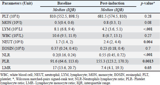
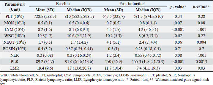
Fig. 2. Tongue histology of rats that died due to mortality. (a) Moderate dysplasia with hyperkeratosis and papillomatous appearance (b) Moderate dysplasia with surface ulceration and extensive keratin pearl formation within the ulcer bed. (c) Well-differentiated SCC with deeply infiltrating malignant squamous cells and connective tissue with numerous keratin pearls. (d) Well-differentiated SCC with prominent epithelial nests and keratin pearls confirming the presence of invasive cancer. Hematological changesAt baseline, the lymphocyte counts markedly exceeded the neutrophil counts. However, following TSCC induction, neutrophil counts increased, whereas lymphocyte counts declined, resulting in a pronounced shift in the neutrophil–lymphocyte balance. Changes in neutrophil and lymphocyte counts reached statistical significance among CBC parameters, leading to corresponding alterations in derived inflammatory ratios (Table 2). Table 2. Effect of tongue cancer induction on hematological parameters and biomarkers (N=20).
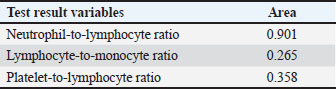
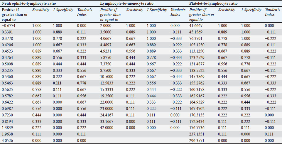
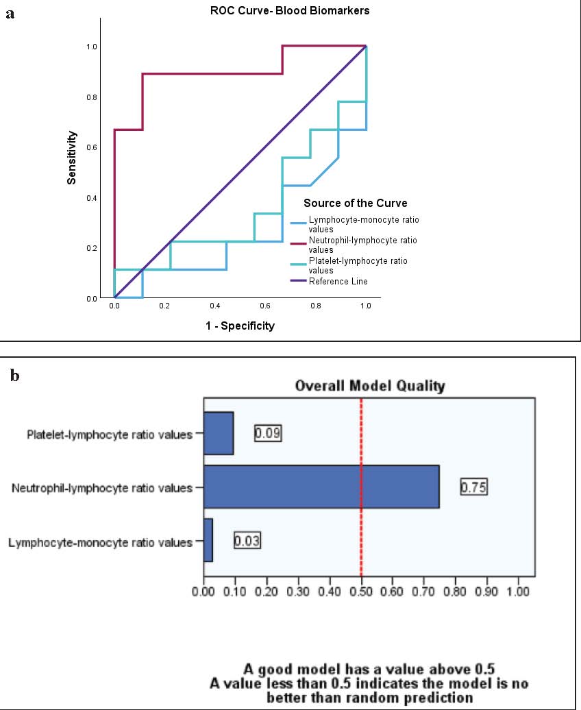
Receiver operating characteristic analysis was performed to assess the discriminatory ability of post-induction inflammatory indices for 42-day mortality. Of the evaluated biomarkers, NLR demonstrated strong discrimination (AUC=0.901), whereas PLR (AUC=0.358) and LMR (AUC=0.265) showed poor model performance. The optimal NLR threshold identified using Youden’s index was 0.5463, yielding a sensitivity of 0.889, a specificity of 0.889, and a Youden’s index of 0.778. Based on this cut-off, the animals were stratified into low (≤ 0.55) and high (> 0.55) NLR groups (Fig. 3, Tables 3 and 4). Table 3. Area under the receiver operating characteristic curve.
Table 4. Receiver operator curve (ROC) performance for blood biomarkers.
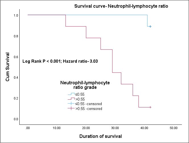
Fig. 3. ROC analysis of post-induction inflammatory blood biomarkers for 42-day survival.) a) Receiver operating characteristic curves comparing the discriminative performance of neutrophil-to-lymphocyte ratio (NLR), platelet-to-lymphocyte ratio (PLR), and lymphocyte-to-monocyte ratio (LMR) for mortality within 42 days following tumor induction. The NLR demonstrated strong discrimination (AUC=0.901), whereas the PLR (AUC=0.358) and LMR (AUC=0.265) showed poor performance, indicating that the approximation was random. (b) Overall model quality for each biomarker, illustrating that only the NLR exceeded the threshold for acceptable predictive performance (>0.5), supporting its selection for subsequent survival analyses. Kaplan–Meier survival analysis (Fig. 4) demonstrated substantially reduced survival in rats with a post-induction NLR > 0.55 compared with those with NLR ≤ 0.55, with eight deaths observed in the high-NLR group versus one death in the low-NLR group during the 42-day follow-up period (log-rank p < 0.001). The mean survival time was shorter in the high-NLR group (29.3 days) than in the low-NLR group (41.9 days). In univariate Cox proportional hazards analysis, high NLR was associated with a significantly increased risk of mortality (HR, 3.03), with the association remaining robust following bootstrap resampling.
Fig. 4. Kaplan–Meier survival analysis based on the post-induction neutrophil-to-lymphocyte ratio Kaplan–Meier curves comparing 42-day survival in rats with low (≤0.55) versus high (>0.55) post-induction NLR following 4NQO-induced tongue carcinogenesis. Rats with elevated NLR demonstrated significantly reduced survival compared with those with lower NLR values (log-rank p < 0.001). DiscussionThe prognostic value of biomarkers is vital for cancer research and management. Additionally, they should be readily available, inexpensive, and easily interpreted. In this study, we investigated the effects of chemically induced tongue cancer on hematological parameters and blood biomarkers in an animal model. The effect of higher and lower values of these blood biomarkers on the overall survival of the animals was also explored. White blood cells, predominantly lymphocytes and neutrophils, are important for the body’s defense against pathological and inflammatory conditions. Although lymphocytes mediate adaptive immunity, neutrophils are essential for maintaining the innate immune response. In addition to being important for innate defenses, neutrophils are also observed in proinflammatory conditions, including cancers, where they have been reported to possess both cancer-promoting and inhibiting properties (Song et al., 2021). In laboratory rats, the complete blood count profile varies with age; however, lymphocytes consistently outnumber neutrophils across all age groups from 1 month to >18 months (Wolford et al., 1986; Jacob Filho et al., 2018; Patel et al., 2024). However, we observed a significant shift in this balance after cancer induction, with the average counts of both cell types becoming almost equal (Supplementary Table 1). Isolated increases in neutrophil counts have been reported in cancers (Lee et al., 2021), and a significant increase in neutrophil counts following oral TSCC induction was also observed. We also observed a significant decrease in the lymphocyte count, indicating that the proinflammatory nature of oral TSCC exceeds the adaptive imbalance in rats. An increase in neutrophil count and a decrease in lymphocyte count lead to a significant increase in NLR, which is reported to be increased in cancer and other proinflammatory conditions such as obesity and stress (Hickman, 2017; Natsar et al., 2024). Singh et al. (2021) attempted to determine the diagnostic efficacy of NLR by comparing pretreatment NLR values in normal individuals, patients with oral cancer, and patients with oral potentially malignant disorders (OPMDs), such as oral leucoplakia. They reported that patients with oral cancer have higher NLR, followed by those with OPMD. However, healthy individuals exhibited lower NLR values. A reduction in the lymphocyte counts significantly changed the PLR and LMR. While we reported a decrease in LMR compared with the baseline, we also observed an increase in PLR. Several studies have shown that cancers are often accompanied by elevated NLR, PLR, and MLR, or conversely reduced LMR, and that these alterations are consistently associated with poorer clinical outcomes (Liu et al., 2016; Ramos-Esquivel et al., 2017; Wang et al., 2018; Dertli et al., 2021; Pingdong et al., 2022). To objectively assess the prognostic relevance of these post-induction inflammatory biomarkers, model performance analyses demonstrated that NLR was the only marker with adequate discriminatory capacity, whereas PLR and LMR showed poor sensitivity. Using the optimal cut-off derived from this model, an NLR value > 0.55 was strongly associated with adverse outcomes, with rats in this category exhibiting significantly poorer OS compared with those with lower NLR values (log-rank p < 0.001). Univariable Cox proportional hazards analysis consistently indicated that elevated NLR was associated with a significantly increased hazard of death during follow-up. This report is in tandem with a meta-analysis report by Mariani et al. (2022), in which 19 studies were examined to investigate whether pretreatment NLR is an independent prognostic factor for HNSCC. They reported that higher NLR values were significantly associated with poor OS and lower disease-free survival in patients with HNSCC. Approximately 90% of the rats lost during this evaluation period had well-differentiated SCC; hence, we can conclude that the prognostic values of NLR reported in this study are due to the successful induction of TSCC in rats. In a retrospective study by Mireștean et al. (2023) on patients with HNSCC, those with high NLR were reported to have poor OS, and we obtained similar results in rats with TSCC. Although survival analysis was not the primary aim of this study, the observed trends offer preliminary insight and may serve as a pilot for future studies. The limited sample size and statistical power underscore the need for larger, adequately powered investigations to validate these findings. ConclusionThis study assessed the prognostic significance of systemic inflammatory blood biomarkers in a 4NQO-induced rat model of TSCC. NLR was the only biomarker with sufficient discriminatory and prognostic performance. In this experimental TSCC, elevated post-induction NLR, especially values greater than 0.55, is significantly associated with lower OS, highlighting its potential usefulness as a practical prognostic indicator. Given the small sample size and secondary nature of the survival analysis, the findings should be interpreted with caution and are not generalizable. Further research using larger numbers of animals is recommended to validate these findings and establish optimal cutoff values for preclinical applications. AcknowledgmentThe authors express their heartfelt gratitude to The Ministry of Higher Education Malaysia (MOHE) for funding this study. The authors extend their gratitude to Normayazi Mohemed Ali, Ahmad Talha Mohamad Yunus, and Adani Shafwan of The Pathology Research Laboratory of Hospital USM for their invaluable assistance in preparing the histology slides. The authors also extend their appreciation to the staff of The Animal Research and Services Center, USM, for their support with blood collection and anesthesia administration, which were crucial for this study’s success. Their contributions are greatly appreciated and have been instrumental in advancing this research. Conflict of interestThe authors have no conflicts of interest to declare. FundingThis study was funded by the My MATCH grant fund of The Ministry of Higher Education Malaysia (MOHE), project code 203.PPSP.6782007. Authors' contributionsAll authors contributed to the study conception and design. Akeem Moradehun Bamiro and Luqman Afiq Mohamad Ishak prepared the materials, collected and analyzed data. The first draft of the manuscript was written by Akeem Moradehun Bamiro, Luqman Afiq Mohamad Ishak, Zul Izhar Mohd Ismail, Sharifah Emilia Tuan Sharif, Tengku Ahmad Damitri Al-Astani Tengku Din, and Nasibah Mohamad, and all authors commented on previous versions of the manuscript. Samir Acherar, Philippe Arnoux, Céline Frochot, and Norhafiza Mat Lazim provided supervision, resources, and funding. All authors contributed to the methodology, conceptualization, and interpretation of data. All authors have read and approved the final version of the manuscript. Data availabilityThe data underlying this study can be obtained from the first author or the corresponding author upon reasonable request. ReferencesAshwini, R., Narayan, M. and Rajkumar, K. 2024. Diagnostic and prognostic markers of oral squamous cell carcinoma- a detailed review. Oral. Oncol. Rep. 10, 100321; doi:10.1016/j.oor.2024.100321 Dertli, R., Asil, M., Biyik, M., Karakarcayildiz, A., Keskin, M., Göktepe, H.M., Kayar, Y. and Ataseven, H. 2021. Can hematological inflammatory parameters predict mortality in hepatocellular carcinoma?. J. Gastrointestinal. Cancer. 52(2), 666–675; doi:10.1007/s12029-020-00448-2 Feng, J.F., Huang, Y. and Chen, Q.X. 2014. Preoperative platelet lymphocyte ratio (PLR) is superior to neutrophil lymphocyte ratio (NLR) as a predictive factor in patients with esophageal squamous cell carcinoma. World J. Surgical Oncol. 12(1), 58; doi:10.1186/1477-7819-12-58 Han, F., Shang, X., Wan, F., Liu, Z., Tian, W., Wang, D., Liu, Y., Wang, Y., Zhang, B. and Ju, Y. 2018. Clinical value of the preoperative neutrophil-to-lymphocyte ratio and red blood cell distribution width in patients with colorectal carcinoma. Oncol. Lett. 15(3), 3339–3349; doi:10.3892/ol.2017.7697 Hickman, D.L. 2017. Evaluation of the neutrophil: lymphocyte ratio as an indicator of chronic distress in the laboratory mouse. Lab. Anim. 46(7), 303–307; doi:10.1038/laban.1298 Hu, X., Tian, T., Sun, Q. and Jiang, W. 2022. Prognostic value of the neutrophil-to-lymphocyte ratio and platelet-to-lymphocyte ratio in laryngeal cancer: what should we expect from a meta-analysis?. Front. Oncol. 12, 945820; doi:10.3389/fonc.2022.945820 Jacob Filho, W., Lima, C.C., Paunksnis, M.R.R., Silva, A.A., Perilhão, M.S., Caldeira, M., Bocalini, D. and De Souza, R.R. 2018. Reference database of hematological parameters for growing and aging rats. Aging Male 21(2), 145–148; doi:10.1080/13685538.2017.1350156 Khozeimeh, F., Ziaei, S., Khalesi, S., Allameh, M. and Jahanshahi, G. 2019. The efficacy of photodynamic therapy in rat tongue dysplasia. J. Clin. Exp. Dentistry. 11(7), e587–e592; doi:10.4317/jced.54425 Lee, P.Y., Oen, K.Q.X., Lim, G.R.S., Hartono, J.L., Muthiah, M., Huang, D.Q., Teo, F.S.W., Li, A.Y., Mak, A., Chandran, N.S., Tan, C.L., Yang, P., Tai, E.S., Ng, K.W.P., Vijayan, J., Chan, Y.C., Tan, L.L., Lee, M.B., Chua, H.R., Hong, W.Z., Yap, E.S., Lim, D.K., Yuen, Y.S., Chan, Y.H., Aminkeng, F., Wong, A.S.C., Huang, Y. and Tay, S.H. 2021. Neutrophil-to-lymphocyte ratio predicts development of immune-related adverse events and outcomes from immune checkpoint blockade: a case-control study. Cancers 13(6), 1–14; doi:10.3390/cancers13061308 Lin, L., Song, C., Wei, Z., Zou, H., Han, S., Cao, Z., Zhang, X., Zhang, G., Ran, J., Cai, Y. and Han, W. 2022. Multifunctional photodynamic/photothermal nano-agents for the treatment of oral leukoplakia. J. Nanobiotechnol. 20(1), 1–17; doi:10.1186/s12951-022-01310-2 Liu, B., Huang, Y., Sun, Y., Zhang, J., Yao, Y. and Shen, Z. 2016. Prognostic value of inflammation-based scores in patients with osteosarcoma. Nature. Publishing. Group. 600(1), 1–9; doi:10.1038/srep39862 Mariani, P., Russo, D., Maisto, M., Troiano, G., Caponio, V.C.A., Annunziata, M. and Laino, L. 2022. Pre-treatment neutrophil-to-lymphocyte ratio is an independent prognostic factor in head and neck squamous cell carcinoma: meta-analysis and trial sequential analysis. J. Oral. Pathol. Med. 51(1), 39–51; doi:10.1111/jop.13264 Miguelanez-Medran, B., Pozo-Kreilinger, J., Cebrian-Carretero, J., Martinez-Garcia, M. and Lopez-Sanchez, A. 2019. Oral squamous cell carcinoma of tongue: histological risk assessment. A pilot study. Medicina. Oral. Patologia. Oral. Y. Cirugia. Bucal. 24(5), e603–e609; doi:10.4317/medoral.23011 Mireștean, C.C., Stan, M.C., Iancu, R.I., Iancu, D.P.T. and Bădulescu, F. 2023. The prognostic value of platelet–lymphocyte ratio, neutrophil–lymphocyte ratio, and monocyte–lymphocyte ratio in head and neck squamous cell carcinoma (HNSCC)—a retrospective single Center Study and a literature review. Diagnostics 13(22), 3396; doi:10.3390/diagnostics13223396 Natsar, A.M.N.A., Safitri, A., Basri, R.P.L., Julyani, S. and Kartika, I.D. 2024. Effect of Dangke on changes in neutrophil to lymphocyte ratio in obese white rats. Green. Med. J. 6(1), 27–33; doi:10.33096/gmj.v6i1.158 Olfert, E., Bhasin, J., Latt, R., Macallum, E., McCutcheon, K., Rainnie, D. and Schunk, M. 1998. CCAC guidelines on: choosing an appropriate endpoint in experiments using animals for research, teaching and testing. Teaching and Testing., 1–33. Ott Joslin, J. 2009. Blood collection techniques in exotic small mammals. J. Exotic Pet Med. 18(2), 117–139; doi:10.1053/j.jepm.2009.04.002 Patel, S., Patel, S., Kotadiya, A., Patel, S., Shrimali, B., Joshi, N., Patel, T., Trivedi, H., Patel, J., Joharapurkar, A. and Jain, M. 2024. Age-related changes in hematological and biochemical profiles of Wistar rats. Lab. Anim. Res. 40(1), 1–12; doi:10.1186/s42826-024-00194-7 Pingdong, L., Haiyang, L., Shuo, D. and Jing, Z. 2022. NLR, PLR, LMR and MWR as diagnostic and prognostic markers for laryngeal carcinoma. Am. J. Translational. Res. 14(5), 3017–3027. Port Louis, L.R., Nagarajan, P. and Balakrishnan Ramajayam, A. 2023. Blood collection guidelines and techniques in small laboratory animals for biomedical research: a systematic review. Int. J. Life. Sci. Pharma. Res. 13(2), L153–L166; doi:10.22376/ijlpr.2023.13.2.l153-l166 Ramos-Esquivel, A., Rodriguez-Porras, L. and Porras, J. 2017. Neutrophil-lymphocyte ratio and platelet-lymphocyte ratio as prognostic factors in non-metastatic breast cancer patients from a Hispanic population. Breast. Dis. 37(1), 1–6; doi:10.3233/BD-160251 Ravindranathan, D., Master, V.A. and Bilen, M.A. 2021. Inflammatory markers in cancer immunotherapy. Biology 10(4), 1–12; doi:10.3390/biology10040325 Ribeiro, D.A. and Salvadori, D.M.F. 2007. Gingival Changes in Wistar Rats after Oral Treatment with 4-Nitroquinoline 1-Oxide. Eur. J. Dentistry. 01(03), 152–157; doi:10.1055/s-0039-1698331 Sexton, G.P., Hintze, J.M., Walsh, P., Moriarty, F., Lennon, P., Fitzgerald, C.W.R. and O’Neill, J.P. 2024. Epidemiology and management of oral cavity squamous cell carcinoma in Ireland. Am. J. Otolaryngol. Head. Neck. Med. Surg. 45(3), 104235; doi:10.1016/j.amjoto.2024.104235 Singh, S., Singh, J., Ganguly, R., Chandra, S., Samadi, F.M. and Suhail, S. 2021. Diagnostic efficacy of neutrophil to lymphocyte ratio (NLR) in oral potentially malignant disorders and oral cancer. Indian J. Pathol. Microbiol. 64(2), 243–249; doi:10.4103/IJPM.IJPM_114_20 Song, M., Graubard, B.I., Rabkin, C.S. and Engels, E.A. 2021. Neutrophil-to-lymphocyte ratio and mortality in the United States general population. Scientific Rep. 11(1), 1–9; doi:10.1038/s41598-020-79431-7 Song, S., Li, C., Li, S., Gao, H., Lan, X. and Xue, Y. 2017. Derived neutrophil to lymphocyte ratio and monocyte to lymphocyte ratio may be better biomarkers for predicting overall survival of patients with advanced gastric cancer. OncoTargets Therapy 10, 3145–3154; doi:10.2147/OTT.S138039 Tripathi, S., Sharma, Y. and Kumar, D. 2025. Unveiling the link between chronic inflammation and cancer. Metab. Open 25, 100347; doi:10.1016/j.metop.2025.100347 Wang, S., Ma, Y., Sun, L., Shi, Y., Jiang, S., Yu, K. and Zhou, S. 2018. Prognostic Significance of Pretreatment Neutrophil/Lymphocyte Ratio and Platelet/Lymphocyte Ratio in Patients with Diffuse Large B-Cell Lymphoma. BioMed. Res. Int. 2018, 9651254; doi:10.1155/2018/9651254 Wolford, S.T., Schroer, R.A., Gohs, F.X., Gallo, P.P., Brodeck, M., Falk, H.B. and Ruhren, R. 1986. Reference range database for serum chemistry and hematology values in laboratory animals. J. Toxicol. Environ. Health 18(2), 161–188; doi:10.1080/15287398609530859 Zhou, X., Du, Y., Huang, Z., Xu, J., Qiu, T., Wang, J., Wang, T., Zhu, W. and Liu, P. 2014. Prognostic value of PLR in various cancers: a meta-analysis. PLos One 9(6), e101119; doi:10.1371/journal.pone.0101119 Zigmundo, G.C.D.O., Schuch, L.F., Schmidt, T.R., Silveira, F.M., Martins, M.A.T., Carrard, V.C., Martins, M.D. and Wagner, V.P. 2022. 4-nitroquinoline-1-oxide (4NQO) induced oral carcinogenesis: a systematic literature review. Pathol. Res. Pract. 236, 0–2; doi:10.1016/j.prp.2022.153970 Supplementary Table 1. Complete table summarising the effect of tongue cancer induction white blood cells and differential, platelet and, inflammatory blood biomarkers (N=20).
| ||
| How to Cite this Article |
| Pubmed Style Bamiro AM, Ismail ZIM, Sharif SET, , Mohamad N, Ishak LAM, Acherar S, Arnoux P, Frochot C, Lazim NM. Neutrophil-to-Lymphocyte Ratio as a Prognostic Biomarker in Experimental Tongue Squamous Cell Carcinoma Induced by 4-Nitroquinoline-1-Oxide—A Pilot Study. Open Vet. J.. 2026; 16(5): 3155-3165. doi:10.5455/OVJ.2026.v16.i5.56 Web Style Bamiro AM, Ismail ZIM, Sharif SET, , Mohamad N, Ishak LAM, Acherar S, Arnoux P, Frochot C, Lazim NM. Neutrophil-to-Lymphocyte Ratio as a Prognostic Biomarker in Experimental Tongue Squamous Cell Carcinoma Induced by 4-Nitroquinoline-1-Oxide—A Pilot Study. https://www.openveterinaryjournal.com/?mno=286546 [Access: June 26, 2026]. doi:10.5455/OVJ.2026.v16.i5.56 AMA (American Medical Association) Style Bamiro AM, Ismail ZIM, Sharif SET, , Mohamad N, Ishak LAM, Acherar S, Arnoux P, Frochot C, Lazim NM. Neutrophil-to-Lymphocyte Ratio as a Prognostic Biomarker in Experimental Tongue Squamous Cell Carcinoma Induced by 4-Nitroquinoline-1-Oxide—A Pilot Study. Open Vet. J.. 2026; 16(5): 3155-3165. doi:10.5455/OVJ.2026.v16.i5.56 Vancouver/ICMJE Style Bamiro AM, Ismail ZIM, Sharif SET, , Mohamad N, Ishak LAM, Acherar S, Arnoux P, Frochot C, Lazim NM. Neutrophil-to-Lymphocyte Ratio as a Prognostic Biomarker in Experimental Tongue Squamous Cell Carcinoma Induced by 4-Nitroquinoline-1-Oxide—A Pilot Study. Open Vet. J.. (2026), [cited June 26, 2026]; 16(5): 3155-3165. doi:10.5455/OVJ.2026.v16.i5.56 Harvard Style Bamiro, A. M., Ismail, . Z. I. M., Sharif, . S. E. T., , Mohamad, . N., Ishak, . L. A. M., Acherar, . S., Arnoux, . P., Frochot, . C. & Lazim, . N. M. (2026) Neutrophil-to-Lymphocyte Ratio as a Prognostic Biomarker in Experimental Tongue Squamous Cell Carcinoma Induced by 4-Nitroquinoline-1-Oxide—A Pilot Study. Open Vet. J., 16 (5), 3155-3165. doi:10.5455/OVJ.2026.v16.i5.56 Turabian Style Bamiro, Akeem Moradehun, Zul Izhar Mohd Ismail, Sharifah Emilia Tuan Sharif, Tengku Ahmad Damitri Al-astani Tengku Din, Nasibah Mohamad, Luqman Afiq Mohamad Ishak, Samir Acherar, Philippe Arnoux, Céline Frochot, and Norhafiza Mat Lazim. 2026. Neutrophil-to-Lymphocyte Ratio as a Prognostic Biomarker in Experimental Tongue Squamous Cell Carcinoma Induced by 4-Nitroquinoline-1-Oxide—A Pilot Study. Open Veterinary Journal, 16 (5), 3155-3165. doi:10.5455/OVJ.2026.v16.i5.56 Chicago Style Bamiro, Akeem Moradehun, Zul Izhar Mohd Ismail, Sharifah Emilia Tuan Sharif, Tengku Ahmad Damitri Al-astani Tengku Din, Nasibah Mohamad, Luqman Afiq Mohamad Ishak, Samir Acherar, Philippe Arnoux, Céline Frochot, and Norhafiza Mat Lazim. "Neutrophil-to-Lymphocyte Ratio as a Prognostic Biomarker in Experimental Tongue Squamous Cell Carcinoma Induced by 4-Nitroquinoline-1-Oxide—A Pilot Study." Open Veterinary Journal 16 (2026), 3155-3165. doi:10.5455/OVJ.2026.v16.i5.56 MLA (The Modern Language Association) Style Bamiro, Akeem Moradehun, Zul Izhar Mohd Ismail, Sharifah Emilia Tuan Sharif, Tengku Ahmad Damitri Al-astani Tengku Din, Nasibah Mohamad, Luqman Afiq Mohamad Ishak, Samir Acherar, Philippe Arnoux, Céline Frochot, and Norhafiza Mat Lazim. "Neutrophil-to-Lymphocyte Ratio as a Prognostic Biomarker in Experimental Tongue Squamous Cell Carcinoma Induced by 4-Nitroquinoline-1-Oxide—A Pilot Study." Open Veterinary Journal 16.5 (2026), 3155-3165. Print. doi:10.5455/OVJ.2026.v16.i5.56 APA (American Psychological Association) Style Bamiro, A. M., Ismail, . Z. I. M., Sharif, . S. E. T., , Mohamad, . N., Ishak, . L. A. M., Acherar, . S., Arnoux, . P., Frochot, . C. & Lazim, . N. M. (2026) Neutrophil-to-Lymphocyte Ratio as a Prognostic Biomarker in Experimental Tongue Squamous Cell Carcinoma Induced by 4-Nitroquinoline-1-Oxide—A Pilot Study. Open Veterinary Journal, 16 (5), 3155-3165. doi:10.5455/OVJ.2026.v16.i5.56 |


