


| Short Communication | ||
Open Vet J. 2023; 13(5): 663-667 Open Veterinary Journal, (2023), Vol. 13(5): 663–667 Short Communication Evaluation of proximodistal patellar alignment in small breed dogs with or without patellar luxation using the Insall–Salvati, Caton–Deschamps, and Blackburne–Peel indicesRadka S. Garnoeva*Department of Veterinary Surgery, Faculty of Veterinary Medicine, Trakia University, Stara Zagora, Bulgaria *Corresponding Author: Radka S. Garnoeva. Department of Veterinary Surgery, Faculty of Veterinary Medicine, Trakia University, Stara Zagora, Bulgaria. Email: dr.garnoeva [at] abv.bg. Submitted: 10/02/2023 Accepted: 19/04/2023 Published: 25/05/2023 © 2023 Open Veterinary Journal
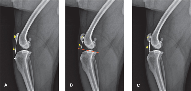
AbstractBackground: Patellar luxation in dogs is thought to be related to proximally (patella alta) or distally (patella baja) positioned patella in the femoral trochlea. Aim: The aim of the present study was to calculate and compare the values of Insall–Salvati (ISI), Caton–Deschamps (CDI), and Blackburne–Peel indices (BPI) in orthopedically healthy dogs and dogs with various grades of medial patellar luxation (MPL) from small breeds on mediolateral radiographs. Methods: The study included 87 dogs (138 stifles) from four breeds (Mini-Pinschers, Pomeranians, Chihuahuas, and Yorkshire terriers). Seventy joints (53 dogs) were diagnosed with various grades of MPL and another 68 joints from 34 dogs, free of orthopedic or neurological disorders, were used as controls. Receiver operating characteristic (ROC) analysis was performed to evaluate the diagnostic value of the three indices. Results: The CDI and BPI showed no significant difference between healthy and MPL joints. The ROC analysis showed that all three studied indices of proximodistal patellar position had poor diagnostic values with low sensitivity and specificity of respective cutoff values. Conclusion: In dogs from the studied four small breeds, the proximodistal ISI, CDI, and BPI patellar indices could not discriminate reliably between healthy stifle joints and joints with MPL. Keywords: Medial patellar luxation, Insall–Salvati index, Caton–Deschamps index, Blackburne–Peel index, Small dog breeds. IntroductionProximally (patella alta) and distally (patella baja) positioned patella in relation to the trochlear groove is supposedly involved in the pathophysiology of canine patellar luxation (Johnson et al., 2006). The increased patellar height (patella alta) is reported as an important correlate of patellofemoral maltracking disorders. It has a role in the occurrence of medial patellar luxation (MPL) especially in dogs from large breeds, whereas patella baja is associated with lateral patellar luxation (Bound et al., 2009; Wangdee and Torwattanachai, 2010; Edwards and Jackson, 2012; Miles et al., 2012). In medium- and large-size dogs, MPL is characterized by a relatively long patellar ligament and patella alta (Mostafa et al., 2008). However, in small breeds, the role of the patellar ligament length in MPL pathophysiology remains disputable. Several studies in these breeds have not associated patellar luxation with abnormal proximodistal patellar position based on the original and modified Insall–Salvati index (mISI) (Towle et al., 2005; Mortari et al., 2009, Wangdee et al., 2015; Ševčík et al., 2019; Feldmane and Theyse, 2021). Patellotibial measurements are widely accepted for the evaluation of patellar sagittal position because of the readily identifiable osseous landmarks. In human medicine, the most preferred parameters for evaluation of the proximodistal patellar malalignment in the trochlea (high-riding and low-riding patellae) are the Insall–Salvati index (ISI), Caton–Deschamps index (CDI), and Blackburne–Peel index (BPI). The Blackburne–Peel (BP) ratio has been introduced as an alternative to Insall–Salvati (IS) ratio, whereas the CDI relies upon the length of the patellar articular surface and its distance from the tibia, reducing errors due to long patellas (Biedert and Tscholl, 2017). In dogs, the cited indices have been discussed in different research reports, but the ISI is still the most commonly reported measure (Mostafa et al., 2008; Ševčík et al., 2019). The BPI and the ISI were determined in 78 dogs before and after tibial tuberosity advancement (TTA) and tibial plateau leveling osteotomy (TPLO) surgery on mediolateral stifle radiographs (Lorinson et al., 2022). Guénégo et al. (2020) have evaluated patellar position by means of ISI, the mIS, and CDI in Labrador Retrievers and Golden Retrievers with/without ruptured cranial cruciate ligament (CCL). Data about the mIS, de Carvalho, patellotrochlear, and BPI were found reliable in healthy Greenland sled dogs (Miles et al., 2012). The “ideal” clinical index is still not defined - the simplest index, ISI, is considered not quite appropriate for postoperative evaluations of the corrected patellar position, while the most angle-stable alternative (BPI) requires more drawing, measurements, and calculation (Miles et al., 2012). No previous studies have reported the values of Caton–Deschamps and BP ratios in small breed dogs, both healthy and affected with MPL. In addition, no study has assessed and compared the diagnostic value of those indices in dogs for the distinction of healthy stifle joints from joints with MPL. That is why the aim of the present study was to calculate and compare the values of ISI, CDI, and BPI in orthopedically healthy dogs and dogs with various grades of MPL in four small breeds and to determine their diagnostic value through receiver operating characteristic (ROC) analysis. Materials and MethodsDogs and measurementsThe study involved 87 dogs from four small breeds (Mini-Pinschers, Pomeranians, Chihuahuas, and Yorkshire terriers). After a physical examination and radiography, 70 joints from 53 dogs were diagnosed with various grades of MPL by the routine clinical classification of Putnam (1968): grade 1: the patella can be manually luxated and returns to a normal position when released; grade 2: the patella luxates during stifle flexion or manual manipulation and remains luxated until stifle extension or manual replacement occurs; grade 3: the patella is permanently luxated, can be manually replaced, but reluxates spontaneously after manual pressure is removed; and grade 4: the patella is permanently luxated and cannot be replaced. Another 34 dogs from the same breeds (68 stifles) without orthopedic or neurological problems were used as controls. After sedation of patients with 0.075 mg/kg medetomidine hydrochloride (Dorbene vet®) and 7.5 mg/kg ketamine hydrochloride (Anaket®), radiographs were obtained in mediolateral view using a Bucky Diagnost CS4 stationary X-ray equipment with iQ-CR ACE acquisition station. Dogs were positioned in lateral recumbency, and the central X-ray beam was focused on the medial femoral condyle and the patella. All measurements (in millimeters) were carried out on radiographs using the iQ-VIEW/PRO version 2.7. software. For the determination of the ISI, the lengths of the patellar ligament and the patella were determined, as described by Mostafa et al. (2008). The patellar ligament length (a) was measured from the distal patellar pole to the point where the ligament inserts onto the tibial tuberosity (Fig. 1A). The length of the patella (b) was measured between its proximal and distal poles. The ISI was calculated as the ratio a/b (Insall and Salvati, 1972). For the measurement of the BPI, a line passing through the tibial plateau was initially drawn (Blackburne and Peel, 1977). BPI is calculated as a ratio of the height of the distal pole of the articular surface above this tibial plateau line (a) to the patellar articular surface length (b) (Fig. 1B).
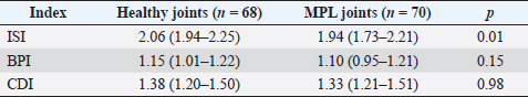
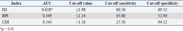
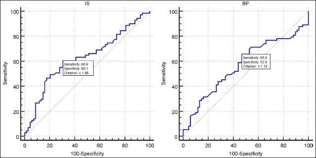
Fig. 1. Measurement of indices of proximodistal patellar position. A. The ISI is measured as the ratio of the patellar tendon length (a) to the distance between proximal and distal patellar poles (b); B. The BPI is the ratio of the height of the distal pole of the articular surface above the tibial plateau line (a) to the patellar articular surface length (b); and C. The CDI is determined as a ratio of the distance between the distal point of the patellar articular surface (a) and the cranioproximal margin of the tibia, divided by the patellar articular surface length (b). For the calculation of the CDI, the distance from the distal edge of the patellar articular surface to the cranioproximal margin of the tibia (a) was divided by the patellar articular surface length (b) (Caton et al., 1982). Statistical analysisThe measurements were reported as median values and interquartile ranges. The Mann–Whitney U-test and Hodges–Lehmann estimator were used to compare the difference in measurements between control joints and joints with MPL. The population mean effect size was estimated using Cohen’s d. ROC curve analysis was run to calculate optimal cut-off values of parameters distinguishing healthy and MPL joints based on the Youden J statistic. The areas under the ROC curves (AUCs) were used as a measure of diagnostic parameter accuracy, sensitivity, and specificity of classifiers. Interpreting the AUC as a measure of test accuracy was 0.90–1: excellent diagnostic test; 0.80–0.90: good diagnostic test; 0.70–0.80: fair diagnostic test; 0.60–0.70: poor diagnostic test; and 0.50–0.60: fail (Nahm, 2022). All computations were performed using MedCalc 15.8 (Belgium). Ethical approvalAll procedures described in this study were carried out with prior consent from dogs’ owners. Results and DiscussionThe results from measured indices of vertical patellar position in healthy and MPL stifles are presented in Table 1. The median ISI values were significantly greater (p=0.01) in healthy joints than in luxation joints, 2.06 versus 1.94 [Hodges–Lehmann (95% CI)=2.01 (1.96–2.07)]. However, the CDI and BPI values in healthy and luxation joints were not considerably different: 1.38 versus 1.33 [Hodges–Lehmann (95% CI)=1.35 (1.31–1.38)] for CDI and 1.15 versus 1.10 [Hodges–Lehmann (95% CI)=1.11 (1.08–1.15)] for BPI. The effect size assessed by Cohen’s d values was moderate for ISI (0.41), and small - 0.12 for CDI and 0.06 for BPI. The ROC analysis data confirmed that the ISI had a poor diagnostic value (AUC=0.628), whereas AUC values of BPI and CDI were close to 0.5, that is, had a predictive value no better than chance. The sensitivity and specificity of the cutoff values of the three ratios were low (Table 2; Fig. 2). The indices of ISI, CDI, and BPI reflect various anatomical relationships between the patella and the proximal tibia. In human medicine, they are among the most commonly used objective methods for the evaluation of patellar vertical position on mediolateral radiographs (Askenberger et al., 2017; Biedert and Tscholl, 2017). The BPI was claimed superior for differentiation of patella alta and patella baja than both ISI and CDI (Seil et al., 2000). The authors also showed that the BPI was more reliable for the determination of patellar vertical position, especially in medial deviation of the tibial tuberosity in chronic MPL than the ISI. Philips et al. (2010) also affirmed that the BPI was one of the more consistent imaging parameters for this purpose. The BPI is influenced by the tibial slope (Mortensen et al., 2021)–increased tibial inclination reduces the BPI values, which is important for its interpretation both in humans and dogs from small breeds. The ISI is one of the most frequently used parameters of proximodistal patellar position in dogs (Mostafa et al., 2008; Łojszczyk-Szczepaniak et al., 2017). In healthy dogs from large breeds, ISI is relatively constant (1.68 ± 0.18) and could serve as a basis for comparison in case of pathological deviations (Johnson et al., 2002). A later study found that the ISI of stifles from large breed dogs with MPL exceeded 1.97 (patella alta) versus 1.71 in healthy stifles (Johnson et al., 2006). Studies in small breed dogs with MPL reported no significant deviation in ISI values in dogs with MPL (Towle et al., 2005; Mortari et al., 2009; Wangdee et al., 2015; Ševčík et al., 2019). For example, there was no statistically significant difference in the relative patellar ligament length between healthy stifles with a mean ± SD ISI of 1.82 ± 0.20 and MPL stifles with a mean of 1.78 ± 0.18 (Feldmane and Theyse, 2021). In dogs with body weight <15 kg, Murakami et al. (2021) found that the proximodistal patellar position in the MPL group did not differ significantly from that in controls despite the tendency to a more distally positioned patella (p=0.073). The reference range of the IS ratio in small breed dogs varies from 1.04 to 2.16 (Ocal et al., 2020). Based on these values, the median ISI values of the two groups in our study were within this range; therefore, patella alta was not present in the studied four small breeds. Recent data in humans showed that IS and mIS methods had very good diagnostic performance and had high AUC values of 0.91 and 0.96, respectively. The ISI and its modification are independent of the tibial slope; therefore, these methods mainly reflect the lengths of the patella and patellar tendon. The BP and CD ratios also had relatively high AUC values (0.72 and 0.86, respectively) (Yamashita et al., 2023). The authors concluded that, while the BP and CD indices might be useful, their ability to predict the incidence of patellar dislocation was lower. Table 1. Patellar proximodistal alignment parameters in both groups of stifle joints. Values are presented as median (interquartile range).
Table 2. AUC, cut-off values, sensitivity, and specificity of patellar proximodistal alignment parameters.
Fig. 2. ROC curves of IS and BP indices in relation to MPL based on 138 stifle joints from Pinschers, Pomeranians, Chihuahuas, and Yorkshire Terriers. The results from the ROC analysis in the present study confirmed that in small breed dogs, the three indices of vertical patellar position had no diagnostic value and, therefore, could not identify stifles affected with MPL. The AUCs and low sensitivity and specificity of cutoff values suggested that in these breeds, the proximodistal position of the patella in the trochlea could not be considered as a factor predisposing to MPL. As confirmed by recent data, the horizontal position of the patella and trochlear morphology seem more important for MPL development (Garnoeva, 2021; Matchwick et al., 2021). ConclusionIn conclusion, the ISI, CDI, and BPI had low sensitivity and specificity, poor diagnostic value, and therefore, could not discriminate reliably between healthy stifle joints and joints with MPL in Mini-Pinschers, Pomeranians, Chihuahuas, and Yorkshire terriers based on proximodistal patellar position. The study findings confirmed that, in these four breeds, the proximodistal position of the patella within the trochlear groove was not a factor predisposing to the development of MPL. AcknowledgmentsThis research is supported by the Bulgarian Ministry of Education and Science under the National Program “Young Scientists and Postdoctoral Students-2”. Conflict of interestThe author declares that there is no conflict of interest. ReferencesAskenberger, M., Janarv, P.M., Finnbogason, T. and Arendt, E.A. 2017. Morphology and anatomic patellar instability risk factors in first-time traumatic lateral patellar dislocations: a prospective magnetic resonance imaging study in skeletally immature children. Am. J. Sports. Med. 45, 50–58. Biedert, R.M. and Tscholl, P.M. 2017. Patella alta: a comprehensive review of current knowledge. Am. J. Orthop. 46, 290–300. Blackburne, J.S. and Peel, T.E. 1977. A new method of measuring patellar height. J. Bone. Joint. Surg. Br. 59, 241–242. Bound, N., Zakai, D., Butterworth, S.J. and Pead, M. 2009. The prevalence of canine patellar luxation in three centres. Clinical features and radiographic evidence of limb deviation. Vet. Comp. Orthop. Traumatol. 1, 32–37. Caton, J., Deschamps, G., Chambat, P., Lerat, J.L. and Dejour, H. 1982. Les rotules basses. A propos de 128 observations. Rev. Chir. Orthop. Repar. Appar. Mot. 68, 317–325. Edwards, G.A. and Jackson, A.H. 2012. Use of a TT plate for correction of severe patella baja in Chihuahua. JAAHA 48, 113–117. Feldmane, L. and Theyse, L. 2021. Proximodistal and caudocranial position of the insertion of the patellar ligament on the tibial tuberosity and patellar ligament length of normal stifles and stifles with grade II medial patellar luxation in small-breed dogs. Vet. Surg. 50, 1017–1022. Garnoeva, R. 2021. Evaluation of trochlear dysplasia in dogs with medial patellar luxation - comparative studies. Acta. Sci. Vet. 49, 1845. Guénégo, L., Serri, P., Charru, P. and Verwaerde, P. 2020. Comparison of the tibial anatomical-mechanical axis angle and patellar positions between Labrador retrievers and Golden Retrievers with and without cranial cruciate ligament rupture. J. Vet. Sci. Res. 5, 000199. Insall, J. and Salvati, E. 1972. Patella position in the normal knee joint. Radiology 101, 101–104. Johnson, A.L., Broaddus, K.D., Hauptman, J.G., Marsh, S., Monsere, J. and Sepulveda, G. 2006. Vertical patellar position in large-breed dogs with clinically normal stifles and large-breed dogs with medial patellar luxation. Vet. Surg. 35, 78–81. Johnson, A.L., Probst, C.W., De Camp, C.E., Rosenstein, D.S., Hauptman, J.G. and Kern, T.L. 2002. Vertical position of the patella in the stifle joint of clinically normal large-breed dogs. Am. J. Vet. Res. 63, 42–46. Łojszczyk-Szczepaniak, A., Silmanowicz, P., Komsta, R. and Osiński, Z. 2017. Determination of reference values and frequency of occurrence of patella alta in German shepherd dogs: a retrospective study. Acta. Vet. Scand. 59, 36. Lorinson, K., Winkler, M., Tichy, A. and Lorinson, D. 2022. Radiologische evaluierung der prä- und postoperativen Patellahöhe bei TPLO und TTA patienten. Wiener Tierärzt. Monatsschrift 109, 1–8. Matchwick, A., Bridges, J.P., Mielke, B., Pead, M.J., Phillips, A. and Meeson, R.L. 2021. Computed tomographic measurement of trochlear depth in three breeds of brachycephalic dogs. Vet. Comp. Orthop. Traumatol. 34, 124–129. Miles, J.E., Dickow, M., Nielsen, D.H., Jensen, B.R., Kirpensteijn, J. and Svalastoga, E.L. 2012. Five patellar proximodistal positioning indices compared in clinically normal 41 Greenland sled dogs. Vet. J. 193, 529–534. Mortari, A.C., Rahal, S.C., Vulcano, L.C., da Silva, V.C. and Volpi, R.S. 2009. Use of radiographic measurements in the evaluation of dogs with medial patellar luxation. Can. Vet. J. 50, 1064. Mortensen, A.J., Todd, B.S., Ludwig, T.C., Adam, B.M., Adeyemi, T.F. and Aoki, S.K. 2021. The blackburne–peel index for determining patellar height is affected by tibial slope. Arthrosc. Sports. Med. Rehabil. 3, e359–e365. Mostafa, A.A., Griffon, D.J., Thomas, M.W. and Constable, P.D. 2008. Proximodistal alignment of the canine patella: radiographic evaluation and association with medial and lateral patellar luxation. Vet. Surg. 31, 201–211. Murakami, S., Shimada, M., Harada, Y. and Hara, Y. 2021. Examination of the proximodistal patellar position in small dogs in relation to anatomical features of the distal femur and medial patellar luxation. PloS One 16, e0252531. Nahm, F.S. 2022. Receiver operating characteristic curve: overview and practical use for clinicians. Korean. J. Anesthesiol. 75, 25–36. Ocal, M.K., Seyrek-Intas, D. and Cagatay, S. 2020 Comparison of insall-salvati index and its modification in normal dogs from four different body weight groups. Vet. Comp. Orthop. Traumatol. 33, 110–115. Philips, C.L., Silver, D.A.T., Schranz, P.J. and Mandalia, V. 2010. The measurement of patellar height: a review of the methods of imaging. J. Bone. Joint. J. 92, 1045–1053. Seil, R., Müller, B., Georg, T., Kohn, D. and Rupp, S. 2000. Reliability and interobserver variability in radiological patellar height ratios. Knee. Surg. Sports. Traumatol. Arthrosc. 8, 231–236. Ševčík, K., Hluchý, M. and Huňáková, K. 2019. Proximodistal alignment of the canine patella in small breeds of dogs. Anim. Physiol. Nutr. Welf. 1, 148–154. Towle, H.A., Griffon, D.J. and Thomas, M.W. 2005. Pre- and postoperative radiographic and computed tomographic evaluation of dogs with medial patellar luxation. Vet. Surg. 34, 265–272. Wangdee, C., Theyse, L.F. and Hazewinkel, H.A. 2015. Proximo-distal patellar position in three small dog breeds with medial patellar luxation. Vet. Comp. Orthop. Traumatol. 28, 270–273. Wangdee, C. and Torwattanachai, P. 2010. Lateral patellar luxation in three Pomeranian dogs: a case report. Thai. J. Vet. Med. 40, 227–231. Yamashita, S., Ishizuka, S., Sakai, T., Oba, H., Sakaguchi, T., Mizuno, T., Kawashima, I., Tsukahara, T., Takahashi, S., Kurokouchi, K. and Imagama, S. 2023. Potential of patellar height measurement methods in predicting recurrent patellar dislocation incidence: a case-control study. Preprint (Version 1) available at Research Square (https://doi.org/10.21203/rs.3.rs-2461090/v1). | ||
| How to Cite this Article |
| Pubmed Style Radka S. Garnoeva. Evaluation of proximodistal patellar alignment in small breed dogs with or without patellar luxation using the Insall-Salvati, Caton-Deschamps and Blackburne-Peel indices. Open Vet J. 2023; 13(5): 663-667. doi:10.5455/OVJ.2023.v13.i5.20 Web Style Radka S. Garnoeva. Evaluation of proximodistal patellar alignment in small breed dogs with or without patellar luxation using the Insall-Salvati, Caton-Deschamps and Blackburne-Peel indices. https://www.openveterinaryjournal.com/?mno=143380 [Access: May 01, 2024]. doi:10.5455/OVJ.2023.v13.i5.20 AMA (American Medical Association) Style Radka S. Garnoeva. Evaluation of proximodistal patellar alignment in small breed dogs with or without patellar luxation using the Insall-Salvati, Caton-Deschamps and Blackburne-Peel indices. Open Vet J. 2023; 13(5): 663-667. doi:10.5455/OVJ.2023.v13.i5.20 Vancouver/ICMJE Style Radka S. Garnoeva. Evaluation of proximodistal patellar alignment in small breed dogs with or without patellar luxation using the Insall-Salvati, Caton-Deschamps and Blackburne-Peel indices. Open Vet J. (2023), [cited May 01, 2024]; 13(5): 663-667. doi:10.5455/OVJ.2023.v13.i5.20 Harvard Style Radka S. Garnoeva (2023) Evaluation of proximodistal patellar alignment in small breed dogs with or without patellar luxation using the Insall-Salvati, Caton-Deschamps and Blackburne-Peel indices. Open Vet J, 13 (5), 663-667. doi:10.5455/OVJ.2023.v13.i5.20 Turabian Style Radka S. Garnoeva. 2023. Evaluation of proximodistal patellar alignment in small breed dogs with or without patellar luxation using the Insall-Salvati, Caton-Deschamps and Blackburne-Peel indices. Open Veterinary Journal, 13 (5), 663-667. doi:10.5455/OVJ.2023.v13.i5.20 Chicago Style Radka S. Garnoeva. "Evaluation of proximodistal patellar alignment in small breed dogs with or without patellar luxation using the Insall-Salvati, Caton-Deschamps and Blackburne-Peel indices." Open Veterinary Journal 13 (2023), 663-667. doi:10.5455/OVJ.2023.v13.i5.20 MLA (The Modern Language Association) Style Radka S. Garnoeva. "Evaluation of proximodistal patellar alignment in small breed dogs with or without patellar luxation using the Insall-Salvati, Caton-Deschamps and Blackburne-Peel indices." Open Veterinary Journal 13.5 (2023), 663-667. Print. doi:10.5455/OVJ.2023.v13.i5.20 APA (American Psychological Association) Style Radka S. Garnoeva (2023) Evaluation of proximodistal patellar alignment in small breed dogs with or without patellar luxation using the Insall-Salvati, Caton-Deschamps and Blackburne-Peel indices. Open Veterinary Journal, 13 (5), 663-667. doi:10.5455/OVJ.2023.v13.i5.20 |

