


| Case Report | ||
Open Vet. J.. 2025; 15(9): 4744-4749 Open Veterinary Journal, (2025), Vol. 15(9): 4744-4749 Case report Computed tomography-based thymoma-to-cranial intrathoracic volume ratio predicts the feasibility of video-assisted thoracoscopic surgery in a small dog: A case reportShinya Mizutani1*, Saki Kageyama2, Yoshimichi Goda3, Yasuhiko Okamura1, Ikki Mitsui4, Natsuki Akashi1, Akihiro Ohnishi1, Teppei Kanda1,2 and Taketoshi Asanuma11Department of Small Animal Surgery, Field of Clinical Veterinary Medicine, Faculty of Veterinary Medicine, Okayama University of Science, Imabari, Japan 2Graduate School of Veterinary Science, Okayama University of Science, Imabari, Japan 3Laboratory of Veterinary Diagnostic Imaging, Department of Veterinary Medicine, School of Veterinary Medicine, Rakuno Gakuen University, Ebetsu, Japan 4No Boundaries Animal Pathology, Fuchu, Japan *Corresponding Author: Shinya Mizutani. Department of Small Animal Surgery, Field of Clinical Veterinary Medicine, Faculty of Veterinary Medicine, Okayama University of Science, Imabari, Japan. Email: s-mizutani [at] ous.ac.jp Submitted: 04/07/2025 Revised: 15/08/2025 Accepted: 21/08/2025 Published: 30/09/2025 © 2025 Open Veterinary Journal
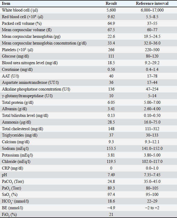
ABSTRACTBackground: Canine thymoma (CT) is the most common neoplastic disease of the cranial intrathoracic cavity. Reports of video-assisted thoracoscopic surgery-thymectomy (VATS-T) in dogs are increasing, but the surgical indication criteria remain unclear. This report highlights the value of a new criterion called the thymoma volume to cranial intrathoracic volume ratio (T/CI ratio) for evaluating the feasibility of VATS-T. Case Description: A neutered 9-year-old Papillon weighing 3.5 kg had a mass of approximately 20 mm in the cranial intrathoracic cavity. The dog was in good general condition and had no clinical symptoms. Computed tomography revealed a solitary mass with clear boundaries and weak contrast enhancement. The T/CI ratio obtained from CT examination was 2.4%. The dog underwent thoracoscopic tumor resection. Pathological examination revealed that the tumor was a thymoma. The dog was discharged without any postoperative complications. The T/CI ratios obtained from CT data of dogs of the same breed and weight, which ranged from 1.1% to 4.1%, were calculated based on previous reports that measured the volume of thymomas. Conclusion: The T/CI ratio may serve as a new objective criterion for evaluating the relationship between cranial intrathoracic volume and thymoma volume for VATS-T, regardless of dog body size. Although this case demonstrates a promising correlation between the CT-based T/CI ratio and the feasibility of VATS-T, the findings are limited to a single case. Further studies involving a larger number of patients are necessary to validate this observation. Keywords: Computed tomography, Dog, Thoracoscopy, Video-assisted thoracoscopic surgery, Thymoma. IntroductionCanine thymoma is a rare neoplastic disease and is considered a common tumor in the cranial intrathoracic cavity (Mayhew and Friedberg, 2008; Zitz et al., 2008). Surgical resection is the primary treatment, and the available approaches are intercostal thoracotomy, median sternotomy, and Video-assisted thoracoscopic surgery-thymectomy (VATS-T) (Mayhew and Friedberg, 2008; Zitz et al., 2008). Median sternotomy provides a wide surgical field but is highly invasive. In contrast, the narrow surgical fields of intercostal thoracotomy and thoracoscopic approaches make them less suitable for large or infiltrative thymomas (Atwater et al., 1994; MacIver et al., 2017). Cases of thymoma associated with tumor-associated syndromes (especially myasthenia gravis) in humans are managed with minimally invasive approaches whenever possible, as this improves prognosis (Wang et al., 2019). VATS-T is recommended when feasible (Wang et al., 2019). VATS-T has been increasingly reported in the field of veterinary medicine in recent years (Mayhew & Friedberg, 2008; MacIver et al., 2017; Alwen et al., 2015; Carroll et al., 2024). However, its indications have not been established. We report a case of a thymoma in a small dog weighing 3.5 kg that was removed using VATS-T. Case DetailsA neutered 9-year-old male Papillon weighing 3.5 kg was referred to the Okayama University of Science Veterinary Medical Teaching Hospital for examination and treatment of an incidentally discovered mass in the cranial intrathoracic cavity during regular medical checkups. The dog had no clinical signs and was in good general condition. However, a mass in the cranial thoracic region was detected by chest radiography. Blood tests showed no hypercalcemia, and arterial blood gas analysis showed no problems (Table 1). Polycythemia, slight hypernatremia, and hyperchloremia, which may be related to dehydration, were also observed. A chest X-ray revealed a mass of approximately 20 mm in the cranial intrathoracic cavity and mild expansion of the mediastinum (Fig. 1). The tumor-associated syndromes, such as megaesophagus and myasthenia gravis, were not observed. Computed tomography (CT) was performed under general anesthesia (Aquilion Lightning; Canon Medical Systems Co., Tokyo, Japan). Iopamidol (Oypalomin 300, Fuji Pharma, Japan) was used as a contrast medium (injection volume, 2.5 ml/kg [750 mgI/kg]; injection time, 15 seconds). Precontrast, arterial phase, venous phase, and equilibrium phase scans were obtained. The cranial intrathoracic mass (length, width, and height of 18.3, 16.0, and 18.6 mm, respectively) was a solitary lesion within the cranial mediastinum (Fig. 2). The mass was well demarcated, and there was no evidence of invasion into the surrounding blood vessels. The CT values of the mass were 43.2, 50.6, 113.1, and 102.2 HU for the precontrast, arterial, venous, and equilibrium phases, respectively (Fig. 2). These contrast enhancement effects were a preliminary diagnosis that the mass was a thymoma (Von Stade et al., 2019). Enlarged surrounding lymph nodes or distant metastasis were not detected. Due to the small size of the mass and its absence from the thoracic cavity margin, preoperative pathological examination could not be performed. Based on the CT results, the mass was provisionally diagnosed as a thymoma. The thymoma volume was measured from the length, width, and height by computed tomography using the elliptical volume formula. The cranial intrathoracic volume was measured using a 3D image analysis software (Ahmics-VAZE, PetCommunications Co., Ltd., Osaka, Japan). The cranial intrathoracic region between the first and fourth sternum at the beginning of the cranial intrathoracic cavity was assumed to be the surgical space for VATS-T (Fig. 3). The dorsal cephalic end was designated as the point where the first sternum extends perpendicularly and joins the thoracic vertebrae, and the dorsal caudal end was designated as the point where the fourth sternum extends perpendicularly and joins the thoracic vertebrae. This region was extracted and its volume was measured using the 3D volume rendering function by Ahmics-VAZE. The ratio of thymoma volume to cranial intrathoracic volume (T/CI ratio) was calculated as follows: thymoma volume/cranial intrathoracic volume × 100). The thymoma volume was 2.3 cm3, the cranial intrathoracic volume was 97.7 cm3, and the T/CI ratio was 2.4%. The thymoma volume was calculated using the approach in a previous report of two cases that described the thoracoscopic resection of thymomas (Mayhew and Friedberg, 2008). We also calculated the cranial intrathoracic volume for the dog, as it was of the same breed and weight as the two reported cases. Subsequently, we calculated the T/CI ratio. In previous reports, the thymoma volumes were 9.5 cm3 and 36.6 cm3, respectively, and the cranial intrathoracic volume was 892.8 cm3. The T/CI ratios were 1.1% and 4.1%, respectively. Table 1. Blood test results and arterial blood gas analysis.
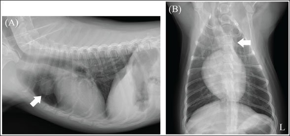
Fig. 1. Chest X-ray imaging. (A) Right lateral view. (B) Ventrodorsal view. X-ray image: a tumor-like lesion measuring approximately 20 mm was observed on the cranial side of the heart (white arrow) (L): Left side.
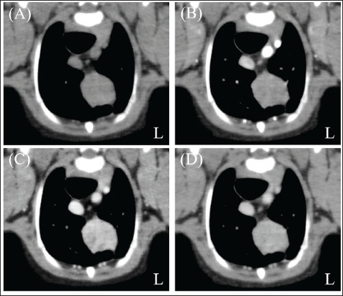
Fig. 2. Chest computed tomography (CT) imaging. (A) Pre-contrast phase. (B) Arterial phase. (C) Venous phase. (D) The equilibrium phase. The cranial chest mass (length, width, and height of 18.3, 16.0, and 18.6 mm, respectively) was a solitary lesion within the cranial mediastinum. The lesion was well demarcated and showed no signs of invasion into the surrounding blood vessels. The CT values of the lesion were 43.2 HU for the pre-contrast phase (A), 50.6 HU for the arterial phase (B), 113.1 HU for the venous phase (C), and 102.2 HU for the equilibrium phase (D) (L): Left side. VATS-T was performed under general anesthesia. The dog was placed in the supine position. The left fourth intercostal space was cut approximately 5 cm dorsoventrally, and the patient’s chest was opened as usual. A silicone endoscopic incision device (LAP PROTECTER [LP] FF0707; HAKKO Co., Ltd., Nagano, Japan) was placed after thoracotomy, and the wound was opened. A 5-mm 30° laparoscopic telescope (OLYMPUS WA50373B Telescope; OLYMPUS Medical Systems Corporation, Tokyo, Japan) attached to a video camera (VISERA ELITE OTV-S190; OLYMPUS Medical Systems Corporation, Tokyo, Japan) and light source (VISERA ELITE CLV-S190; OLYMPUS Medical Systems Corporation, Tokyo, Japan) was used to record the manipulations while adjusting the light intensity. The soft tissue and blood vessels surrounding the thymoma were dissected, coagulated, and repeatedly incised using an endoscopic surgical ultrasonic coagulation and cutting device (SONICBEAT; OLYMPUS Medical Systems Corporation, Tokyo, Japan). This exposure of the tumor led to its resection. The excised mass measured 2.5 × 1.7 × 1.3 cm and consisted of multiple firm, smooth, white to light tan nodules (Fig. 4A). On cut sections, the mass showed mottled white, light tan, and dark red coloration (Fig. 4B). After LP removal, a chest drain was inserted, and the wound was closed according to the standard procedure. The total surgical time was approximately 150 minutes. The dog had no problems during the postoperative period and was discharged on postoperative day 2. The excised mass was histologically evaluated by a board-certified veterinary anatomic pathologist (I. M.). The mass was composed of multiple solid sheet coalescing nodules or rare tubular/microcystic structures of oval, polyhedral, or short spindle cells without atypia. The tumor cells had distinct cell boundaries, scant pale eosinophilic lacy cytoplasm, round to ovoid hypochromatic minimally anysokaryotic nucleus, and indistinct nucleolus. The mitotic count was 2 in 2.37 mm2. The interstitium comprised scant fibrovascular tissue with occasional congestion and invasion by a few small lymphocytes. No node metastasis or vascular invasion by tumor cells was observed. Based on these findings, the patient was diagnosed with type A thymoma (Fig. 5).
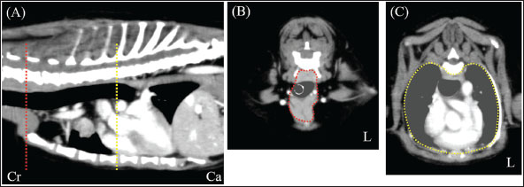
Fig. 3. CT imaging of the cranial intrathoracic cavity. (A) Sagittal section. (B) Transverse section of the first sternum. (C) Transverse section of the fourth sternum. The surgical space for VATS was assumed to be the cranial intrathoracic cavity between the first and fourth sternum (A; red and yellow dotted line, respectively). Cross-sections (B and C) of the two dotted lines. The circular areas enclosed by dotted lines within the cross-sections were continuously extracted and designated as the intracranial thoracic cavity, and their volumes were measured. (L): Left side, (Cr): Cranial, (Ca): Caudal.
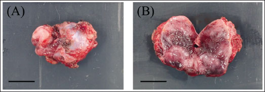
Fig. 4. Gross morphology of thymoma before formalin fixation. Bar=1 cm. (A) The mass consisted of multiple firm, white to light tan nodules with smooth surfaces. (B) On cut sections, the mass had mottled white, light tan, and dark red areas.
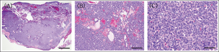
Fig. 5. Histologic findings of the thymoma. (A) Subgross image of the mass. The excised mass comprises multiple coalescing nodules. Bar=2 cm. (B) Low magnification of the mass. The tumor consists of a solid sheet or rare tubular/microcystic tumor cells. Bar=200 µm. (C) High magnification image of the mass. No atypia is observed in ovoid, polyhedral, or short spindle tumor cells Bar=100 µm. DiscussionIn recent years, VATS-T has been performed in veterinary medicine (Mayhew and Friedberg, 2008; Alwen et al., 2015; MacIver et al., 2017; Carroll et al., 2024). However, the criteria for its application have not yet been established. Two previous reports evaluated the indications for VATS-T. The first report established the following criteria: dogs weighing >20 kg, non-invasive tumors with a tumor volume of less than 300 cm³, and a tumor diameter of 8 cm (MacIver et al., 2017). The first criteria are only applicable to large-breed dogs and cannot be applied to small-breed dogs, such as those in our case. In large breed dogs, relatively small thymomas may allow sufficient thoracic cavity space for VATS-T (Mayhew and Friedberg, 2008; Alwen et al., 2015; MacIver et al., 2017; Carroll et al., 2024). However, small thymomas may not have sufficient space in the cranial intrathoracic cavity in small-breed dogs. Another report established the criterion of tumors with a maximum diameter-to-body weight ratio of approximately 0.02 (Carroll et al., 2024). The second criterion is based solely on the maximum tumor diameter and does not account for the 3D tumor volume. Additionally, the chest shape can vary significantly depending on the breed, even in dogs of the same weight. Therefore, body weight-based evaluation may not be appropriate. The feasibility of VATS-T in small-breed dogs ultimately depends on the judgment of the surgeon. In this study, the T/CI ratio obtained from CT data may provide an objective means of evaluating the relationship between thymomas and the CI in individual cases. Thymoma resection via VATS-T is feasible in small breeds of dogs. The T/CI ratio for this case (2.4%) was similar to those for previous VATS-T cases (1.1% and 4.1%, respectively). VATS-T may be feasible for cases with a T/CI ratio of approximately 5% or less. Cranial intrathoracic cavity opened during thymectomy has no specific definition. In this study, we defined the cranial intrathoracic region using the T/CI ratio to represent the area from the tip of the first sternum to the caudal end of the fourth sternum. The tip of the first sternum is located in the cervicothoracic part and is not included in the surgical space. The actual intrathoracic cavity begins around the first sternum’s center. However, the region designated as “around the center” of the first sternum may be ambiguous when measuring the cranial intrathoracic volume. Therefore, the tip of the first sternum was designated as the cranial end. The caudal end of the fourth sternum was assumed to be the most caudal point where the actual VATS space could be used. Nowadays, histologic diagnosis of thymoma in animals is based on the World Health Organization classification for humans, which includes five histologic subtypes defined by detailed microscopic findings (Valli et al., 2016). The present case was diagnosed as type A thymoma because of the predominant component of characteristic spindle/oval cells without nuclear atypia, with few non-neoplastic background lymphocytes (Valli et al., 2016). This study has some limitations. Based on our T/CI ratio, a complete thoracoscopic thymoma resection may have been possible in this dog. However, these procedures have been performed in larger dogs. We considered performing complete thoracoscopy but opted for VATS-T due to the small size of the dog (3.5 kg), which has not been previously reported. The criteria for determining the suitability of thoracoscopic surgery based on the T/CI ratio remain unclear and warrant further evaluation in larger case series. ConclusionThis report demonstrates the T/CI ratio as an objective marker for determining the feasibility of VATS-T in dogs. The T/CI ratio may be used to assess the relationship between VATS-T cranial intrathoracic volume and thymoma volume in dogs, regardless of their body size. This case highlights the potential utility of the T/CI ratio measured by CT in preoperative planning. However, broader studies with statistically adequate sample sizes are required to draw generalized conclusions and confirm the predictive value of this parameter, given the limitation of a single case. Conflict of interestThe authors have no conflicts of interest to declare. FundingThis study received no specific grant. Authors’ contributionsConceptualization: S. M., S. K., Y. G., Y. O., I. M., N. A., A. O., T. K., and T. A.; methodology: S. M.; formal analysis: S. M.; investigation: S. M., S. K., Y. G., Y. O., I. M., N. A., A. O., T. K., and T. A.; data curation: S. M., S. K., Y. G.; writing—original draft preparation: S. M.; writing—review and editing: S. M., S. K., Y. G., Y. O., I. M., N. A., A. O., T. K., and T. A.; supervision: S. M. All authors have read and approved the published version of the manuscript. Data availabilityAll data supporting this study’s findings are available within the manuscript. ReferencesAlwen, S.G.J., Culp, W.T.N., Szivek, A., Mayhew, P.D. and Eckstrand, C.D. 2015. Portal site metastasis after thoracoscopic resection of a cranial mediastinal mass in a dog. J. Am. Vet. Med. Assoc. 247, 793–800. Atwater, S.W., Powers, B.E., Park, R.D., Straw, R.C., Ogilvie, G.K. and Withrow, S.J. 1994. Thymoma in dogs: 23 cases (1980–1991). J. Am. Vet. Med. Assoc. 205, 1007–1013. Carroll, K.A., Mayhew, P.D., Culp, W.T.N., Massari, F., Peláez, M.J., Steffey, M.A., Giuffrida, M., Balsa, I.M., Gibson, E.A., Farrell, M.A., Singh, A., Buote, N., Scharf, V.F., Brissot, H. and Thomson, C. 2024. Thoracoscopic removal of cranial mediastinal masses in dogs is associated with a low conversion rate, excellent survival to discharge, and good long-term outcome. J. Am. Vet. Med. Assoc. 262(1), 1–8. MacIver, M.A., Case, J.B., Monnet, E.L., Hunt, G.B., Mayhew, P.D., Oblak, M.L., Runge, J.J., Singh, A., Smeak, D.D., Steffey, M.A. and Boston, S.E. 2017. Video-assisted extirpation of cranial mediastinal masses in dogs: 18 cases (2009–2014). J. Am. Vet. Med. Assoc. 250, 1283–1290. Mayhew, P.D. and Friedberg, J.S. 2008. Video-assisted thoracoscopic resection of noninvasive thymomas using one-lung ventilation in two dogs. Vet. Surg. 37, 756–762. Valli, V.E.O., Kiupel, M. and Bienzle, D. 2016. Chapter 2: hematopoietic system. In Pathology of domestic animals, 6th ed,. Ed., Maxie, M.G. St. Louis, MO: Elsevier, pp: 151–158. Von Stade, L., Randall, E.K., Rao, S. and Marolf, A.J. 2019. CT imaging features of canine thymomas. Vet. Radiol. Ultrasound 60, 659–667. Wang, G.W., Tao, T., Li, C.K., Li, Q.C., Duan, G.X., Sang, H.W., Dong, H.J. and Wang, Z.Y. 2019. Comparison between thoracoscopic and open approaches in thymoma resection. J. Thorac. Dis. 11, 4159–4168. Zitz, J.C., Birchard, S.J., Couto, G.C., Samii, V.F., Weisbrode, S.E. and Young, G.S. 2008. Results of excision of thymoma in cats and dogs: 20 cases (1984–2005). J. Am. Vet. Med. Assoc. 232, 1186–1192. | ||
| How to Cite this Article |
| Pubmed Style Mizutani S, Kageyama S, Goda Y, Okamura Y, Mitsui I, Akashi N, Ohnishi A, Kanda T, Asanuma T. Computed tomography-based thymoma-to-cranial intrathoracic volume ratio predicts the feasibility of video-assisted thoracoscopic surgery in a small dog: A case report. Open Vet. J.. 2025; 15(9): 4744-4749. doi:10.5455/OVJ.2025.v15.i9.79 Web Style Mizutani S, Kageyama S, Goda Y, Okamura Y, Mitsui I, Akashi N, Ohnishi A, Kanda T, Asanuma T. Computed tomography-based thymoma-to-cranial intrathoracic volume ratio predicts the feasibility of video-assisted thoracoscopic surgery in a small dog: A case report. https://www.openveterinaryjournal.com/?mno=268892 [Access: January 24, 2026]. doi:10.5455/OVJ.2025.v15.i9.79 AMA (American Medical Association) Style Mizutani S, Kageyama S, Goda Y, Okamura Y, Mitsui I, Akashi N, Ohnishi A, Kanda T, Asanuma T. Computed tomography-based thymoma-to-cranial intrathoracic volume ratio predicts the feasibility of video-assisted thoracoscopic surgery in a small dog: A case report. Open Vet. J.. 2025; 15(9): 4744-4749. doi:10.5455/OVJ.2025.v15.i9.79 Vancouver/ICMJE Style Mizutani S, Kageyama S, Goda Y, Okamura Y, Mitsui I, Akashi N, Ohnishi A, Kanda T, Asanuma T. Computed tomography-based thymoma-to-cranial intrathoracic volume ratio predicts the feasibility of video-assisted thoracoscopic surgery in a small dog: A case report. Open Vet. J.. (2025), [cited January 24, 2026]; 15(9): 4744-4749. doi:10.5455/OVJ.2025.v15.i9.79 Harvard Style Mizutani, S., Kageyama, . S., Goda, . Y., Okamura, . Y., Mitsui, . I., Akashi, . N., Ohnishi, . A., Kanda, . T. & Asanuma, . T. (2025) Computed tomography-based thymoma-to-cranial intrathoracic volume ratio predicts the feasibility of video-assisted thoracoscopic surgery in a small dog: A case report. Open Vet. J., 15 (9), 4744-4749. doi:10.5455/OVJ.2025.v15.i9.79 Turabian Style Mizutani, Shinya, Saki Kageyama, Yoshimichi Goda, Yasuhiko Okamura, Ikki Mitsui, Natsuki Akashi, Akihiro Ohnishi, Teppei Kanda, and Taketoshi Asanuma. 2025. Computed tomography-based thymoma-to-cranial intrathoracic volume ratio predicts the feasibility of video-assisted thoracoscopic surgery in a small dog: A case report. Open Veterinary Journal, 15 (9), 4744-4749. doi:10.5455/OVJ.2025.v15.i9.79 Chicago Style Mizutani, Shinya, Saki Kageyama, Yoshimichi Goda, Yasuhiko Okamura, Ikki Mitsui, Natsuki Akashi, Akihiro Ohnishi, Teppei Kanda, and Taketoshi Asanuma. "Computed tomography-based thymoma-to-cranial intrathoracic volume ratio predicts the feasibility of video-assisted thoracoscopic surgery in a small dog: A case report." Open Veterinary Journal 15 (2025), 4744-4749. doi:10.5455/OVJ.2025.v15.i9.79 MLA (The Modern Language Association) Style Mizutani, Shinya, Saki Kageyama, Yoshimichi Goda, Yasuhiko Okamura, Ikki Mitsui, Natsuki Akashi, Akihiro Ohnishi, Teppei Kanda, and Taketoshi Asanuma. "Computed tomography-based thymoma-to-cranial intrathoracic volume ratio predicts the feasibility of video-assisted thoracoscopic surgery in a small dog: A case report." Open Veterinary Journal 15.9 (2025), 4744-4749. Print. doi:10.5455/OVJ.2025.v15.i9.79 APA (American Psychological Association) Style Mizutani, S., Kageyama, . S., Goda, . Y., Okamura, . Y., Mitsui, . I., Akashi, . N., Ohnishi, . A., Kanda, . T. & Asanuma, . T. (2025) Computed tomography-based thymoma-to-cranial intrathoracic volume ratio predicts the feasibility of video-assisted thoracoscopic surgery in a small dog: A case report. Open Veterinary Journal, 15 (9), 4744-4749. doi:10.5455/OVJ.2025.v15.i9.79 |

