


| Research Article | ||
Open Vet. J.. 2025; 15(9): 4569-4577 Open Veterinary Journal, (2025), Vol. 15(9): 4569-4577 Research Article Histological study on the prenatal development of compartment 1 of the stomach of the camel (Camelus dromedarius)Abdelhay Mohamed Ali*Department of Anatomy, College of Veterinary Medicine, King Faisal University, Al-Ahsa, Saudi Arabia *Corresponding Author: Abdelhay Mohamed Ali. Department of Anatomy, College of Veterinary Medicine, King Faisal University, Al-Ahsa, Saudi Arabia. Email: arhamallha [at] kfu.edu.sa Submitted: 22/03/2025 Revised: 05/08/2025 Accepted: 24/08/2025 Published: 30/09/2025 © 2025 Open Veterinary Journal
ABSTRACTBackground: The largest portion of the dromedary camel’s stomach, known as Compartment 1, plays a vital role in the microbial fermentation of ingested food. Aim: This study aimed to investigate the prenatal development of Compartment 1 in camel fetuses during the three gestational trimesters. Methods: Stomach samples were collected from 21 healthy camel fetuses of both sexes at various gestational ages (79–390 days). The samples were fixed in 10% buffered formalin and histologically examined. Results: The primitive stomach wall consisted of three layers in the early first trimester (79 days): epithelial, pluripotent blastemic tissue, and myoblastic. By 87–115 days, the wall displayed smooth to undulating surface regions and comprised four layers: epithelium, lamina propria-submucosa, tunica muscularis, and serosa. At 120–128 days, the smooth surface region of Compartment 1 was lined with stratified epithelium comprising a basal layer of cuboidal cells and 2–3 layers of lightly stained polyhedral cells. In contrast, the undulating surface was lined with pseudostratified columnar epithelium. During the second trimester (163–234 days), the smooth surface region differentiated into a non-glandular zone composed of three layers: stratum basale, spinosum, and granulosum. The folded surface developed into a glandular region characterized by multiple folds and deep pits forming chamber-like structures. These folds exhibited distinct epithelial linings: stratified epithelium at the apices, pseudostratified epithelium on the lateral sides, and simple columnar epithelium in the chambers. In the third trimester (270–390 days), a superficial layer of squamous cells forming the stratum corneum appeared above the three epithelial strata in the non-glandular region. Additionally, the glandular region’s lamina propria contained simple tubular glands lined by simple columnar epithelium. Conclusion: Compartment 1 of the dromedary camel undergoes marked histological differentiation throughout gestation, evolving from a simple three-layered wall to a highly specialized organ. Notably, the apical folds develop lateral vertical projections in the glandular region where the stratified epithelium transitions into the pseudostratified epithelium, demonstrating regional specialization and functional differentiation. Keywords: Compartment 1, Dromedary camel, Glandular and non-glandular regions, Histology. IntroductionThe dromedary camel’s stomach, uniquely adapted for arid environments, plays a central role in digestive system adaptation through its large size, high cellulose-digesting capacity, specialized motility, active microbial flora, and efficient food mixing (Ouajd and Kamel, 2009). The morphology of the adult camel’s stomach has been extensively investigated (Hegazi, 1950; Hansen and Schmid-Nielsen, 1957; Purohit and Rathor, 1962; Dougbag and Berg, 1981; Lechner-Doll et al., 1995; Eerdunchaolu et al., 1999; Osman, 1999; Osman et al., 2001a,b; Abuagla et al., 2014; Allouch, 2016; Ibrahim and Almundarij, 2023). However, controversy remains regarding the nomenclature of its compartments. Ruminant-based terminology, such as rumen, reticulum, omasum, and abomasum, has been adopted by some researchers (Hegazi, 1950; Hansen and Schmid-Nielsen, 1957; Smuts and Bezuidenhout, 1987). Others have referred to the divisions as the first, second, and third compartments (Dougbag and Berg, 1981), whereas others have described them as four compartments, namely, Compartments 1, 2, 3, and a hind stomach or Compartment 4 (Lechner-Doll et al., 1995; Osman, 1999; Osman et al., 2001b; Abuagla et al., 2014). In contrast to adult studies, only a few researchers have addressed the developmental anatomy of the dromedary stomach (Mayhew and Cruz-Orive, 1974; Bello et al., 2014; Abuagla et al., 2014; Ibrahim et al., 2017). The fetal camel stomach consists of four compartments: the rumen, reticulum, omasum, and abomasum. This classification was based on observations during development (Mayhew and Cruz-Orive, 1974). In contrast, another classification system refers to the cavities as Compartments 1, 2, 3, and 4 (Abuagla et al., 2014). The fetal stomach reportedly comprises a voluminous smooth compartment (rumen), a relatively small bean-shaped reticulum, and a tubular abomasum (Bello et al., 2014). Compartment 1 consists of a dorsally located smooth oval part, along with the cranioventral and caudodorsal sacs. The cranioventral sac is oval and relatively smooth, whereas the caudodorsal sac is irregularly shaped, larger, and more sacculated (Abuagla et al., 2014). Histologically, the glandular sacs of Compartment 1 are lined with two types of epithelia; the bands and folds are lined by folded non-keratinized stratified squamous epithelium, whereas the epithelium changes abruptly to simple columnar epithelium at the bottom of the chambers (Osman, 1999; Osman et al., 2001a). The mucous membrane of the smooth dorsal part of the rumen of the adult dromedarius stomach is lined with a keratinized stratified squamous epithelium (Hansen and Schmidt-Nielsen, 1957; Purohit and Rathor, 1962; Lechner Doll et al., 1995; Osman, 1999; Osman et al., 2001a; Abuagla et al., 2014). The ventral part of compartment 1 is lined with simple columnar epithelium (Lechner Doll et al., 1995). The area of the glandular sacs of the rumen is lined with simple columnar epithelium (Hansen and Schmidt-Nielsen, 1957; Purohit and Rathor, 1962; Singh et al., 1996). Additionally, the epithelium of the glandular region in a 250-day-old camel fetus developed as a simple columnar layer (Naghani and Akradi, 2011). However, the water sacs are lined with keratinized stratified squamous epithelium (Shahrasbi and Radmehr, 1974). Compartment 1 in the Bactrian camel also contains glandular and non-glandular regions. The glandular epithelium comprises tall cells rich in mucous granules, whereas the non-glandular surface is covered with thick keratinized epithelium (Amasaki et al., 1988). Only a limited number of studies have examined the histology of the fetal camel stomach (Abuagla et al., 2014; Ibrahim et al., 2017; Ibrahim and Siddig, 2022). These authors reported that except for the dorsal part, Compartment 1 is primarily lined by glandular mucosa. The stomach wall differentiates into four layers in the first trimester: epithelium, propria-submucosa, tunica muscularis, and serosa. At this stage, the epithelium is undifferentiated and stratified with polyhedral cells. Later in the first trimester, the non-glandular region mucosa becomes lined by stratified epithelium. Compartment 1 is lined predominantly by non-keratinized stratified squamous epithelium from the second trimester until birth (Abuagla et al., 2014; Ibrahim et al., 2017; Ibrahim and Siddig, 2022). There is still no clear agreement on the histological features and epithelial transitions in Compartment 1 despite extensive research on the morphology of the dromedary camel stomach. Variations in the epithelial lining of glandular sacs have been reported in several studies (e.g., Osman, 1999; Abuagla et al., 2014; Aiyan et al., 2018). Some authors have noted a sudden change from stratified squamous to simple columnar epithelium (Naghani and Akradi, 2011), while others have noted pseudostratified columnar types (Ibrahim and Siddig, 2022). Additionally, due to discrepancies in the identification and classification of the glandular and non-glandular zones, especially during fetal development, there is a substantial knowledge gap on the exact chronology and structural differentiation of this compartment. Therefore, the present study aims to address the existing knowledge gap by providing a detailed histological investigation of the prenatal development of Compartment 1 in dromedary camel fetuses throughout the three gestational trimesters. Materials and MethodsStomach samples were collected from 21 healthy dromedary camel fetuses of both sexes and gestational ages (79–390 days) with CVRL ranging from 5 to 115 cm. Fetuses were obtained from Omran Slaughterhouse in Al-Ahsa, Saudi Arabia. Fetuses were categorized into three groups: first trimester (0–130 days), second trimester (131–260 days), and third trimester (261–423.5 days). The GA of each fetus was estimated using the following equation: GA=(CVRL + 23.99)/0.366, GA is the gestational age in days, and CVRL is the length of the crown vertebral rump in centimeters (Elwishy et al., 1981). Small tissue samples (approximately 1 cm³ or less) were taken from Compartment 1, including the dorsal smooth non-sacculated, cranioventral sac, and caudodorsal sacculated parts. Histologically, the samples were fixed in 10% buffered formalin. After fixation, the samples were dehydrated in ascending grades of ethanol, cleared in xylene, and infiltrated with melted paraffin wax using a tissue processor (Histokintte, Leica, Germany). The samples were then embedded in paraffin blocks using a paraffin dispenser (Histokinte, Leica, Germany) and cooled on a 6°C plate until hardening. Sections were cut at 5-μm thickness using a Leica rotary microtome. They were floated in a warm water bath at 41°C to ensure proper stretching, then mounted onto Marienfeld microscope slides and dried on a hot plate. The slides were deparaffinized in xylene, rehydrated in descending grades of ethanol, rinsed in distilled water, and stained with hematoxylin and eosin (H&E) and Masson’s trichrome according to the procedure described by Bancroft and Gamble (2008). The slides were dehydrated in ascending ethanol grades, cleared in xylene, and coverslipped using a DPX mounting medium after staining. The slides were examined under a light microscope (Leica DM6000 B, Germany) equipped with a digital camera (Leica DFC420, Germany), and photomicrographs were taken for documentation. Ethical approvalAll procedures were approved by the Institutional Animal Ethics Committee of King Faisal University, Saudi Arabia (Ref. No. KFU-REC-2022-FEB-ETHICS517) dated: 22 February 2022. ResultsThis study adopted the naming convention for the camel stomach compartments (Compartments 1, 2, 3, and 4) (Fig. 1a).
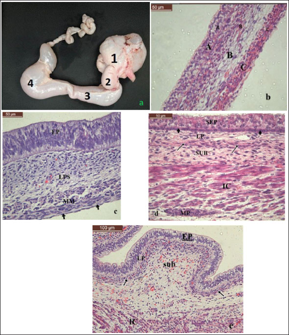
Fig. 1. Photomicrographs of the fetal camel stomach. a) Gross image showing the four distinct compartments of the fetal stomach: Compartments 1–4. b) Section of the primitive stomach wall at 79 days of gestation, showing an undifferentiated stratified epithelium (A), underlying pluripotent blastemic tissue (B), and an outer myoblastic layer (C). H&E. c) Section of Compartment 1 in fetuses aged 87–115 days of gestation, demonstrating four histological layers: epithelial layer (EP), lamina propria-submucosa (LPS), tunica muscularis (M), and serosa (arrows). H&E. d) Section of Compartment 1 in fetuses aged 120–128 days of gestation, showing a stratified epithelium (SEP), basal layer (arrowheads), lamina propria (LP), submucosa (SUB), muscularis mucosa (arrows), inner circular layer of tunica muscularis (IC), and the myenteric plexus (MP). H&E. e) Section of Compartment 1 from fetuses aged 120–128 days showing a folded epithelium (EP), lamina propria-submucosa (sub), a denser band of lamina propria-submucosa (arrows), and the inner circular layer of the tunica muscularis (IC) H&E. First trimesterAt 79 days of gestation (CVRL: 5 cm), the primitive stomach wall consisted of three layers: epithelial, pluripotent blastemic tissue, and myoblastic (Fig. 1b). The innermost layer consisted of undifferentiated stratified squamous to cuboidal epithelial cells with varying levels of cytoplasmic vacuoles and nuclei. The middle layer comprised undifferentiated mesenchymal cells and angioblasts, and the outermost layer comprised myoblastic cells. As the embryo developed, between 87 and 115 days of gestation (CVRL: 8–18 cm), the inner surface of Compartment 1 exhibited both smooth and undulating regions (Fig. 1c). Both areas of the wall comprised four distinct layers: epithelium, lamina propria-submucosa, tunica muscularis, and serosa. The epithelial layer was a stratified columnar epithelium consisting of 3–4 layers of cells. The submucosal lamina propria comprises mesenchymal cells and loose connective tissue. The tunica muscularis comprised myoblastic cells, whereas the serosa was composed of thin fibroblastic connective tissue and externally covered by a single layer of flat mesothelial cells. By 120–128 days of gestation (CVRL: 20–23 cm), the smooth-surfaced region of Compartment 1 was lined with stratified epithelium consisting of a darkly stained basal layer of cuboidal cells with rounded nuclei and a lightly stained 2–3 layer of polyhedral cells (Fig. 1d). The undulating region became more folded and was lined with pseudostratified columnar epithelium (Fig. 1e). The lamina propria-submucosa in both regions comprised connective tissue containing fibroblasts, reticular fibers, blood vessels, and lymphatics. However, the lamina propria was separated from the submucosa in the smooth surface region by a thin layer of smooth muscle fibers forming the muscularis mucosa (Fig. 1d). In contrast, the folded region’s lamina propria-submucosa was highly vascularized, and the connective tissue formed a dense band beneath the epithelial base (Fig. 1e). The tunica muscularis comprises an inner thick circular layer and an outer thin longitudinal layer of smooth muscle cells. The serosa comprised loose connective tissue and was externally covered by the mesothelium. Second trimesterAt this stage, Compartment 1 externally exhibited a dorsal smooth (non-sacculated) part, which internally corresponded to the smooth surface region that later developed into the non-glandular area. The cranioventral and caudodorsal sacs were associated with the internal folded regions, eventually forming the glandular region. In fetuses aged 163–234 days (CVRL: 36–62 cm), the stratified epithelium of the smooth surface (non-glandular region) increased in thickness due to the presence of more epithelial cells. These cells were organized into three distinct layers: stratum basale, spinosum, and granulosum (Fig. 2a).
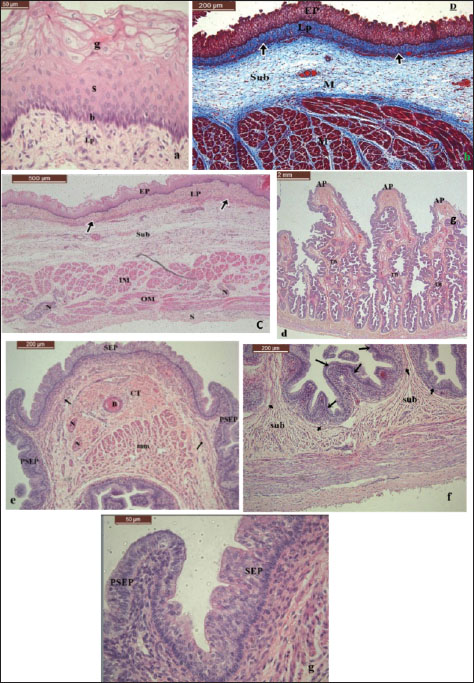
Fig. 2. Photomicrographs of the fetal camel stomach during the second trimester (163–234 days of gestation). a) Non-glandular region of Compartment 1 showing stratified epithelium composed of three layers: stratum basale (b), stratum spinosum (s), and stratum granulosum (g), with underlying lamina propria (LP). H&E. b) Non-glandular region of Compartment 1 showing dense irregular connective tissue in the lamina propria (LP), separated from the submucosa (Sub) by a thin muscularis mucosa (arrows) and tunica muscularis (M). Masson’s trichrome stain. c) Non-glandular region of Compartment 1 with stratified squamous epithelium (EP), LP, submucosa (Sub), inner circular (IM) and outer longitudinal (OM) layers of the tunica muscularis, serosa (S), myenteric plexus (N), and muscularis mucosa (arrows). H&E. d–g) Glandular region of Compartment 1. d) TB and AP of the fold. The tubular gland (g) is seen in the lamina propria. e) The apical part of the fold is lined with SEP, transitioning laterally to PSEP; the central blood vessel (B) is surrounded by connective tissue (CT), nerve fibers (N), and muscular tissue (MM) at the tip of the fold; muscularis mucosa (arrows). f) The chambers are lined with simple columnar epithelium (arrows), supported by submucosa (Sub) and muscularis mucosa (arrowheads). g) The transition zone shows a change from SEP at the apex to PSEP laterally H&E. The stratum basale comprised columnar cells with oval to elongated nuclei and basophilic cytoplasm resting on the basement membrane. The stratum spinosum contained 3–4 layers of polyhedral cells with oval nuclei and moderately stained cytoplasm. The stratum granulosum consisted of 2–3 layers of flattened cells, with vesicular nuclei and unstained cytoplasm in the uppermost cells (Fig. 2a). In the same age group (163–234 days), the folded (glandular) region of Compartment 1 exhibited deep pits dividing the mucosa into numerous folds, with some transverse bands connecting the large folds (Fig. 2d). The apical parts of the large folds were lined with stratified columnar epithelium. The lateral sides were lined with pseudostratified columnar epithelium (Fig. 2e and g). The chambers at the fold base were lined with simple columnar epithelium (Fig. 2f). Lateral projections at the apices of the folds marked the transition zone where the epithelial lining changed from stratified to pseudostratified (Fig. 2e and g). The lamina propria was composed of a thin layer of dense, irregular connective tissue containing collagen and reticular fibers, fibroblasts, and blood and lymphatic vessels in both non-glandular and glandular regions. A thin layer of muscularis mucosa separated it from the submucosa. Simple branched tubular glands were seen in the lamina propria of the glandular region (Fig. 2d). The submucosa consisted of loose connective tissue, blood vessels, diffuse lymphatic cells, and occasional lymphoid nodules (Fig. 2b and c). The tunica muscularis became more prominent, comprising a thick inner circular and a thin outer longitudinal smooth muscle layer, with nerve fibers and neurons forming the myenteric (Auerbach’s) plexus between them (Fig. 2c). The serosa contained loose connective tissue, blood vessels, and nerves and was externally covered by mesothelium. Additionally, the muscularis mucosa, submucosa, and fibers from the inner circular layer extended into the large mucosal folds in the glandular region (Fig. 2f). A dense aggregation of muscular tissue, nerve fibers, and a centrally located blood vessel, all encased in a thick connective tissue capsule, was observed at the fold tip (Fig. 2e). Third trimesterIn fetuses aged 270–390 days (CVRL: 75–115 cm), the non-glandular region epithelium remained as stratified squamous epithelium, as observed in the second trimester. Additionally, a superficial layer of flattened squamous cells formed the stratum corneum. However, the stratum granulosum became noticeably thinner (Fig. 3a). The epithelial organization in the glandular region remained similar to that in earlier stages. The apical portions of the mucosal folds were lined with stratified columnar epithelium, the lateral aspects were covered with pseudostratified columnar epithelium, and the chambers at the base of the folds were lined by simple columnar epithelium (Fig. 3b).
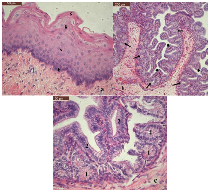
Fig. 3. Photomicrographs of fetal camel Compartment 1 at 270–390 days of gestation. a) Non-glandular region showing stratified epithelium composed of stratum basale (b), stratum spinosum (s), stratum granulosum (G), and stratum corneum (C). b) Glandular region with deep chambers lined by simple columnar epithelium (arrowheads); the lamina propria contains simple branched tubular glands (arrows). c) High magnification of the glandular region showing: (1) tubular glands with narrow lumens lined by simple columnar epithelium with basally located round nuclei and (2) chambers lined by simple columnar epithelium with basally located oval nuclei H&E. In both the non-glandular and glandular regions, the thickness of the lamina propria and muscularis mucosa increased. The lamina propria comprised dense connective tissue. Furthermore, the glandular region’s lamina propria contained simple branched tubular glands (Fig. 3b), each with a narrow lumen and simple columnar epithelium with basally located nuclei (Fig. 3c). The submucosa comprised loose connective tissue that progressively became denser near the inner circular layer of the tunica muscularis. Although the tunica muscularis was well developed and organized into inner circular and outer longitudinal smooth muscle layers, a thickened serosa surrounded it externally. DiscussionThe present study demonstrated that the wall of the primitive stomach in all camel compartments consisted of three layers during the first trimester, at 79 days of gestation: an inner stratified epithelial layer with cytoplasmic vacuoles, a middle layer of blastemic tissue composed of undifferentiated mesenchymal cells, and an outer layer of myoblastic cells. Franco et al. (2011) reported that the developing rumen in sheep and red deer at 30 and 60 days of gestation comprised three distinct layers: inner epithelium, middle pluripotent blastemic tissue, and outer myoblastic layer. Similarly, the ruminal wall of the primitive gastric tube in goats at 38 days of gestation was composed of an internal epithelial layer, a middle layer of pluripotent blastemic tissue, and an external serosal layer (García et al., 2012; Wojtasiak and Stankiewicz, 2021). A similar three-layered organization was observed in bovine fetuses as early as day 23 of gestation (Vivo et al., 1990). Compartment 1 exhibited both smooth and undulating surface regions between 87 and 115 days of gestation. Histologically, both regions of the wall comprised four layers: a stratified epithelial layer, lamina propria-submucosa, tunica muscularis, and a thin serosa formed of fibroblasts and covered by a single layer of flat mesothelial cells. These findings are consistent with those reported for camel fetuses between 50 and 140 days of gestation (Naghani and Akradi, 2011), 70 and 89 days of gestation (Ibrahim and Siddig, 2022), and between 23 and 42 days of gestation in bovine (Vivo et al., 1990). In this study, at 120–128 days, the smooth region of Compartment 1 was lined by stratified epithelium consisting of a basal layer of darkly stained cuboidal cells with rounded nuclei and an overlying 2–3 layers of lightly stained polyhedral cells. A thin layer of smooth muscle cells separated the lamina propria from the submucosa that formed the muscularis mucosa. In contrast, the undulating region developed prominent folds lined by pseudostratified columnar epithelium. The lamina propria-submucosa formed a dense band directly under the epithelial base, which transitioned into looser connective tissue below. No distinct muscularis mucosa was observed in this region. Similar epithelial differentiation and layering were observed in the goat rumen on day 53 of gestation (Isbilir et al., 2024). During the second trimester (145–234 days), the non-glandular region of Compartment 1 exhibited three characteristic epithelial strata: basale, spinosum, and granulosum—indicating increased epithelial thickness in the smooth region. The glandular (folded) region was subdivided into multiple mucosal folds, separated by deep pits, and ended in chamber-like structures. Transverse bands connecting the larger folds and tubular glands in the lamina propria were observed. These features are consistent with the division of Compartment 1 into glandular and non-glandular regions, which reportedly begins at 140–160 days of gestation, according to Naghani and Akradi (2011). In contrast, the division was observed as early as 100–110 days of gestation, with the discrepancy possibly attributed to breed-related variation (Ibrahim and Siddig, 2022). By the third trimester (270–390 days), the epithelium of the non-glandular region consisted of stratified squamous layers: basale, spinosum, and granulosum. A superficial stratum corneum layer of flattened squamous cells, indicating terminal differentiation, was also present. The lamina propria in the glandular region contained simple branched tubular glands with narrow lumens lined by simple columnar epithelium with basally located nuclei. These results are consistent with findings regarding the stomach of late-stage fetal camels (Naghani and Akradi, 2011; Ibrahim and Siddig, 2022). Similar glandular structures have been documented in adult dromedaries (Abdel-Magied and Taha, 2003; Abuagla et al., 2014; Osman et al., 2001a). In conclusion, this study demonstrated that Compartment 1 of the dromedary camel undergoes progressive histological differentiation throughout gestation. Initially, the primitive gut wall consists of three main layers. By the end of the first trimester, it differentiated into four layers with distinct smooth and folded regions. Throughout the second and third trimesters, Compartment 1 developed into a well-organized structure with distinct non-glandular and glandular regions. These regions undergo epithelial specialization, gland formation, and thickening of the lamina propria, muscularis mucosa, and tunica muscularis, indicating functional maturation for postnatal digestion. The non-glandular region develops into keratinized stratified squamous epithelium. The glandular region and the apical parts of the mucosal folds are flanked by lateral vertical projections, where the epithelium transitions from stratified columnar to pseudostratified columnar. These histogenetic features reflect structural adaptation for postnatal digestive function. AcknowledgmentsThe author would like to thank the Scientific Research Deanship of King Faisal University in Saudi Arabia for funding this study. Conflict of interestThe author has no conflicts of interest to disclose. FundingThe research was funded by the Scientific Research Deanship of King Faisal University in Saudi Arabia. Authors’ contributionsThere is one author for this manuscript. Data availabilityThe data used to support the findings of this study are included in the article and will be made available on request. ReferencesAbdel-Magied, E.M. and Taha, A.A. 2003. Morphological, morphometric, and histochemical characterization of the gastric mucosa of the camel (Camelus dromedarius). Anat. Histol. Embryol. 32, 42–47; doi:10.1046/j.1439-0264.2003.00436.x Abuagla, I.A., Ibrahim, Z.H. and Ali, H.A. 2014. Gross anatomical and histometric studies on the stomach glandular sacs of the dromedary camel (Camelus dromedarius). Sudan J. Sci. Technol. 15, 46–56. Aiyan, A.A., Richardson, K., Shawaf, T., Abdullah, S., Barigye, R., A., A.A., Richardson, K., Shawaf, T., Abdullah, S. and Barigye, R. 2018. A histologic and histomorphometric study of the first compartment of stomach in the dromedary (Camelus dromedarius). J. Camel Pract. Res. 25(2), 221–230. Allouch, G. 2016. Anatomical study of the water cells area in the dromedary camel’s rumen (Camelus dromedarius). Nova J. Med. Biol. Sci. 5(1), 1–4. Amasaki, H., Rung, G., Matsumoto, S. and Daigo, M. 1988. Scanning and electron microscopic observation on the epithelial surface of the forestomach in Bactrian camel (Camelus bactrianus). Jpn. J. Zootech. Sci. 59(6), 527–531. Bancroft, J.D. and Gamble, M. 2008. Theory and practice of histological techniques: Elsevier health sciences. Churchill Livingstone, China: Elsevier. Bello, A., Onyeanusi, B.I., Sonfada, M.L., Adeyanju, J.B., Umaru, M.A. and Onu, J.E. 2014. Gross embryonic differentiation of the stomach of the one-humped camel (Camelus dromedarius). J. Anat. Physiol. 4(1), 2–4. Dougbag, A.S. and Berg, R. 1981. Histological and histochemical studies on the pyloric mucosa of the camel’s stomach (Camelus dromedarius). Zbl. Vet. Med. C Anat. Histol. Embryol. 10, 187–192; doi:10.1111/j.1439-0264.1981.tb00517.x Eerdunchaolu, K., Takehana, K., Kobayashi, A., Baiyin, G.F.C., Andrew, A., Iwasa, K. and Abe, M. 1999. Morphological characterization of gland cells of glandular sac area in the complex stomach of the Bactrian camel (Camelus bactrianus). Anat. Histol. Embryol. 28, 183–191; doi:10.1046/j.1439-0264.1999.00185.x Elwishy, A.B., Hemeida, N.A., Omar, M.A., Mobarak, A.M. and El Sayed, M.A. 1981. Functional changes in the pregnant camel with special reference to foetal growth. Br. Vet. J. 137(5), 527–537; doi:10.1016/s0007-1935(17)31592-0 Franco, A., Masot, J. and Redondo, E. 2011. Ontogenesis of the rumen: a comparative analysis of the Merino sheep and Iberian red deer. Anim. Sci. J. 82, 107–116; doi:10.1111/j.1740-0929.2010.00814.x García, A., Masot, J., Franco, A., Gázquez, A. and Redondo, E. 2012. Histomorphometric and immunohistochemical study of the goat rumen during prenatal development. Anatomical Rec. 295, 776–785; doi:10.1002/ar.22431 Hansen, A. and Schmidt-Nielsen, K. 1957. On the stomach of the camel with special reference to the structure of its mucous membrane. Acta Anat. 13(3), 353–375; doi:10.1159/000141291 Hegazi, A.H. 1950. The stomach of the camel. Br. Vet. J. 106, 209–213; doi:10.1016/s0007-1935(17)52827-4 Ibrahim, M.I.A. and Siddig, R.S.A. 2017. Prenatal development of compartment 1 of the stomach in the one-humped camel (Camelus dromedarius): topography and gross anatomy. J. Vet. Med. Anim. Prod. 8, 76–83. Ibrahim, M.I.A. and Siddig, R.S.A. 2022. Histological and histometric study on compartment 1 of the one-humped camel (Camelus dromedarius) during prenatal development. J. Vet. Med. Anim. Prod. 1(2), 62–71. Ibrahim, Z.H. and Almundarij, T.I. 2023. Morphology of the dromedary camel stomach with reference to physiological adaptation. Slov. Vet. Res. 60(Suppl 25), 341–352. Isbilir, F., Isbilir, I., Atli, M., Yavas, E.S., Yavas, O. and Guzel, B. 2024. Macroscopic and microscopic development of the stomach in Hamdani crossbred sheep fetuses (Ovis aries). J. Res. Vet. Med. 43(1), 85–92. Lechner-Doll, M., Engelhardt, W.V., Abbas, A.M., Mousa, H.M., Luciano, L. and Reale, E. 1995. Particularities in forestomach anatomy, physiology, and biochemistry of camelids compared to ruminants. Center Int. De Hantes 13, 19–32. Mayhew, T.M. and Cruz-Orive, L.M. 1974. Caveat on the use of the Delesse principle of areal analysis for estimating component volume densities. J. Microsc. 102(2), 195–207; doi:10.1111/j.1365-2818.1974.tb03979.x Naghani, E.S. and Akradi, L. 2011. Histogenesis of the rumen in one-humped camel (Camelus dromedarius). Pak. Vet. J. 32(2), 29–272. Osman, E.E. 1999. Morphological and some immunohistochemical observations on the stomach of the camel (Camelus dromedarius). MSc thesis, University of Khartoum, Sudan. Osman, E.E., Osman, D.I. and Ali, A.M. 2001a. Gross anatomy of the stomach of the dromedary camel (Camelus dromedarius). Camel Newslett. 18, 53–61. Osman, E.E., Osman, D.I. and Ali, A.M. 2001b. Histological observations of the stomach of the dromedary camel (Camelus dromedarius). Camel Newslett. 18, 44–52. Ouajd, S. and Kamel, B. 2009. Physiological particularities of dromedary (Camelus dromedarius) and experimental implications. Scand. J. Lab. Anim. Sci. 36, 19–29. Purohit, M.S. and Rathor, S.S. 1962. Stomach of the camel in comparison to that of the ox. Indian Vet. J. 39, 604–608. Shahrasbi, H. and Radmehr, B. 1974. Studies on the anatomy and histology of rumen water sacs in camel (Camelus dromedarius). Iran. J. Vet. Med. 30(3), 14–25. Singh, M., Nagpal, S. and Singh, Y. 1996. Histomorphological studies on the glandular mucosa of rumen, reticulum, and omasum in camel (Camelus dromedarius). Indian J. Anim. Sci. 66(9), 881–884. Smuts, M.S. and Bezuidenhout, A.J. 1987. Anatomy of the dromedary. Clarendon Press, Oxford, pp. 124–177. Vivo, J.M., Robina, A., Regodón, S., Guillén, M.T., Franco, A. and Mayoral, A.I. 1990. Histogenetic evolution of bovine gastric compartments during the prenatal period. Histology Histopathology 5, 461–476. Wojtasiak, N. and Stankiewicz, T. 2021. Prenatal development of the stomach in the goat (Capra hircus). Rocz. Nauk. Pol. Tow. Zootech. 1, 23–32. | ||
| How to Cite this Article |
| Pubmed Style Abdelhay Mohamed Ali. Histological study on the prenatal development of compartment 1 of the stomach of the camel (Camelus dromedarius). Open Vet. J.. 2025; 15(9): 4569-4577. doi:10.5455/OVJ.2025.v15.i9.63 Web Style Abdelhay Mohamed Ali. Histological study on the prenatal development of compartment 1 of the stomach of the camel (Camelus dromedarius). https://www.openveterinaryjournal.com/?mno=248758 [Access: January 25, 2026]. doi:10.5455/OVJ.2025.v15.i9.63 AMA (American Medical Association) Style Abdelhay Mohamed Ali. Histological study on the prenatal development of compartment 1 of the stomach of the camel (Camelus dromedarius). Open Vet. J.. 2025; 15(9): 4569-4577. doi:10.5455/OVJ.2025.v15.i9.63 Vancouver/ICMJE Style Abdelhay Mohamed Ali. Histological study on the prenatal development of compartment 1 of the stomach of the camel (Camelus dromedarius). Open Vet. J.. (2025), [cited January 25, 2026]; 15(9): 4569-4577. doi:10.5455/OVJ.2025.v15.i9.63 Harvard Style Abdelhay Mohamed Ali (2025) Histological study on the prenatal development of compartment 1 of the stomach of the camel (Camelus dromedarius). Open Vet. J., 15 (9), 4569-4577. doi:10.5455/OVJ.2025.v15.i9.63 Turabian Style Abdelhay Mohamed Ali. 2025. Histological study on the prenatal development of compartment 1 of the stomach of the camel (Camelus dromedarius). Open Veterinary Journal, 15 (9), 4569-4577. doi:10.5455/OVJ.2025.v15.i9.63 Chicago Style Abdelhay Mohamed Ali. "Histological study on the prenatal development of compartment 1 of the stomach of the camel (Camelus dromedarius)." Open Veterinary Journal 15 (2025), 4569-4577. doi:10.5455/OVJ.2025.v15.i9.63 MLA (The Modern Language Association) Style Abdelhay Mohamed Ali. "Histological study on the prenatal development of compartment 1 of the stomach of the camel (Camelus dromedarius)." Open Veterinary Journal 15.9 (2025), 4569-4577. Print. doi:10.5455/OVJ.2025.v15.i9.63 APA (American Psychological Association) Style Abdelhay Mohamed Ali (2025) Histological study on the prenatal development of compartment 1 of the stomach of the camel (Camelus dromedarius). Open Veterinary Journal, 15 (9), 4569-4577. doi:10.5455/OVJ.2025.v15.i9.63 |

