


| Case Report | ||
Open Vet. J.. 2025; 15(5): 2265-2269 Open Veterinary Journal, (2025), Vol. 15(5): 2265-2269 Case Report Clear cell hepatocellular carcinoma incidentally detected in an 11-year-old female Tibetan Terrier dog: A case reportAlan Maksimović1*, Selma Filipović1, Muamer Obhođaš1, Bianca Pehar1, Nermina Spahija1, Kenan Tabaković2, Alma Šeho-Alić1, Jovana Dervović1 and Amer Alić11Department of Clinical Sciences, University of Sarajevo – Veterinary Faculty, Sarajevo, Bosnia and Herzegovina 2Private Veterinary Clinic, Sarajevo, Bosnia and Herzegovina *Corresponding Author: Alan Maksimović. Department of Clinical Sciences, University of Sarajevo – Veterinary Faculty, Sarajevo, Bosnia and Herzegovina. Email: alan.maksimović [at] vfs.unsa.ba Submitted: 06/02/2025 Revised: 11/04/2025 Accepted: 20/04/2025 Published: 31/05/2025 © 2025 Open Veterinary Journal
ABSTRACTBackground: Hepatobiliary tumors are uncommon in dogs, and metastatic liver tumors are diagnosed more frequently than primary liver tumors. Hepatocellular carcinoma is the most prevalent primary liver malignant tumor. Case Description: An 11-year-old spayed female Tibetan Terrier was referred to the University of Sarajevo Veterinary Teaching Hospital due to recurrent painful urination attempts, straining urination, hematuria, and decreased appetite. Abdominal ultrasonography revealed a moderately distended bladder containing urolith, bladder wall thickening, and sediment. Incidentally, a hyperechoic hepatic mass on the left medial liver lobe and hepatomegaly were detected along with gallbladder sludge. Surgical cystolithotomy and partial liver lobectomy were performed. Histopathological examination confirmed the hepatic mass as clear cell hepatocellular carcinoma (CCHCC). Conclusion: Although this rare histological subtype has been documented, its biological behavior and clinical features remain poorly understood due to the scarcity of cases. A recent publication by Jung et al. (2021). described the first cytological, histological, and clinical case presentation of CCHCC in dogs, suggesting that obesity and hyperlipidemia may be potential risk factors. However, these proposed risk factors were not detected in the present case, implying that CCHCC in dogs is a rare and poorly understood condition that warrants further attention in veterinary research. Keywords: Clear cell hepatocellular carcinoma, Dog, Case report. IntroductionHepatobiliary neoplasia is relatively uncommon in dogs, accounting for approximately 0.6%–1.3% of all cancers diagnosed through necropsy studies. Tumors of the hepatobiliary system can originate either as primary tumors arising from the liver, gallbladder, or bile ducts or secondary tumors originating from other organs and subsequently metastasizing to the liver. Notably, in dogs, metastatic liver tumors are more frequently diagnosed than primary tumors (Selmic, 2017). Hepatocellular carcinoma (HCC) is the most prevalent primary malignant liver tumor in dogs. It originates from hepatocytes and exhibits diverse biological and morphological characteristics. The classification of HCC in dogs is crucial for understanding its pathogenesis, guiding clinical management, and predicting prognosis. Unlike in humans, where HCC is often associated with predisposing conditions such as hepatitis or cirrhosis, canine HCC typically arises spontaneously, with no discernible association with underlying liver diseases (Liptak et al., 2004; Leela-Arporn et al., 2019). Canine HCC is generally categorized into three primary forms based on gross morphology: massive, nodular, and diffuse. The massive form is the most prevalent and frequently manifests as a solitary, large, well-defined mass, predominantly localized to a single liver lobe. This form is associated with the most favorable prognosis owing to the feasibility of surgical resection. Nodular HCC is characterized by multiple distinct tumors affecting multiple lobes, while the diffuse form involves extensive infiltration throughout the liver parenchyma and is associated with poor prognosis due to limited treatment options and aggressive behavior (Patnaik and Hurvitz, 1980) HCCs exhibit a highly variable histological appearance influenced by distinct cellular arrangements. The four primary histological variants were trabecular, pseudoglandular, scirrhous, and solid. In certain instances, most neoplastic hepatocytes may manifest prominent cytoplasmic clearing or vacuolation, leading to the designation of these neoplasms as clear cell hepatocellular carcinomas (CCHCCs) (Liptak et al., 2004; Cullen, 2017). Although this rare histological subtype has been documented, its biological behavior and clinical features remain poorly understood due to the scarcity of cases (Jung et al., 2021). Recent advancements in molecular biology and cell line development have significantly expanded our understanding of the histopathological diversity of HCC in dogs. Lee et al. (2024) established six canine HCC cell lines and demonstrated the variability in tumor architecture and genetic profiles, which may correlate with histopathological patterns. Nevertheless, gaps remain in the literature, particularly regarding rare variants such as CCHCC. Case DetailsAn 11-year-old spayed female Tibetan Terrier was referred to the University of Sarajevo Veterinary Teaching Hospital with stranguria, hematuria, and inappetence. The dog’s medical history included severe atopic dermatitis (AD) since the age of four, which was managed according to current recommendations for long-term control of AD, including allergen avoidance, skin barrier care, and systemic therapies based on the severity of clinical signs (Miller et al., 2023). On clinical examination, the patient was alert and cooperative, with a body condition score of 5/9. Frequent attempts to urinate were observed, accompanied by signs of discomfort. Abdominal palpation revealed a tense and distended bladder. Radiography revealed no significant findings, while abdominal ultrasonography (Fig. 1) revealed a moderately distended bladder containing a hyperechoic urolith, sediment, and a thickened bladder wall. Hepatomegaly was detected with a hyperechoic lesion on the left medial liver lobe, alongside echogenic gallbladder sludge (Fig. 2). All other abdominal organs were normal. Hematology revealed lymphopenia (0.97 × 10^9/l), and biochemical analysis showed hypophosphatemia (0.70 mmol/l) and elevated liver enzyme levels (ALKP 266 U/l, GGT 22 U/l). Other parameters were within normal limits. Urinalysis was performed, revealing dark brown, turbid urine with a specific gravity of 1.034, pH of 6.5, protein of 500 mg/dl, and erythrocytes 500 Ery/μl. Following diagnostic evaluation, surgical cystolithotomy and partial lobectomy of the left medial liver lobe were performed (Fig. 3).
Fig. 1. Ultrasonographic image demonstrating hyperechoic urolith measuring 1.11 × 0.61 cm.
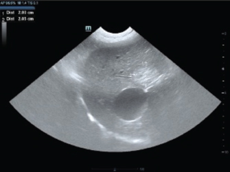
Fig. 2. Ultrasonographic image of the liver lobe showing a hyperechoic lesion (CCHCC) measuring 2.00 × 2.05 cm.
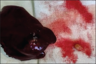
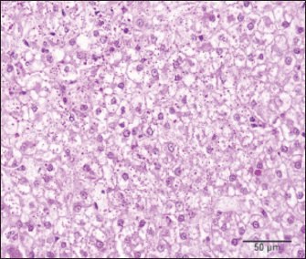
Fig. 3. Macroscopic appearance of a CCHCC solitary nodule in the excised liver tissue and removed urolith. For histopathology, tissue samples were placed overnight in 10% neutral buffered formalin, routinely processed through different grades of alcohols and xylene in a tissue processor (STP 120, Microm, Spain), and embedded in paraffin blocks in a tissue embedding center (EC 500, Myr, Spain). A thin section (4 micrometers) cut at a microtome (RM 2145, Leica, Germany) and stained with hematoxylin and eosin and periodic acid-shiff (PAS) stains was examined under the light microscope (BX-51, Olympus, Germany) by two pathologists. Microscopic examination revealed a multinodular, well-demarcated mass characterized by a solid growth of neoplastic hepatocytes consistent with CCHCC. The neoplastic cells were oval-to-polygonal with clear-to-highly vacuolated cytoplasm and mostly central round nuclei with one small nucleolus (Fig. 4). Moderate anisocytosis, a small number of larger nuclei, and rare mitotic figures were observed. Multifocally, small nodular neoplastic foci were present near the main neoplastic nodules. The remaining hepatic parenchyma surrounding the neoplastic nodules exhibited varying degrees of hepatocyte vacuolation and sinusoidal congestion. Numerous small purple droplets of glycogen were visible in the cytoplasm of neoplastic cells on slides stained with PAS staining (Fig. 5).
Fig. 4. Liver, dog: solid nodular growth of clear neoplastic cells with central round nucleus effaces adjacent vacuolated hepatocytes. Hematoxylin and eosin, 40×.
Fig. 5. Liver dog: numerous PAS-positive glycogen droplets in the cytoplasm of neoplastic cells. PAS, 40×. The patient was found to have good general health and behavior 3 months post-surgery. DiscussionCCHCC is a rare variant of HCC in dogs. The condition is characterized by hepatocytes with clear cytoplasm due to glycogen or lipid accumulation (Jung et al., 2021). Although HCC is the most prevalent primary liver tumor in dogs, comprising up to 77% of primary hepatobiliary neoplasms (Patnaik et al., 1981), reports on the clear cell variant are scarce. Therefore, the current understanding of CCHCC clinical behavior, prognosis, and treatment options in dogs remains limited. Jung et al. (2021) reported an 11-year-old obese dog with hyperlipidemia who exhibited no apparent clinical signs of disease. Subsequently, the dog was diagnosed with CCHCC after detection of a liver mass. Obesity and hyperlipidemia are potential risk factors for CCHCC in dogs (Jung et al., 2021). In the present case, a hepatic mass was incidentally detected during an ultrasound examination of the abdominal cavity, which was conducted as part of the evaluation of the primary complaint of urinary system issues. In contrast to the case reported by Jung et al. (2021), obesity and hyperlipidemia were absent, necessitating further investigation to identify the risk factors associated with CCHCC. In our case, based on anamnestic data, the only prominent concurrent disorder observed was severe AD, which was treated over an 8-year period using oclacitinib, cyclosporine, corticosteroids, and lokivetmab. AD is a prevalent chronic inflammatory skin disease in dogs. It is characterized by dysregulated immune responses and systemic cytokine release. Chronic inflammation is a known contributor to cancer development across species. In humans, links between immune-mediated disorders and hepatic carcinogenesis have been attributed to persistent inflammatory signaling (Sasco et al., 2004). In comparison, recent recentings of Wan et al. (2023) suggest a lack of substa substantialelation between AD and malignant tumor. However, the findings exhibited variations across age groups, severity of AD, and specific cancer types. There is no strong, consistent evidence directly linking AD and HCC; however, some studies have explored potential indirect correlations through shared inflammatory and immunological pathways, as well as overlapping risk factors. Chronic systemic inflammation in AD, marked by elevated interleukins such as IL-6, IL-17, IL-22, and IL-33 expression, is also implicated in HCC progression. These cytokines are known to influence tumor development and may bridge inflammatory skin disorders with liver carcinogenesis (Huang et al., 2023; Laska et al., 2024). Furthermore, the presence of IL-22-producing cells in the tumor microenvironment is correlated with poor outcomes in HCC (Laska et al., 2024). The potential correlation between AD and malignant tumor in dogs has not yet been investigated. Regarding the anamnestic data, no other remarkable findings or concurrent disorders were observed. HCC in dogs is histopathologically classified into three main architectural patterns: trabecular, pseudoglandular, and solid, based on the structural arrangement of tumor cells and their resemblance to normal hepatic tissue. A rarer subtype, the clear cell variant, is characterized by tumor cells with clear cytoplasm due to glycogen or lipid accumulation. Although histologically distinct, its biological behavior in dogs remains poorly understood due to the limited number of documented cases (Patnaik and Hurvitz, 1980; Liptak et al., 2004; Jung et al., 2021). Human studies have demonstrated that a specific cell population within hepatic tumors can be utilized for survival time prognosis. A higher proportion of clear cells within the CCHCC is associated with a more favorable prognosis, with patients exhibiting longer survival rates when clear cells constitute ≥70% of the tumor (Chen et al. 2016; Chen et al., 2016; Deng et al., 2021). Similar investigations are lacking in veterinary medicine. Histopathological classification not only aids in distinguishing HCC from other hepatic neoplasms, such as biliary carcinoma and metastatic tumors, and provides prognostic value. For instance, well-differentiated trabecular HCCs are associated with better prognosis, particularly when they are confined to a single liver lobe and amenable to surgical excision (Liptak et al., 2004). Surgical resection remains the cornerstone of HCC treatment in dogs, including rare subtypes like CCHCC. Although general data on massive HCC provides a strong foundation for understanding the potential outcomes of surgical therapy, specific information on CCHCC is scarce, limiting definitive conclusions regarding prognosis. In their study of 48 cases of massive HCC, Liptak et al. (2004) reported that surgical excision resulted in a median survival time exceeding 1,460 days in dogs with complete tumor resection. Recurrence was rare, suggesting that HCCs, even in large and invasive forms, often demonstrate low metastatic potential when resected entirely. Although no specific survival data are available for CCHCC, the tumor’s presumed similarities in behavior to other HCC variants suggest that timely surgical removal may yield comparable outcomes. Incomplete excision or the presence of metastases at the time of diagnosis can worsen outcomes. However, considering that CCHCC cases often present with nonspecific signs leading to delayed diagnosis, early surgical intervention is particularly challenging. In this case, the hepatic mass was incidentally identified during the examination of the primary complaint of urinary tract issues. The general survival times of dogs with CCHCC are virtually nonexistent because of the limited number of documented cases. In our case, the owners were informed about the absence of robust survival time data and were provided with a prognosis based on a single liver lobe solitary carcinoma node, complete tumor excision, and available data regarding survival time in dogs with HCC. A recent retrospective study conducted by Leela-Arporn et al. (2019) shed light on the critical epidemiological insights into the prevalence of massive HCC in dogs over a 4-year period. The study revealed that Welsh Corgis and Beagles exhibit a predisposition to HCC, and hyperadrenocorticism is a potential risk factor. Several other studies have investigated potential risk factors for HCC in dogs, identifying an overrepresentation of specific breeds, such as Miniature Schnauzers and Shih Tzus, as well as male dogs (Patnaik et al., 1980, 1981; Liptak et al., 2004; Hirose et al., 2014). However, these findings remain inconclusive, and definitive risk factors for HCC in dogs have yet to be established. ConclusionCCHCC in dogs is a rare and poorly understood condition that requires further attention in veterinary research. Surgical resection is the primary and most effective therapeutic modality for canine HCC, including the clear cell subtype. However, specific survival data for patients with CCCHCC are lacking in the current literature. General findings from broader studies indicate that dogs with complete tumor excision can achieve extended survival time. Nevertheless, the rarity of CCCHCC underscores the importance of future research and case studies. AcknowledgmentsNone. Conflict of interestThe authors declare no conflict of interest. FundingThis research received no specific grant. Authors’ contributionsAlan Maksimović: Conceptualization, case management, data interpretation, manuscript writing, and final approval of the manuscript as the corresponding author. Nermina Spahija, Selma Filipović, Muamer Obhođaš, Bianca Pehar, and Kenan Tabaković: Case management, clinical data collection, and manuscript review. Alma Šeho-Alić, Jovana Dervović, and Amer Alić: Pathological analysis, histopathological diagnosis, and manuscript review. Data availabilityAll data supporting the findings of this study are available in the manuscript. ReferencesChen, Z.S., Zhu, S.L., Qi, L.N. and Li, L.Q. 2016. Long-term survival and prognosis for primary clear cell carcinoma of the liver after hepatectomy. Onco. Targets Ther. 9, 4129–4135; doi: 10.2147/OTT.S104827. Cullen, J.M. 2017. Tumors of the liver and gallbladder. In Tumors in domestic animals. Ed., Meuten, D.M. Hoboken, NJ: Wiley Blackwell, pp: 602–631. Deng, Y., Zhu, S., Yan, W., Liu, W., Huang, X., Liu, H. and Zhou, J. 2021. Influence of clear cell carcinoma on the post-hepatectomy prognosis of patients with hepatocellular carcinoma. Future Oncol. 18(5), 543–552. Hirose, N., Uchida, K., Kanemoto, H., Ohno, K., Chambers, J. K. and Nakayama, H. 2014. A retrospective histopathological survey on canine and feline liver diseases at the University of Tokyo between 2006 and 2012. J. Vet. Med. Sci. 76(7), 1015–1020; doi: 10.1292/jvms.14-0083 Huang, S., Wang, H., Zheng, H., Fan, W. and Liu, Y. 2023. Association between IL-6 polymorphisms and atopic dermatitis in Chinese Han children. Front. Pediatr. 11, 1156659. Jung, J.H., Kim, H.J., Park, J.H., Kim, S., Song, W., Kim, Y. and Choi, J. 2021. A clear cell hepatocellular carcinoma in an obese dog with hyperlipidemia: a case report. Korean J. Vet. Res. 61(4), e34. Laska, J., Tota, M., Łacwik, J., Sędek, Ł. and Gomułka, K. 2024. IL-22 in atopic dermatitis. Cells 13(16), 1398. Lee, J.Y., Bae, K., Kim, J.H., Kim, E., Han, S. and Park, Y. 2024. Establishment and characterization of six canine hepatocellular carcinoma cell lines. Front. Vet. Sci. 11, 1392728. Leela-Arporn, R., Ohta, H., Nagata, N., Sasaki, N. and Mizutani, M. 2019. Epidemiology of massive hepatocellular carcinoma in dogs: a 4-year retrospective study. Vet. J. 248, 74–78. Liptak, J.M., Dernell, W.S., Monnet, E., Powers, B.E., Bachand, A.M. and Withrow, S.J. 2004. Massive hepatocellular carcinoma in dogs: 48 cases (1992-2002). J. Am. Vet. Med. Assoc. 225(8), 1225–1230. Miller, J., Simpson, A., Bloom, P., White, S. and Thompson, K. 2023. 2023 AAHA management of allergic skin diseases in dogs and cats guidelines. J. Am. Anim. Hosp. Assoc. 59(6), 255–284. Patnaik, A.K. and Hurvitz, A.I. 1980. Canine hepatic neoplasms: a clinicopathologic study. Vet. Pathol. 17(5), 553–565. Patnaik, A.K., Hurvitz, A.I., Lieberman, P.H. and Johnson, G.F. 1981. Canine hepatocellular carcinoma. Vet. Pathol. 18(4), 427–438. Sasco, A.J., Secretan, M.B. and Straif, K. 2004. Cancer and autoimmune diseases: a review. Br. J. Cancer 87(7), 826–832. Selmic, L.E. 2017. Hepatobiliary neoplasia. Vet. Clin. North Am. Small Anim. Pract. 47(3), 725–735. Wan, J., Shin, D.B., Syed, M.N., Gelfand, J.M., Margolis, D.J. and Takeshita, J. 2023. Malignancy risk in patients with atopic dermatitis: a population-based cohort study. Br. J. Dermatol. 189(1), 53–61. | ||


| How to Cite this Article |
| Pubmed Style Maksimović A, Pehar B, Spahija N, Filipović S, Obhodas M, Tabaković K, Seho-alić A, Dervović J, Alić A. Clear cell hepatocellular carcinoma incidentally detected in an 11-year-old female Tibetan Terrier dog: A case report. Open Vet. J.. 2025; 15(5): 2265-2269. doi:10.5455/OVJ.2025.v15.i5.44 Web Style Maksimović A, Pehar B, Spahija N, Filipović S, Obhodas M, Tabaković K, Seho-alić A, Dervović J, Alić A. Clear cell hepatocellular carcinoma incidentally detected in an 11-year-old female Tibetan Terrier dog: A case report. https://www.openveterinaryjournal.com/?mno=241411 [Access: January 25, 2026]. doi:10.5455/OVJ.2025.v15.i5.44 AMA (American Medical Association) Style Maksimović A, Pehar B, Spahija N, Filipović S, Obhodas M, Tabaković K, Seho-alić A, Dervović J, Alić A. Clear cell hepatocellular carcinoma incidentally detected in an 11-year-old female Tibetan Terrier dog: A case report. Open Vet. J.. 2025; 15(5): 2265-2269. doi:10.5455/OVJ.2025.v15.i5.44 Vancouver/ICMJE Style Maksimović A, Pehar B, Spahija N, Filipović S, Obhodas M, Tabaković K, Seho-alić A, Dervović J, Alić A. Clear cell hepatocellular carcinoma incidentally detected in an 11-year-old female Tibetan Terrier dog: A case report. Open Vet. J.. (2025), [cited January 25, 2026]; 15(5): 2265-2269. doi:10.5455/OVJ.2025.v15.i5.44 Harvard Style Maksimović, A., Pehar, . B., Spahija, . N., Filipović, . S., Obhodas, . M., Tabaković, . K., Seho-alić, . A., Dervović, . J. & Alić, . A. (2025) Clear cell hepatocellular carcinoma incidentally detected in an 11-year-old female Tibetan Terrier dog: A case report. Open Vet. J., 15 (5), 2265-2269. doi:10.5455/OVJ.2025.v15.i5.44 Turabian Style Maksimović, Alan, Bianca Pehar, Nermina Spahija, Selma Filipović, Muamer Obhodas, Kenan Tabaković, Alma Seho-alić, Jovana Dervović, and Amer Alić. 2025. Clear cell hepatocellular carcinoma incidentally detected in an 11-year-old female Tibetan Terrier dog: A case report. Open Veterinary Journal, 15 (5), 2265-2269. doi:10.5455/OVJ.2025.v15.i5.44 Chicago Style Maksimović, Alan, Bianca Pehar, Nermina Spahija, Selma Filipović, Muamer Obhodas, Kenan Tabaković, Alma Seho-alić, Jovana Dervović, and Amer Alić. "Clear cell hepatocellular carcinoma incidentally detected in an 11-year-old female Tibetan Terrier dog: A case report." Open Veterinary Journal 15 (2025), 2265-2269. doi:10.5455/OVJ.2025.v15.i5.44 MLA (The Modern Language Association) Style Maksimović, Alan, Bianca Pehar, Nermina Spahija, Selma Filipović, Muamer Obhodas, Kenan Tabaković, Alma Seho-alić, Jovana Dervović, and Amer Alić. "Clear cell hepatocellular carcinoma incidentally detected in an 11-year-old female Tibetan Terrier dog: A case report." Open Veterinary Journal 15.5 (2025), 2265-2269. Print. doi:10.5455/OVJ.2025.v15.i5.44 APA (American Psychological Association) Style Maksimović, A., Pehar, . B., Spahija, . N., Filipović, . S., Obhodas, . M., Tabaković, . K., Seho-alić, . A., Dervović, . J. & Alić, . A. (2025) Clear cell hepatocellular carcinoma incidentally detected in an 11-year-old female Tibetan Terrier dog: A case report. Open Veterinary Journal, 15 (5), 2265-2269. doi:10.5455/OVJ.2025.v15.i5.44 |

