


| Review Article | ||
Open Vet. J.. 2025; 15(4): 1542-1548 Open Veterinary Journal, (2025), Vol. 15(4): 1542-1548 Review Article Anatomical differences in the biliary duct system between canines and felines: A review articleNoor Hussein Yousif1* and Mohammed Sulaiman Dawood21Iraq Natural History Research Center &Museum, University of Baghdad, Baghdad, Iraq 2Departmnet of Anatomy & Histology, College of Veterinary Medicine, University of Baghdad, Baghdad, Iraq *Corresponding Author: Noor Hussein Yousif. Iraq Natural History Researches Center & Museum, University of Baghdad, Baghdad, Iraq. Email: noor.yousif2208p [at] covm.uobaghdad.edu.iq Submitted: 20/01/2025 Accepted: 18/03/2025 Published: 30/4/2025 © 2025 Open Veterinary Journal
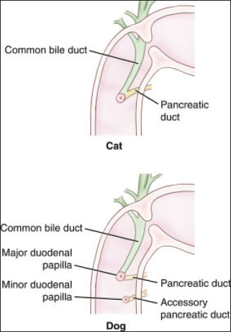
AbstractThe liver is one of the largest glands in the digestive system and performs 13 various functions, including the secretion of hormones and enzymes. The gallbladder serves as a storage reservoir for secretions before they are released into the digestive system through the duodenum. The bile ducts branch from the liver‘s lobes and ultimately connect to the digestive system, making this structure significant and distinct among different animal species. This review focuses on the differences between dogs and cats, highlighting the importance of these differences from both health and pathological perspectives. After conducting a detailed scientific review of the biliary tree in dogs and cats, we concluded that cats are more susceptible to the disease than dogs. We recommend conducting extensive radiological studies to determine which species experience cases of this disease more frequently. Additionally, it would be beneficial to explore crossbreeding with another species with a biliary tree structure that offers protection against certain diseases affecting the liver and bile. Keywords: Anatomy, Bile duct, Billary system, Canine, Feline, Hepatic duct. IntroductionDogs and cats belong to the Canidae and Felidae families, respectively. Carnivores include a group of domestic animals that are considered field animals. Their diet is different and is not limited to meat only (Johnson-Delaney, 2006). The liver is the largest gland in mammals and releases substances using the biliary system, including lipase, which is secreted into the small intestine of the digestive system. The biliary system is considered an advanced system resulting from the merging of a group of ducts reaching from the liver that transport secretory materials and drain into a major hepatic duct, later connects to the cystic duct and then merges to the common bile duct, which comes from merges major pancreatic ducts with it empties into the duodenum (Park et al., 2018; Mahadevan, 2020). The aim of this article is to examine the key differences between two families (Canidae and Felidae), as well as the variation within the species themselves. The study explored the scientific significance of these distinctions. The gross anatomy of the liver and the biliary tree system with the sphincter of Oddi and their interconnection with the pancreatic ducts in some canine species showed that the liver is composed of a group of lobes: a left lobe divided into (lateral and medial), a right lobe divided into (lateral and medial), a quadrate lobe, and a caudate lobe. The gallbladder is located between the right lobe and the lateral and quadrate lobes, and the bile ducts join within the liver, initiating from the intrahepatic ducts to the outside of the hepatic parenchyma, forming the extrahepatic ducts descending from all the lobes, forming a bile duct tree called the biliary system ducts, ducts which in turn form on the right side the major hepatic duct and the left hepatic duct unite to form the common hepatic duct. The common bile duct merges with the cystic duct that emerges from the gallbladder, and together they unite to form the common bile duct, which empties with the pancreatic duct into the duodenum, the first part of the intestine that enters the sphincter of Oddi, or the pancreatic duodenal papilla. This tree differs from one animal to another, especially between dogs and cats, through the union of the major pancreatic ducts with the common bile duct and their effusion into the duodenum. There is a note that the movement of the Oddi sphincter, which enters the ducts, differs from the movement of the intestines and is considered a fundamental movement (<AQ1>Sleight et al., 1970; Grace et al., 1988; Johnson-Delaney, 2006; Mahadevan, 2020). GallbladderPrevious studies have demonstrated that the gallbladder of carnivores (dog and cat) and domestic animals (goat, cattle, and buffalo) exhibits variation in shape and size from one animal to another. In carnivores, the gallbladder has a pear-shaped flexible wall that can be expanded because it stores the bile secreted by the liver, in addition to making some physiological additions (Thune et al., 1990; Aqeel, 2024). The gallbladder‘s function is to store bile and synthesize bile by absorbing water, sodium, chloride, bromide, and carbonate, which are stored in this sac until the time of feeding and then emptied into the duodenum once at the time of digestion (Dyce et al., 2010; Mahadevan, 2020). Anatomically, the gallbladder of carnivores is located on the ventral side of the liver, between the right lobe of the liver and the quadrate lobe. The area in which it resides is called the cystic fossa (Grace et al., 1988; Thune et al., 1990; Park et al., 2018). In addition, it extends from the level of the eighth to tenth intercostal spaces. The depth of the gallbladder varies between dogs and cats; in some, the gallbladder reaches the diaphragm, called the fossa. This fossa is called the biliary fossa. Innervations of the gallbladder through the sympathetic system, which is responsible for its contraction and relaxation, while the parasympathetic system has the opposite effect. The blood supply of the gallbladder is via the cystic artery, while the hepatic ducts are supplied from the vascular plexus, which surrounds the bile duct. Bile is a digestive fluid produced by the liver that flows through a network of branching tubes called the biliary tree. This complex system starts at the microscopic level within each liver cell (hepatocyte). Here, tiny channels named canaliculi collect the bile produced (Thune et al., 1990). These canaliculi converge into slightly larger ducts known as intralobular ducts. Each intralobular duct drains the bile from a single lobule, which is the functional unit of the liver. Finally, the intralobular ducts connect with even larger interlobular ducts positioned between the lobules (Park et al., 2018; Mahadevan, 2020). Biliary duct systemThe gallbladder in mammals stores the bile that is produced in the liver, which flows into the interlobar and extralobar ducts of the liver and empties into the intestine through a small opening called the major duodenal papilla. This substance helps digest food (Mahadevan, 2020). In goat and buffalo, there are many ducts inside the liver that transport the substance secreted by the hepatocytes. The hepatic ducts live in the liver and flow into the gallbladder. These hepatic ducts consisted of 3–6 ducts. The common bile duct is created by the joining of the extra hepatic biliary ducts with the pancreatic duct, forming the large duct that drains into the first part of the intestine, the duodenum. Bile secretion into the intestines occurs during several sequences, not at one time, and remains stored inside the bile sac (AL-Mayahi, 2008; Dyce et al., 2010). In previous studies of the comparative anatomy of the extrahepatic bile duct, scientists illustrated that the extrahepatic ductal parts in domestic animals (including dogs and cats) are similar. The cystic duct begins in the form of a narrow, twirling tube connected to the gallbladder, then begins to expand and connect with the hepatic ducts and pancreatic duct. Despite the similar anatomy, the size and length of the bile duct in domestic animals vary from one species to another (Getty, 1975; Aqeel, 2024). In dogs, the hepatic ducts consist of 3–4 main ducts that drain from the liver into the bile. The cystic duct connects either to the right or central bile duct before emptying into the common bile duct. It has been observed in some canine species that they contain two separate cystic ducts finally, the common bile duct part crosses along the descending part of the small intestine (duodenum) and ends at a small opening called the major duodenal papilla (Breazile, 1971; Aqeel, 2024). The biliary tree begins from the hepatocytes in the following order: canaliculi, interlobarducts, hepatic ducts, and common bile duct (Fig. 1). The common bile duct starts at the junction between the cystic duct and the first hepatic duct. The biliary tree is responsible for transporting bile and begins within liver cells via tiny channels called canalicular ducts. These ducts merge into progressively larger ducts: interlobar ducts, followed by hepatic ducts. Finally, the common bile duct forms where the first hepatic duct meets the cystic duct from the gallbladder. In dogs, the common bile duct and pancreatic duct run close together but remain separate. In some dogs, the accessory pancreatic duct opens at the minor duodenal papilla, 2 cm further down the duodenum. In cats, the common bile duct and pancreatic duct merge before emptying at the major duodenal papilla. Only about 20% of cats have an accessory pancreatic duct opening at the minor papilla (Johnson-Delaney, 2006; Aqeel, 2024).
Fig. 1. The anatomical structure of the biliary tract, with permission from Elsevier, 2021 (Bashir et al., 2024).
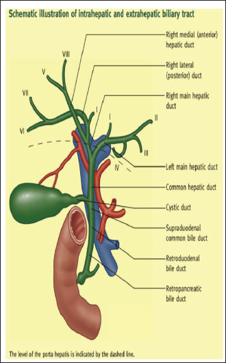
Fig. 2. Comparative veterinary anatomy a clinical approach with permission from Elsevier 2022, Pages 287–291. https://radiopaedia.org/articles/biliary-tree-anatomy Histologically, the hepatic ducts of domestic animals can be categorized into intrahepatic and extrahepatic ducts. The intrahepatic ducts consist of canaliculi, which contain microvilli to increase surface area; intralobular ducts are present all over the hepatic artery and portal vein region. The right hepatic duct drains via the right lobe of the liver, whereas the left hepatic duct drains via the left lobe. The caudate lobe of the liver is drained by small ducts from both the left and right lobes. The extrahepatic ducts were created from the right and left hepatic ducts, and these two ducts joined to form the common hepatic duct. The cystic duct that emerges from the gallbladder joined with the common hepatic duct. Finally, the common bile duct was created. The normal width of the common bile duct should be approximately less than 6 mm, and its length ranges from 6.0 to 8.0 cm (Fig. 3) (McPherson et al.,1984; Kahlil et al., 2005; Mehler, 2011).
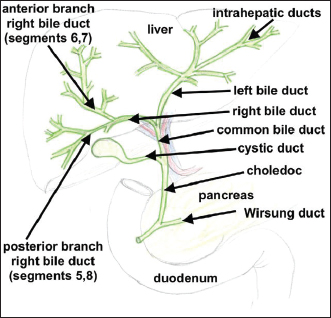
Fig. 3. Division of the biliary tree. In the book: Anatomy of the annexes of the digestive tract. Publisher: AvidScience, June 2017. The researchers employed various techniques to study the biliary tree. This included using radiography to inject a specialized colored substance into the biliary system ducts and biles. Additionally, resin was utilized to study the branching in different animals, with the branching originating from the bile sac and extending to other areas. This method proved to be helpful in identifying differences and facilitating the study (Getty, 1975; Dyce et al. , 2010; Dawood and Khamas, 2017). CanaliculiThe canaliculi originate from the hepatocytes in some goat, cattle, and gazelle. The ductulus merges with the nearest and closest ductulus to form the interhepatic duct of the liver parenchyma. Then, they merge to create the right and left hepatic ducts, and then a large duct emerging from the lobes of the liver is called the hepatic duct, after the transcystic duct is connected to the hepatic duct, the previous studies noted that the biliary tree is originally a complex network that alternates inside the liver, draining from hepatocytes and then descending into small tubes that gradually grow largely, then created the liver duct in the liver parenchyma. These ducts vary in length and size. The ductulus emerges from the right and left lobes of the liver and forms the major hepatic duct. The ducts correspond to the blood supply to the liver in the portal region. The tunicae of the ductulus and bile sac are similar (Neer, 1992; Keplinger and Bloomston, 2014; Kummeling, 2016; Reem et al., 2019).
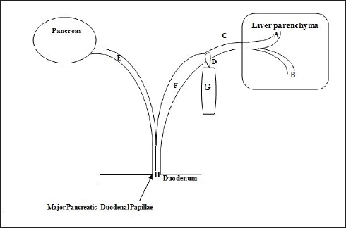
Fig. 4. The biliary system of the liver with gallbladder in a dog showed the united of the common bile duct with the pancreatic duct A: Left interhepatic duct. B: Right interhepatic duct. C: Extrahepatic duct, D: Cystic duct. F: Common bile duct. E: Pancreatic duct. H: Not merged ducts. The interlobar ducts that follow the ducts are lined by cholangiocytes of 4–5 hexogen-shaped cells that interconnect between adjacent hepatocytes and microvilli and then emerge into the bile duct; the bile cells also have villi. These bile cells are considered sensory cells and play an important role in regulating the biological activities of bile cells through secretions and proliferation (Getty, 1975; Kummeling, 2016). Right and left interhepatic ductsIn camel, chinchilla, goat, gazelle, cattle, dog, and cat, the right hepatic duct consists of the flow of the right liver lobes (right lateral and medial ducts), with the duct reaching from the quadrate lobe conducting from inside the liver, then the main right hepatic duct leaves the hepatic lobes. The left hepatic duct consists of the merging of the duct reaching from the left liver lobs (left lateral and medial ducts) with the caudate lobe (caudate prosses and the papilla ducts) then passing outside the hepatic lobes, forming the left main hepatic duct, which in turn fuses with the main right hepatic duct, forming the extrahepatic duct or main hepatic duct, which unites with the bile duct (cystic duct), except the camel, and then they form the common bile duct in (Breazile, 1971; Dyce et al., 2010; Al-Rekabi, 2011; Siddig et al., 2014; Evans and De Lahunta, 2016; Nowak et al., 2016; Song et al., 2020). Right and left extrahepatic ductsAll research on canines and felines indicates that the right hepatic duct extends from the right hepatic lobe toward the abdomen through a small pathway. The length ranges from 2 to 5 cm in different animals and varies with age. The cystic duct of the gallbladder is connected. The left hepatic duct is longer than the right, measuring about 3–6 cm. It runs parallel to the left hepatic artery and connects to the right duct at the hepatic edge. This forms an acute angle from which the participating hepatic duct emerges (Strazzabosco and Fabris, 2008; Morell et al., 2013; Siddig et al., 2014; Dawood and Khamas, 2017; Dawood and Yousif, 2022). The common hepatic ductThe goat, gazelle, and canine are formed in the portal area by connecting the right and left hepatic ducts at an acute angle. The average length is about 5 cm and about 3 mm wide. The duct passes ventrally into the middle bile duct and joins on its right side at an acute angle with the main pancreatic duct to form the common bile duct. The distance from the pylorus to the duodenal papilla is approximately 20 cm in adult dogs compared with that in goats and gazelles (Sleight and Thoford, 1970; Dyce et al., 2010; Al-Rekabi, 2011; Dawood and Khamas, 2017; Dawood and Yousif, 2022). The cystic ductIt is a short, narrow, and twisted duct in immature cats characterized by two cystic ducts, as well as contains two bile sacs. The cystic duct in goats is about 2 cm long and 2 mm wide and arises from the neck of the gallbladder. It extends upward along the lesser omentum and attaches to the right part of the hepatic duct compared with goat, cattle, and gazelle (Sleight and Thoford, 1970; Dyce et al., 2010; Siddig et al., 2014; Evans and De Lahunta, 2016; Dawood and Khamas, 2017; Dawood and Yousif, 2022; Aqeel, 2024).
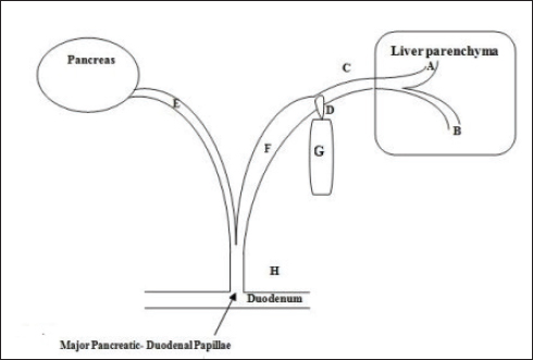
Fig. 5. The biliary system of the liver with gall bladder in a cat shows the unity of the common bile duct with the pancreatic duct. A: Left interhepatic duct. B: Right interhepatic duct. C: Extrahepatic duct. D: Cystic duct. F: Common bile duct. E: Pancreatic duct. H: Merged ducts. Common bile ductAll domestic animals, including dogs, cats, goats, cattle, and gazelles, have the common duct, which branches from the main pancreatic duct into two parts. 1. The retroduodenal portion extends from the confluence of the common hepatic duct with the major pancreatic duct to the duodenal wall. 2. The intraduodenal wall portion (intramural) passes obliquely within the thickness of the duodenal wall and opens from the pylorus. The opening is directed aborally. It is important to note that there is no true duodenal papilla in small ruminants (Getty, 1975; Dyce et al., 2010; Al-Rekabi, 2011; Siddig et al., 2014; Dawood and Khamas, 2017; Dawood and Yousif, 2022; Aqeel, 2024). Differential between dogs and catsScientific studies in this field have agreed unanimously that it is evident that the ductal system that crosses from the pancreas and arrives from the bile differs between cats and dogs before it enters the duodenum through the major pancreatic-duodenal papilla. The distance between them also varies. In dogs, the ducts open via the duodenum, which is open as double openings; the major pancreatic duct leads to the pancreatic duodenal papilla, and the common bile duct leads to the intramural duodenal wall. In contrast, in cats, there is a conjunction between the major pancreatic duct and the common bile duct, which then opens in the duodenum. This distinction explains the causes of some cat diseases that may cause blockage in these ducts. Studies in veterinary medicine have shown that felines often suffer from inflammation and tumors in both the pancreas and duodenum. Therefore, many cases of pancreatic duct obstruction, bile duct inflammation, and even duodenal cancer occur (Nielsen and Bishop, 1954; Tangner et al., 1982; Al-Rekabi, 2011; Al-Saffar and Riyadh, 2020) and (Kummeling, 2016). In (McPherson et al., 1984) study of the extrahepatic biliary system in humans and primate domestic animals (including dogs and cats), it was found that the accessory hepatic biliary system continues as the common hepatic duct, which then becomes the common bile duct. The common bile duct receives both the hepatic and lobar ducts. The study also provided measurements for the common bile duct: in humans, the average length was 7.67 cm and the average diameter was 4.4 mm, gazelle the average length was 4 cm, while the width was about 3 mm, and the wide duct 3 cm length and 4 mm diameter in sheep and 3 cm × 3 mm, respectively, in goat. While in dogs, the measurements were 4.72 cm and 0.6 mm, and in cats, 5.63 cm and 0.4 mm (Fig. 2) and (Figs. 4 and 5) (Kummeling, 2016; Song et al., 2022). ConclusionAfter reviewing all the research and books on the anatomy of the biliary system ducts (biliary tree), it became evident that it is important to recognize the differences between families and species, even within a single species. Studying embryonic development is crucial for understanding the reasons for these differences. For example, some feline families are at risk of bile stones, which may cause obstruction and ultimately lead to many pathological complications. The issue is not limited to stones and extends to various pathological cases. It is recommended to conduct extensive anatomical surgical research on different animals to understand the extent of fetal development, how to protect them, and to take the necessary precautions. AcknowledgmentsThe authors are grateful to the College of Veterinary Medicine and the Iraq Natural History Research Center and Museum staff, University of Baghdad, for their support, information, and facilitation in this research paper. Conflict of interestThe authors declare that they have no conflicts of interest. FundingNon funding. Authors‘ contributionsNoor Hussein Yousif: Conceptualization; Data curation; Investigation; Methodology; Project administration; Resources; Software; Writing - original draft; Writing - review and editing. Mohammed Sulaiman Dawood: Supervision; Validation; Writing - review and editing. Data availabilityData are available within the article. ReferencesAl-Rekabi, S. 2011. Anatomical, radiological and histological study of the gallbladder and biliary duct system (extra and intra hepatic) in indigenous buffalo (Bubalus bubalis). MSc thesis, Veterinary Medicine College, University of Baghdad, Baghdad, Iraq (Anatomy and Histology Department). AL-Mayahi, M.S. 2008. Anatomical, radiological and histological study of the gallbladder and biliary duct system (extra, and entra hepatic) in small ruminants. M.Sc. thesis. College of Veterinary Medicine, University of Baghdad, Baghdad, Iraq. Al-Saffar, F.J. and Riyadh, H.N. 2020. Morphological study of the pancreas and duodenum in adult guinea pigs (Cavia porcellus). Iraqi J. Vet. Med. 44(1), 1–9. Aqeel, A.M. 2024. Anatomical, radiological, and corrosion casting of the liver, gallbladder, and biliary duct system of local breed cattle (Bos ttaurus). Wasit J. Pure Sci. 3(1), 116–127. Bashir, O., Feger, J. and Verikios, N. 2024. Biliary tree anatomy. Reference article, Radiopedia.org Breazile, J.E. 1971. The lower alimentary tract. In Textbook of veterinary physiology. Ed., Breazile, J.E. Philadelphia, PA: Lea & Febiger, pp: 402–405. Dawood, M.S. and Khamas, M.J. 2017. Anatomical features of the liver, gallbladder, and biliary duct system of Indigenous Gazelle (Gazella subgutturosa). Entom. Zool. J. Studies 5(6), 2200–2205. Dawood, M.S. and Yousif, N.H. 2022. Using silicon (luminal cast plastination technique) for the lower respiratory tract in sheep as an alternative to cement. GSC Biol. Pharm. Sci. 18(3), 130–136. Dyce, M., Sack, O. and Wensing, G. 2010. Textbook veterinary anatomy, 4th ed. Amsterdam, Netherlands: Elsevier Health Sciences, pp: 551–220560. <AQ2> Getty, R. 1975. The anatomy of the domestic animals, 5th ed. Philadelphia, PA: W.B. Saunders Co. Grace, P.A., McShane, J. and Pitt, H.A. 1988. Gross anatomy of the liver, biliary tree, and pancreas in the black-tailed prairie dog (Cynomys ludovicianus). Lab. Anim. 22(4), 326–329. Evans, H.E. and De Lahunta, A. 2016. Guide to dissection of the dog-e-book. Amsterdam, Netherlands: Elsevier Health Sciences. Johnson-Delaney, C.A. 2006. Anatomy and physiology of the gastrointestinal system of the ferret and selected exotic carnivores. In Proceedings of the Association of Avian Veterinarians. pp: 29–38. <AQ3> Jones, M.W., Hannoodee, S. and Young, M. 2022. Anatomy, abdomen, and pelvis: gallbladder. Treasure Island, FL: Stat Pearls Publishing. Kahlil, M.M., Islam, M.N., Khlil, M.M., Khan, Z.I., Adiluzzaman, A.A. and Hussein, M. 2005. Topography and morphometry of common bile duct and major duct papilla of man and domesticated animals. Mgmensingh Med. J. 14(1), 6–12. Keplinger, K.M. and Bloomston, M. 2014. Anatomy and embryology of the biliary tract. Surg. Clin. 94(2), 203–217. Kummeling, A. 2016. Hepatic and biliary tract surgery. Complications Small Anim. Surg. 441–445. <AQ4> Mahadevan, V. 2020. Anatomy of the gallbladder and bile ducts. Surgery (Oxford) 38(8), 432–436. McPherson, G.A.D., Benjamin, I.S., Hodgson, H.J.F., Bowley, N.B., Allison, D.J. and Blumgart, L.H. 1984. Pre-operative percutaneous transhepatic biliary drainage: the results of a controlled trial. Br. J. Surg. 71(5), 371–375. Mehler, S.J. 2011. Complications of the extrahepatic biliary surgery in companion animals. Vet. Clin. Small Anim. Pract. 41(5), 949–967. Morell, C.M., Fabris, L. and Strazzabosco, M. 2013. Vascular biology of the biliary epithelium. J. Gastro. Hepatol. 1(1), 26–32. Neer, T.M. 1992. A review of disorders of the gallbladder and extrahepatic biliary tract in the dog and cat. Vet. Int. Med. J. 6(3), 186–192. Nielsen, S.W. and Bishop, E.J. 1954. The duct system of the canine pancreas. Am. J. Vet. Res. 2015, 266–271. Nowak, E. Kuchinka, J. Szczurkowski, A. and Kuder, T. 2016. Extrahepatic biliary tract in chinchilla (Chinchilla laniger, Molina). Anat. Histol. Embryol. 44(3), 236–240. Orsini, J.A., Greager, N.S. and de Lahunta, A. 2017. Comparative veterinary anatomy, a clinical approach with permission from Elsevier, Vol. 2. Cambridge, MA: Academic Press, pp: 287–291. Park, H.Y., Cho, Y.G., Lee, Y.W. and Choi, H.J. 2018. Evaluation of gallbladder and common bile duct size and appearance by computed tomography in dogs. J. Vet. Sci. 19(5), 653–659. Reem, R.T., Maher, M.A., Alaa, H.E. and Farghali, H.A. 2019. Comparative anatomical, ultrasonographic, and radiological studies of the biliary system of rabbits and domestic cats in Egypt. BioRxiv, 834408. Siddig, R.S.A., Abdalla, A.B. and Ali, A.H. 2014 .Gross anatomical study on the liver of the one humped camel (Camelus dromedarius). Vet. Med. J Animal Prod. 5, 2. Sleight, D.R. and Thoford, N.R. 1970. Gross anatomy of the blood supply and biliary drainage of the canine liver Anat. Rec. 166(2), 153–160. Song, G. Zhao, H.Q. Liu, Q. and Fan, Z. 2022. A review on biodegradable biliary stents: materials and future trends. Bioact. Mater. 17, 488–495. Strazzabosco, M. and Fabris, L. 2008. Functional anatomy of normal bile ducts. Anat. Rec (Hoboken). 291(6), 653–660. Tangner, C.H., Turrel, J.M. and Hobson, H.P. 1982. Complications associated with proximal duodenal resection and cholecystoduodenostomy in two cats. Vet. Surg. 11, 60–64. Thune, A. Friman, S., Conradi, N. and Svanvik, J. 1990. Functional and morphological relationships between the feline main pancreatic and bile duct sphincters. Gastroenterology 98(3), 758–765. Watson , P. 2005. Diseases of the liver. In BSAVA Manual of canine and feline gastroenterology. Gloucester, UK: BSAVA Library, pp: 240–268.<AQ5> | ||
| How to Cite this Article |
| Pubmed Style Yousif NH, Dawood MS. Anatomical differences in the biliary duct system between canines and felines: A review article. Open Vet. J.. 2025; 15(4): 1542-1548. doi:10.5455/OVJ.2025.v15.i4.4 Web Style Yousif NH, Dawood MS. Anatomical differences in the biliary duct system between canines and felines: A review article. https://www.openveterinaryjournal.com/?mno=238746 [Access: June 22, 2026]. doi:10.5455/OVJ.2025.v15.i4.4 AMA (American Medical Association) Style Yousif NH, Dawood MS. Anatomical differences in the biliary duct system between canines and felines: A review article. Open Vet. J.. 2025; 15(4): 1542-1548. doi:10.5455/OVJ.2025.v15.i4.4 Vancouver/ICMJE Style Yousif NH, Dawood MS. Anatomical differences in the biliary duct system between canines and felines: A review article. Open Vet. J.. (2025), [cited June 22, 2026]; 15(4): 1542-1548. doi:10.5455/OVJ.2025.v15.i4.4 Harvard Style Yousif, N. H. & Dawood, . M. S. (2025) Anatomical differences in the biliary duct system between canines and felines: A review article. Open Vet. J., 15 (4), 1542-1548. doi:10.5455/OVJ.2025.v15.i4.4 Turabian Style Yousif, Noor Hussein, and Mohammed Sulaiman Dawood. 2025. Anatomical differences in the biliary duct system between canines and felines: A review article. Open Veterinary Journal, 15 (4), 1542-1548. doi:10.5455/OVJ.2025.v15.i4.4 Chicago Style Yousif, Noor Hussein, and Mohammed Sulaiman Dawood. "Anatomical differences in the biliary duct system between canines and felines: A review article." Open Veterinary Journal 15 (2025), 1542-1548. doi:10.5455/OVJ.2025.v15.i4.4 MLA (The Modern Language Association) Style Yousif, Noor Hussein, and Mohammed Sulaiman Dawood. "Anatomical differences in the biliary duct system between canines and felines: A review article." Open Veterinary Journal 15.4 (2025), 1542-1548. Print. doi:10.5455/OVJ.2025.v15.i4.4 APA (American Psychological Association) Style Yousif, N. H. & Dawood, . M. S. (2025) Anatomical differences in the biliary duct system between canines and felines: A review article. Open Veterinary Journal, 15 (4), 1542-1548. doi:10.5455/OVJ.2025.v15.i4.4 |

