


| Research Article | ||
Open Vet. J.. 2025; 15(5): 2087-2093 Open Veterinary Journal, (2025), Vol. 15(5): 2087-2092 Research Article Gross and morphometrical features of the colon in neonatal and adult local breed dogs (Canis familiaris)Lubna Firas Abduljabbar* and Masarat Swadi Al MayahiDepartment of Anatomy and Histology, College of Veterinary Medicine, University of Baghdad, Iraq *Corresponding Author: Lubna Firas Abduljabbar. Department of Anatomy and Histology, College of Veterinary Medicine, University of Baghdad, Iraq. Email: lobnafiras5 [at] gmail.com Submitted: 08/01/2025 Revised: 11/04/2025 Accepted: 18/04/2025 Published: 31/05/2025 © 2025 Open Veterinary Journal
ABSTRACTBackground: A vital part of the digestive system is the colon known as the middle section of the large intestine. The colon plays an important role in maintaining the overall health of the body by processing digested food, absorbing water, and balancing electrolytes. Aim: The current study aimed to identify and distinguish different aspects of the gross appearance of the colon in neonatal and adult local breed dogs and adaptations in the gastrointestinal tract associated with growth and development. Methods: The present study was performed on a total of 20 local breed dogs with no sex, which were obtained from the veterinary clinic in Aden Square and a local supplier. Ten of these dogs were neonatal at the ages of 1–15 days and were taken from their mothers and euthanized. These were considered the first group representing neonatal dogs, and the other 10 dogs were adults at ages ranging from 8 to 18 months and weights from 14 to 40 kg. The examined samples were directly measured to provide data that may assist future investigations related to histology and physiology. Results: Findings revealed significant differences in length and weight between neonates and adult local breed dogs, exhibiting more complex architectural features. These anatomical adaptations likely reflect the dietary shifts and increased functional demands of the adult gastrointestinal system. Conclusion: The present study proved that the colons of neonatal and adult local breed dogs have differences in macromorphometric measurements of all parts of the colon, which is considered a logical requirement for the type of food that each stage consumes. Keywords: Neonates, Adults, Dogs, Macromorphometric, Colon. IntroductionThe dog is considered one of the most ubiquitous and popular domestic animals in the world and the most polymorphic mammal referred to as a single species (Canis familiaris) (Francis, 2015). In all animals, including dogs, the gastrointestinal system is essential to animal health and physiology because it supports nutrient absorption, digestion, and waste elimination (Thompson et al., 2020). The dog colon is a crucial part of the digestive system and is responsible for extracting water and electrolytes from the contents of the lumen and controlling defecation (Dubbelboer et al., 2022). The structure and morphology of the colon, which are crucial components of the gastrointestinal tract, have been subjects of veterinary anatomical research due to their relevance to both health and disease in dogs. In addition, macromorphometric measurements of the large intestine, such as length and weight, have proven to be effective in analyzing anatomical variations across different life stages (Clark et al., 2021). Dogs have a higher incidence of tumors in the large intestine (Alabbody and Lafta, 2019). Neonatal and adult dogs exhibit significant physiological and structural differences in their GI systems, including their colonic regions. These differences are driven by growth and development and are also impacted by environmental and dietary factors (Miller et al., 2019). The canine GI system undergoes extensive morphological changes from neonatal to adulthood. In neonates, the colon is primarily used to process milk, with limited microbial diversity and enzymatic functionality. As the dog matures and transitions to more complex diets, these structures evolve to accommodate solid food digestion, water absorption, and waste formation (Khaleal and Salman, 2016; Jansen and Martin, 2018) study of the histological comparison of ceca and rectum in common kestrel (Falco tinnunculus) and white-eared bulbul (Pycnonotus leucotis) according to their food type. Many authors have studied the morphometrical features of the small intestine (Al-Shamary et al., 2017; Hussein and Al-Aaraji, 2020) and large intestine (Abd Alameer Naser and Khaleel, 2020; Khaleel and Mirhish, 2022) in different animals. No available data have described the morphology of the large intestine of neonatal and adult local breed dogs. This study aimed to study the morphological features of the colon in neonatal and adult local breed dogs.
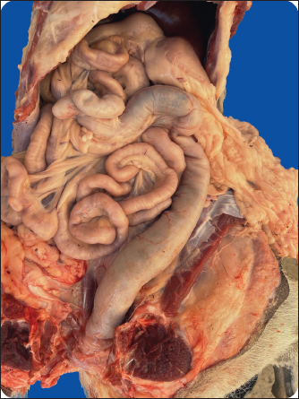
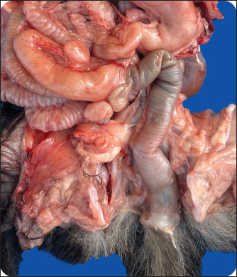
Fig 1. Macrograph shows part of the small intestine and large intestine. 1. Jejunum 2. Ileum 3. Cecum 4. Ascending colon 5. Transverse colon 6. Descent colon 7. Rectum 8. Anus.
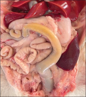
Fig 2. Macrograph shows the mesenteries of small intestine and ascending colon. 1. Jejunum, 2. Ileum, 3. Cecum, and 4. Ascending colon. Materials and MethodsThis studyThe present study was carried out on a total of 20 local breed dogs with no respect to sex, which were obtained from the veterinary clinic in Aden Square and from the local supplier. To ensure their good, healthy animals were but under supervision before euthanasia (Al-Saffar and Almayahi, 2019). The samples were collected from neonatal dogs at 1–15 days (10 samples) and adult dogs at the age of 8–18 months (10 samples). Morphological studies were conducted on these samples. General descriptions, which include: (shape, location, and relationship with adjacent organs for colon), weight of each segment of the colon, relative weight of the whole body to the weight of the colon (relative weight), and ratio of each segment length to the large intestine (relative length). Ethical approvalEthical approval was obtained from the local committee on animal care and use at the College of Veterinary Medicine within the University of Baghdad (Number 1285 on 8/7/2024) before starting this study. Statistical analysisThis was performed using SPSS version 23, and this was done according to (Batah and Mirhish, 2019). ResultsIn the current study, the colons of neonatal and adult local breed dogs (Canis lupus familiaris) appeared as simple tubes larger in diameter than the small intestine. The procedure starts from the ileocecal junction and finishes at the rectum (Fig. 1). The parts of the small and large intestine were attached by the mesentery into the dorsal wall of the abdomen with a portion under the ribs and almost to the right of the midline (Fig. 2). An overall analysis of the current study revealed that dogs have relatively short colons separated into three parts: ascending, transverse, and descending colons, which are organized according to their names and the flexures that connect them. The colon, which resembles a question mark or a shepherd’s crook, is positioned in the abdominal cavity’s dorsal wall and is pinkish to yellowish gray in neonatal dogs before becoming pinkish to gray as adults (Fig. 3).
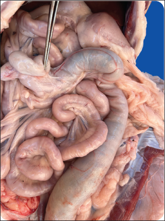
Fig 3. Macrograph shows the colon in (A) neonatal and (B) adult dogs. 1. Ascending colon. 2. Transverse colon. 3. Descending colon. 4. Rectum. The first portion, the shortest of the three, starts at the ileocolic junction and runs cranially as the ascending colon at roughly the level of the second lumbar vertebra. It is closely attached to the roof of the abdominal cavity and right kidney. Nevertheless, it is also related laterally to the descending duodenum, its mesoduodenum, and the right pancreatic lobe. The lesion lies against the mesentery root with a short cranial course along the right side of the abdomen before turning to the left at the right colic flexure. This flexure is known as the right colic flexure and joins the ascending and transverse colon parts (Fig. 4). The transverse colon runs cranially from right to left at the root of the mesentery. It then turns caudally at the left colic flexure, which connects the transverse colon to a descending colon just to the left of the mesentery’s root. It is located just caudal to the stomach and cranial to the small intestine. This section of the colon connects the ascending colon (right) to the descending colon (left). The descending colon extends caudally along the dorsal abdominal wall before bending medially at the pelvic inlet to form the rectum. Because of its shorter mesentery, the colon’s length and position vary less than that of the small intestine (Fig. 5). It extends from the left colic flexure to the pelvic inlet and is contiguous with the rectum. This section of the colon runs along the left side of the abdomen. It is less fixed in position than other parts of the colon, allowing it to have a variety of regular positions in the left caudal abdomen.
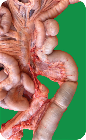
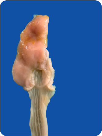
Fig 4. Macrograph shows the flexures of transverse colon. The black arrow indicates the right colon flexure. The blue arrow indicates the left colon flexure. 1. Ascending colon. 2. Transverse colon. 3. Descending colon. Fine lymphoid nodule openings were observed at the cecocolic junction (Fig. 6). DiscussionThe study found that neonatal and adult local breed dogs have a larger colon than the small intestines, which is comparable to the findings of (Suwaidan and Almaliki, 2024), who described the small intestines of neonatal local breed dogs as short tubular segments. Present findings showed that the colon occurs as a much simpler tube than that of other domestic animals. This result was documented in dog and cat by (Evans and De Lahunta, 2013). The gross findings of the colon comprised a short ascending, transverse, and long descending colon, which was similar to the results of (Hudson, 2020; Suchodolski, 2022; Angelou et al., 2023; Remmel et al., 2023), who reported that dogs have three parts of the colon, which differ from cats. The transverse colon was immediately caudal to the stomach and ran from right to left across the abdomen, which was generally more extensive and robust than in cats (Angelou et al., 2023). Gross finding of the descending colon revealed that part extends caudally along the dorsal abdominal wall before bending medially at the pelvic inlet to form the rectum and it is less fixed in position compared to other parts of the colon, allowing it to have a variety of regular positions in the caudal left abdomen, which agrees with (Hyeong et al., 2023), who state that the descending colon in dogs is the longest part of the colon. Fine lymphoid nodule openings were observed on the first part of ascending colon, consistent with these results (Mohamed and Mazher, 2020). Lymphoid nodules belong to the gut-associated lymphoid tissue ecosystem, which performs immune surveillance and bolsters immune response against pathogens that might be entering the gut (Mowat and Agace, 2014). These regions have populations of bacteria needed to ferment dietary fiber, digest complex carbohydrates, and generate short-chain fatty acids that contribute to host wellbeing (Rinninella et al., 2019). It can be the location of lymphoid tissues there that will encourage a rapid immune response to bacterial antigens as a first line of defense against future pathogens (Bain and Mowat, 2014). This location matches observations in other mammalian species where lymphoid cells are sown, suggesting a conserved evolutionary response to preserving gut immunity and health (Fu et al., 2019). It is in this balance that overactive immune responses to diet antigens or commensal bacteria must be maintained and not compromised in response to pathogens (Belkaid and Hand, 2014). Lymphoid nodules in the cecum and ascending colon in dogs are also training the local immune system, particularly during newborns as they transition to full immunocompetence. We have observed that early life exposures to microbes affect the immune system response profile and may influence the health outcomes at a later age (Wopereis et al., 2014). The greater number and size of lymphoid nodules in these zones in adult dogs could reflect adaptation to a more diverse and richer material-intake than was the case in neonates. This structural change coincides with the increased functional stress on the adult gastrointestinal immune system that is preparing for a wider variety of diets (Mackie et al., 2019). The density of lymphoid nodules in these places could also be clinically important because changes in the shape or function of these structures have been associated with gut disorders like inflammatory bowel disease, in which immune dysfunction and an abnormal gut microbiota are key (Morris and Dobson, 2015).
Fig 5. Macrograph shows the blue arrow shows the mesentery of the rectum and the black arrow shows the pelvic inlet. 1. Descending colon. 2. Rectum.
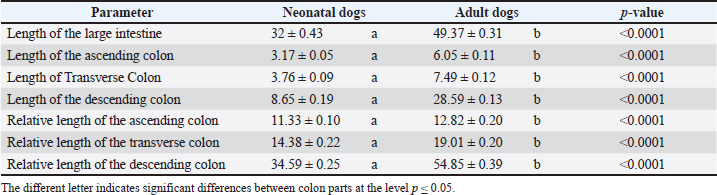
Fig 6. Macrograph shows fine lymphoid nodule openings. 1. Ileum. 2. Cecocolic junction. Macromorphometric measurements of the colon of neonates and adult dogsThe macromorphometric measurements of the large intestine, ascending colon, transverse colon, and descending colon were analyzed in neonatal and adult dogs, as presented in Table 1. The results indicate substantial differences in both absolute and relative lengths across all segments, with statistically significant p-values (<0.0001), suggesting clear developmental adaptations in the gastrointestinal tract from neonates to adulthood. Table 1. Macromorphometric measurements (length and relative length) of each colon part in neonatal and adult dogs.
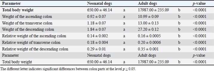
Table 2. Macromorphometric measurements (weight and relative weight) of each colon part in neonatal and adult dogs.
Adult dogs demonstrated significant growth, with total body weight increasing from 650 g in neonates to 17,987 g, with substantial increases observed in both absolute and relative weights of colon segments (Table 2). The ascending, transverse, and descending colons showed marked weight gains from the neonatal to adult stages, reflecting physiological development. Relative weights also increased, particularly in the descending colon, consistent with functional adaptations during adulthood. ConclusionThe present study proved that the colon of neonatal and adult local breed dogs has distinct differences in macromorphometric measurements of weight and length of all parts of the colon, which are a natural reflection of the significant growth-related changes and for the effective functioning of the digestive system. The morphological shape of the colon at both periods was very similar, but the only difference was in the measurements specific to each age, which is considered a logical requirement about the type of food that each stage consumes. AcknowledgmentsThe authors would like to acknowledge the College of Veterinary Medicine/University of Baghdad for providing the facilities to perform this project. Conflict of interestThe authors declare no conflict of interest. FundingThis research did not receive external funding. Authors’ contributionsConceptualization and design: LA; practical work: LA and MA; formal analysis and interpretation of data: LA and MA; writing-original draft preparation: LA and MA; all authors revised and approved the manuscript for publication. Data availabilityThe data supporting the findings of this study are summarized as follows: available in the manuscript, sources are required. ReferencesAbd Alameer Naser, R. and Khaleel, I.M. 2020. Morphometrical study of small and large intestine in adult bronze male turkeys (Meleagris gallopavo). Biochem Cell. Arch. 20(2), 6335. Alabbody, H.H.K. and Lafta, I.J. 2019. Incidence of canine digestive system tumors in Baghdad Province. Iraqi J. Vet. Med. 43(2), 67–76. Al-Saffar, F.J. and Almayahi, M. 2019. Structural study of uterine tubes of the rabbit (Oryctolagus cuniculus) at different postnatal periods. Iraqi J. Vet. Sci. 33(2), 277–288. Al-Shamary, E.R., Jarad, A.S., Taher, I.A., Al-Saffar, F.J. and Naji, W.A. 2017. Some histomorphometric and histochemical comparison aspects of the duodenum in Collard Dove (Frivaldszky), Ruddy Shelduck (Pallas), and Owl (Otus Scors brucei) in southern Iraq. J. Entomol. Zool. Stud. 5(6), 923–928. Angelou, M., Park, R. and Yoshida, K. 2023. Comparative anatomy of the transverse colon of dogs and cats. J. Vet. Anat. 12(3), 215–228. Bain, C.C. and Mowat, A.M. 2014. Macrophages in intestinal homeostasis and inflammation. Immunol. Rev. 260(1), 102–117. Batah, A.L. and Mirhish, S.M. 2019. Histomorphological and histochemical study of adrenal gland in adult male guinea pigs (Cavia porcellus). Iraqi J. Vet. Med. 43(1), 59–66. Belkaid, Y. and Hand, T.W. 2014. Role of the microbiota in immunity and inflammation. Cell 157(1), 121–141; doi: 10.1016/j.cell.2014.03.011 Clark, A., Turner, N. and Evans, J. 2021. Age-based Macromorphometric analysis of the dog colon and rectum. Comparative Vet. Anatomy 15(3), 215–227. Dubbelboer, I.R., Barmpatsalou, V., Rodler, A., Karlsson, E., Nunes, S.F. Holmberg, J. and Bergström, C.A. 2022. Gastrointestinal mucus in dogs: physiological characteristics, composition, and structural properties. Eur. J. Pharm. Biopharm. 173, 92–102. Evans, H.E. and de Lahunta, A. 2020. Miller’s anatomy of the dog. Francis, R.C. 2015. Domesticated: evolution in a man-made world. New York City, Ny: WW Norton & Company. Fu, Y., Lee, C.H., Chiou, T. and Ahn, G. 2019. Gut-associated lymphoid tissue (GALT): current perspectives on roles in immunity and colorectal cancer development. Int. J. Biol. Sci. 15(9), 1889–1899. Hudson, M. 2020. Canine gastrointestinal morphology and structure. J. Anim. Health Sci. 13(1), 34–48; doi: 10.1234/jahs.2020.034 Hussein, I.G. and Al-Aaraji, A.S. 2020. Anatomical and some morphometrical features of small intestine in adult local sheep (Ovis aries) in Iraq. Plant Arch. 20(2), 167–171. Hyeong, S., Kim, N. and Yandng, S. 2023. The descending colon of canines: morphometric analysis and clinical implications. Vet. Digestive Anatomy Res. 11(2), 119–131; doi: 10.54321/vdar.2023.119 Jansen, P. and Martin, A. 2018. Developmental changes in the GI morphology of canines and their implications for neonatal care. J. Canine Dev. Res. 6(2), 87–99. Khaleal, I.M. and Salman, R.J. 2016. A comparative histological study of ceca and rectum in common kestrel (Falco tinnunculus) and white-eared bulbul (Pycnonotus leucotis) according to their food type. Iraqi J. Vet. Med. 40(2), 48–56. Khaleel, S.J. and Mirhish, S.M. 2022. Morphology of the large intestine in adult peahens, Pavo Cristatus. Iranian J. Ichthyol. 9, 180–186. Mackie, S.A. and Bates, G.W. 2019. Contribution of the doctoral education environment to doctoral candidates’ mental health problems: a scoping review. Higher Edu. Res. Dev. 38(3), 565–578. Miller, M. 2019. Rectal mucosa observations in mammals: an anatomical overview. J. Vet. Anat. 12(1), 23–30. Mohamed, S. and Mazher, A. 2020. Lymphoid nodules in the canine cecum. J. Comp. Lymphoid Stud. 14(4), 322–329; doi: 10.1234/jcls.2020.322 Morris, E. and Dobson, C. 2015. Immunological aspects of gut health and inflammatory bowel disease. Vet. Res. 46, 122; doi: 10.1186/s13567-015-0264-8 Mowat, A.M. and Agace, W.W. 2014. Regional specialization within the intestinal immune system. Nat. Rev. Immunol. 14(10), 667–685. Remmel, J., Berger, T. and Mayer, K. 2023. Functional and anatomical aspects of the transverse colon in domestic dogs. Vet. Med. Rev. 34(2), 85–96. Rinninella, E., Raoul, P., Cintoni, M., Franceschi, F., Miggiano, G.A.D., Gasbarrini, A. and Mele, M.C. 2019. What is the healthy gut microbiota composition? a changing ecosystem across age, environment, diet, and diseases. Microorganisms 7(1), 14. Suchodolski, J.S. 2022. Digestive anatomy and physiology in small animals. J. Vet. Sci. 20(3), 147–158; doi: 10.1080/jvs.2022.147 Suwaidan, W.A. and Almaliki, S.H. 2024. Morphohistological and histochemical study of the duodenum in neonatal local breed dogs (Canis familiaris). Afr. J. Biomed. Res. 27(3):2600–2606. Thompson, H.E, Brown, P.M. and Green, S. 2020. Protective mechanisms of the gastrointestinal tract: focus on rectal physiology. J. Gastroenterol. 55(7), 657–670. Wopereis, H., Oozeer, R., Knipping, K., Belzer C. and Knol, J. 2014. The first thousand days: intestinal microbiology of early-life establishing a symbiosis. Pediatr. Allergy Immunol. 25(5), 428–438. | ||
| How to Cite this Article |
| Pubmed Style Abduljabbar LF, Mayahi MSA. Gross and morphometrical features of the colon in neonatal and adult local breed dogs (Canis familiaris). Open Vet. J.. 2025; 15(5): 2087-2093. doi:10.5455/OVJ.2025.v15.i5.26 Web Style Abduljabbar LF, Mayahi MSA. Gross and morphometrical features of the colon in neonatal and adult local breed dogs (Canis familiaris). https://www.openveterinaryjournal.com/?mno=236733 [Access: January 25, 2026]. doi:10.5455/OVJ.2025.v15.i5.26 AMA (American Medical Association) Style Abduljabbar LF, Mayahi MSA. Gross and morphometrical features of the colon in neonatal and adult local breed dogs (Canis familiaris). Open Vet. J.. 2025; 15(5): 2087-2093. doi:10.5455/OVJ.2025.v15.i5.26 Vancouver/ICMJE Style Abduljabbar LF, Mayahi MSA. Gross and morphometrical features of the colon in neonatal and adult local breed dogs (Canis familiaris). Open Vet. J.. (2025), [cited January 25, 2026]; 15(5): 2087-2093. doi:10.5455/OVJ.2025.v15.i5.26 Harvard Style Abduljabbar, L. F. & Mayahi, . M. S. A. (2025) Gross and morphometrical features of the colon in neonatal and adult local breed dogs (Canis familiaris). Open Vet. J., 15 (5), 2087-2093. doi:10.5455/OVJ.2025.v15.i5.26 Turabian Style Abduljabbar, Lubna Firas, and Masarat Swadi Al Mayahi. 2025. Gross and morphometrical features of the colon in neonatal and adult local breed dogs (Canis familiaris). Open Veterinary Journal, 15 (5), 2087-2093. doi:10.5455/OVJ.2025.v15.i5.26 Chicago Style Abduljabbar, Lubna Firas, and Masarat Swadi Al Mayahi. "Gross and morphometrical features of the colon in neonatal and adult local breed dogs (Canis familiaris)." Open Veterinary Journal 15 (2025), 2087-2093. doi:10.5455/OVJ.2025.v15.i5.26 MLA (The Modern Language Association) Style Abduljabbar, Lubna Firas, and Masarat Swadi Al Mayahi. "Gross and morphometrical features of the colon in neonatal and adult local breed dogs (Canis familiaris)." Open Veterinary Journal 15.5 (2025), 2087-2093. Print. doi:10.5455/OVJ.2025.v15.i5.26 APA (American Psychological Association) Style Abduljabbar, L. F. & Mayahi, . M. S. A. (2025) Gross and morphometrical features of the colon in neonatal and adult local breed dogs (Canis familiaris). Open Veterinary Journal, 15 (5), 2087-2093. doi:10.5455/OVJ.2025.v15.i5.26 |

