


| Research Article | ||
Open Vet. J.. 2025; 15(8): 3631-3637 Open Veterinary Journal, (2025), Vol. 15(8): 3631-3637 Research Article Histomorphological and ultrastructural determination of the adrenal gland in swan geese (Anser cygnoides)Khalid Ibrahim Abd Alkhazraji1, Salah Hasan Almaliki2, Rabab Abdulameer Naser3, Ali Ibrahim Ali Al-Ezzy 4* and Fatimah Swadi Zghair 51Department of Anatomy, College of Medicine, University of Diyala, Baqubah, Iraq 2Department of Anatomy and Histology, College of Veterinary Medicine, University of Baghdad, Baghdad, Iraq 3Department of Anatomy and Histology, College of Veterinary Medicine, University of Diyala, Baqubah, Iraq 4Department of Pathology, College of Veterinary Medicine, University of Diyala, Baqubah, Iraq 5Department of Anatomy and Histology, College of Veterinary Medicine, University of AL-Qadisiyah, Al-Diwaniyah, Iraq. *Corresponding Author: Ali Ibrahim Ali Al-Ezzy, College of Veterinary Medicine, University of Diyala, Baqubah, Iraq. Email: alizziibrahim [at] gmail.com Submitted: 11/11/2024 Revised: 20/06/2025 Accepted: 03/07/2025 Published: 31/08/2025 © 2025 Open Veterinary Journal
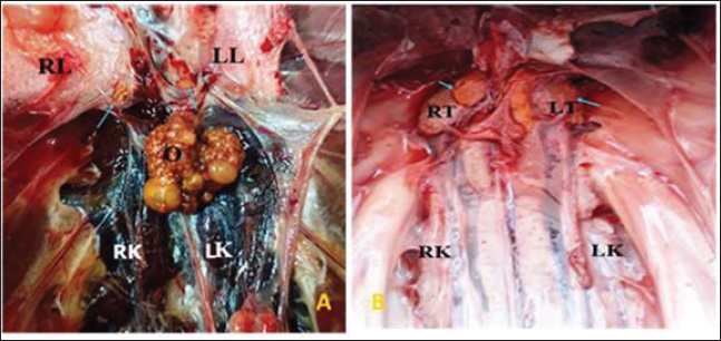
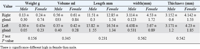
ABSTRACTBackground: The adrenal gland of the avian is considered a critical organ in the body because its hormones coordinate and control many pivotal physiological actions. Aim: The current research aimed to describe the ultrastructural, macroscopical, and histological characteristics of the adrenal gland in swan geese using electron and light microscopy, as well as perform a gross investigation. Methods: Twelve healthy swans were purchased from a bird market in Baghdad. After the swans were euthanized, the adrenal glands were immediately examined morphologically and histologically using standard histological techniques. Results: Morphological findings clarified that the adrenals were a yellowish structure, found close to the gonads, and were cranial to medial for each kidney. A significant variation in length and weight was observed between the two glands. High significance difference in female vs. male microscopically, they were covered with a thin capsule containing blood capillaries scattered between their fibers. In addition to several blood sinusoids, two intermingled types of tissue constituted the gland parenchyma: the medullary and cortical tissues. The layer is subdivided into the central zone, peripheral zone, and subcapsular layer. Small islets of chromaffin cells representing medullary tissue were distributed throughout the cortical portion. Ultrastructurally, the cortical cells showed numerous globules, curved cords, and blebs. Conclusion: The adrenal structure in swans exhibits that the amount of cortical tissue is higher than that in the medullary gland, indicating the physiological need for cortical tissue secretions since they are migratory birds. Keywords: Cords, Electron microscopy, Swan, Medulla. IntroductionSince ancient times, geese have been part of the history of multiple countries in the world, and they constitute one of the oldest domesticated birds (Ali and Zghair, 2022). In northeast China, central and western Mongolia are the sites of the breeding and distribution of the swan goose (Zhang et al., 2011). In all animals, the adrenal glands are indispensable structures that play a necessary role in various kinds of stress responses, and they help to preserve homeostasis. The glands work by liberating various hormones that are required in different aspects of daily life (Gjuen and ensen, 2022). The adrenal glands of birds liberate catecholamine hormones from the medullary portion, and the mineralocorticoids and glucocorticoids from the cortical part. Bird lives were threatened by experimental elimination of adrenals, and ultimately, death resulted (Fathima and Lucy, 2014). The left and right adrenals in aves are often yellowish small size. The environment, health, age, breed, and species are considered important factors affecting the anatomical measures of adrenals in birds (Humayun et al., 2012). The boundaries between the medulla and cortex in birds are unclear; thus, the tissues of the two parts of the gland are intermixed in most bird species. Ghosh et al., 2001). Three layers comprise the chicken adrenal gland (Humayun et al., 2012). Previous ultrastructural studies have indicated that Egyptian geese contain two types of cells in the adrenal gland, which are differentiated depending on the number of lipid droplets and mitochondria, in addition to containing secretory granules (Kot et al., 2023). The medulla of the adrenal glands of local domestic pigeons is composed of two types of secretory cells, which differ in origin: chromaffin cells and sympathetic ganglion cells (Sadoon, 2018). During ultrastructure scanning of the adrenal glands in Egyptian chickens by using electron microscopic observed that the cortex was enveloped by the capsule, which has different types of fibers, was observed (Moawad and Randa, 2017). The adrenal glands have also been examined microscopically and grossly in a number of mammals, such as hamsters and squirrels (Kadhim and Khalil, 2022) and in dogs (Alkhazarji et al., 2023) in order to identify the histological structures of these important glands in those animals. Due to the scarcity of data about the structures of adrenal glands in swan geese, this experiment aimed to provide a description of the macroscopical and microscopical structures of this gland in swan geese with the assistance of ultrastructural, histological, and gross investigations, and those contribute to providing an understanding of the gland’s function to physiologists. Materials and MethodsBirdsTwelve healthy geese, weighing 2.8–3.6 kg, of both sexes were studied in the current experiment. The birds were purchased from a bird market in Baghdad and housed in an animal farm at Vet. Med. College/Diyala University. All swans used in the current study had a standard diet ad libitum and free access to water for 3 weeks prior to the procedure. Study designA total of 12 swan geese were divided into three equal groups: Group 1: Four swan geese were allocated to the anatomical examination of the adrenal glands. Group 2: Four swan geese were used to assess the histological composition of adrenals, which was accomplished using a light microscope. Group 3: Four swan geese were assigned for ultrastructure examination of adrenals by using scanning electron microscopy. All experimental swan geese in the current study were euthanized using an overdose of xylazine (10mg/Kg) and Ketamine (100 mg/Kg) (Hess, 2005). The adrenal glands, after sacrifice of the swan geese, were removed carefully from their abdomen and underwent anatomical, histological, and scanning electron microscopy examinations. Anatomical studyMacroscopic examination of the adrenal gland site and shape. Morphometric measurements, including weight, thickness, wide, and length, were measured by use of a sensitive scale and caliper (Alkhazarji et al., 2023). Histological studyTissue samples of the adrenal glands were immersed in a fixative solution containing 10% formalin for 24 hours (Alkhazraji et al., 2021). After fixation, dehydration of specimens was achieved using a series upgrading of alcohol from 50% to 100%. The dehydrated samples were immersed in a clearing agent of xylene. Paraffin blocks were prepared by infiltration of paraffin into cleared samples. By using a microtome, the blocks were cut in serial sections of a 5 µm thickness, which were mounted on a glass slide for routine staining (hematoxylin and eosin), and special stains included Masson’s Trichrom, PAS, and verrhove stain (Alkhazarji et al., 2021; Alshammary, 2021; Alkhazarji and Naser 2024; Alkhazraji et al., 2024). Scanning electron microscopyAdrenal samples for ultrastructure examination were placed in a solution of glutaraldehyde (4%) at 4 °C for 1 hour for fixation. Then rinsed 2–3 times in phosphate buffer at pH 7.4 and postfixed with 1% osmium teroxide. Next, the specimens were dehydrated using a series of graded alcohols. The samples were dried in air, mounted on aluminum stubs, and covered with gold. The samples were then examined and photographed using a scanning electron microscope (a JEOL/EO-JSM-6510) (Tawfiek and Mahmoud, 2021). Data analysisThe values are represented in (mean ± SE) (Hameed et al., 2024). SPSS program (version 24) was used to analyze the data using the t-test (Ahmad et al., 2019; Hameed et al., 2020). The significance level was (p < 0.05) (Hameed and Al-Ezzy, 2019). Ethical approvalThe current experiment received agreement from the Scientific Ethics Committee of the University of Diyala/College of Veterinary Medicine (approval no. VM325/2023-KR). ResultsAnatomical findingsThe left and right adrenals of swan geese were found close to the gonads, cranio-medially to the kidneys, and on the two sides of the midline (Fig. 1. A and B). The swan goose has pairs of adrenal glands situated in the cranial pole of the apical lobe kidney, caudal to the lung. In males, the gland lies against the testes (Fig. 1 B), and in females, the left ovary fully covers the surface of the left gland (Fig. 1A). Currently, the right gland is shaped like a triangle, while the left one is elongated. The glands appeared yellow in color (Fig. 1A). The morphometry of the adrenal gland in the present study revealed variations in structural measurements between the right and left glands. The morphometric data for each adrenal gland are shown in Table 1. Length and weight measures revealed a significant increase in the left side at the level of (p < 0.05). In addition, the width and thickness measurements showed nonsignificant differences between the left and right gland (in female and male) at the level (p < 0.05) (Table 1).
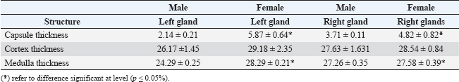
Fig. 1. photography illustrate the morphology of the adrenal gland in swan geese A: female, (B):male, RK,LK; right and left kidney, O (ovary), RT,LT; right and left tests, LL(left lung and the Adrenal gland (blue arrow). Table 1. Morphological measurement of the adrenal glands in female and male swan geese.
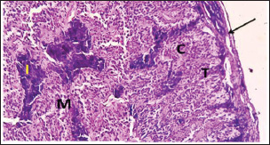
Histological findingsThe adrenal glands of swan geese are covered with thin capsules of connective tissue rich in blood capillaries. Thin and short trabecular ions were sent from the capsule to the interior of the gland (Fig. 2). There were two intermingled types of tissue constituting the gland parenchyma: the medullary and cortical tissues (Figs. 3 and 4). The capsule in females is thicker than in males (Table 2). During the histological examination, no boundaries between the cortex and medulla were observed. The adrenal glands were subdivided into the central zone, the peripheral zone, and the subcapsular layer. The main composition of the central and peripheral zones was cortical cells, whereas the subcapsular layer mostly contained medullary cells. Within the examinations, columnar acidophilic cells in the cortex portions were organized as cords that were cylindrically shaped and irregularly orientated. The cords of the central zone were smaller than those found in the peripheral zone and were mostly curved and straight in arrangement. The cells of cortical cords were small but more proportion to chromaffin medullary cells. The medullary portion of the adrenal gland in swan geese is represented by small islets of chromaffin cells that are distributed irregularly between the cords of the cortex. The chromaffin cells were large, basophilic, and polygonal. The current investigation showed that the cortical tissue was in greater proportion in the adrenal of swan geese than the medullary tissues. The blood sinusoid was scattered within the gland between the cortical or medullary tissues, but those located in the peripheral zone were narrower than those located in the central zone (Fig. 5). Scanning electron microscopy findingsThe adrenal glands of swan geese under the SEM examination show rough surfaces on their external surface (Fig. 6). The cortex cells showed on their surfaces numerous globules, curved cords, and blebs, which were observed as elevated wavy masses interwoven with medulla tissue (Fig. 7). The cells of the medullary portion in the current study were observed as membranous granular bodies. In addition, stellate-shaped cells with cytoplasmic projections were detected in this region (Figs. 8 and 9).
Fig. 2. photomicrograph section in adrenal of swan geese illustrated: the capsule (black arrow) sent trabeculae (T), cortical cords (C), medullary tissue (M), and islets of chromaffin cells (I). (H&E Stain) 100X)
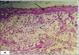
Fig. 3. photomicrograph section in the adrenal of swan geese illustrated: capsule (black arrow) that sent trabeculae (T), cortical cords (C) show mild reaction for PAS due to presence of neutral polysaccharides. (PAS Stain A&B:X 200) DiscussionAnatomical findingsIn the current study, the left and right adrenals of swan geese were found close to the gonads, cranio-medially to the kidneys, and on the two sides of the midline. This outcome was comparable to that stated in ducks (Fathima and Lucy, 2014) and Egyptian geese (El-Zoghby, 2010). However, (Zhang, 1988) reported the entire fusion of adrenals in the woodpecker. In the current study, the adrenal glands were covered with ovaries and testes, which are in accordance with those detected in Egyptian chicken (Moawad and Randa, 2017), but were not compatible with those mentioned in the African ostrich chick by Tang et al. (2009). These differences may be related to age or maturity status.
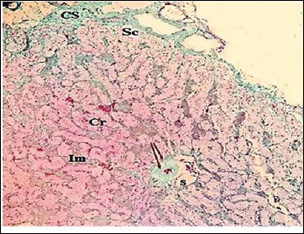
Fig. 4. photomicrograph section in adrenal of swan geese illustrated: the capsule (Cs), subcapsular layer (Sc), cortical cords (Cr), medullary tissue (Im), large sinus (S), and blood vessels (arrow). (Masson’s Trichrom Stain X 100) The right gland was shaped like a triangle, while the left one was elongated, which is not consistent with that noted in Egyptian chicks by Moawad and Randa (2017). Moreover, (Humayun et al., 2012) also observed the elongated shape in the left adrenal glands of chickens. The glands in our study appeared yellow, similar to the color observed in chicken by Moawad and Randa (2017). These results were not corresponded with those showing yellow–brown adrenal in domestic ducks (Prokopenko and Kot, 2021) and creamy to yellowish in pigeons and quails (El-Desoky and Mustafa, 2021; Erdem et al., 2021). In the current study, the morphometry of the adrenal gland revealed variations in structural measurements between the female and male right and left glands, as well as length and weight measures. However, no significant difference in the width and thickness measures showed non-significant differences between the left and right glands, which is in agreement with that reported in chicken by Humayun et al. (2012). Histological findingsThe adrenal glands of swan geese are covered with thin capsules of connective tissue rich in blood capillaries. Thin and short trabecular fibers were sent from the capsule to the interior of the gland and showed significance according to sex, which coincides with that detected in chicken (Moawad and Randa, 2017). There were two intermingled types of tissue constituting the gland parenchyma: the medullary and cortical tissues (Figs. 2 and 4). During the histological examination, no boundaries between the cortex and the medulla were observed, which is in agreement with that observed by El-Zoghby (2010) in geese. These results did not correspond with those observed in the adrenal gland in mammals (Alkhazarji et al., 2023), who reported that the adrenal gland in dogs consists of two clear parts: inner medulla and outer cortex. Table 2. Microscopic data of the adrenal gland measurements in swan goose (Mean ± SE, in µm).
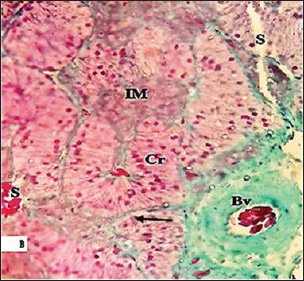
Fig. 5. photomicrograph section in adrenal of swan geese show: cortical cord (Cr), medullary tissue (IM), blood sinusoid (S), and blood vessel (Bv). Masson’s Trichrom Stain X 200
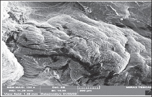
Fig. 6. Scanning electron photomicrography of adrenal swan geese: indicating that the superior part of the gland had a rough surface. The adrenals were subdivided into the central zone, the peripheral zone, and the subcapsular layer. These findings supported the results detected in quail (El-Desoky and Mustafa, 2021). The main composition of the central and peripheral zones was cortical cells, whereas the subcapsular layer mostly contained medullary cells. Within the examinations, columnar acidophilic cells in the cortex portions were organized as cords that were cylindrically shaped and irregularly orientated. The cords of the central zone were smaller than those found in the peripheral zone and were mostly curved and straight in arrangement. The cells of cortical cords were small but more proportionate than chromaffin medullary cells, which are in accordance with that reported in the pigeon (Erdem et al., 2021) and ducks by Al-Jebori et al. (2016) supporting the current findings.
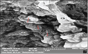
Fig. 7. Scanning electron photomicrography of adrenals swan geese show: the Globules, curved cords (AC), and blebs on the cortical cells surfaces (Arrow), observing as elevated wavy masses interwoven with tissue of the medulla (Me).
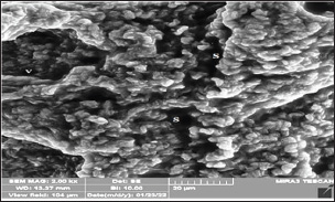
Fig. 8. Scanning electron photomicrography of adrenal swan geese show: the Medullary tissue contains numerous veins (V) and sinuses (S).
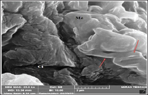
Fig. 9. Scanning electron photomicrography of adrenal swan geese show: the Stellate cells (arrow), medullary cell(Me), and capillary (Ci). The medullary portion of the adrenal gland in swan geese is represented by small islets of chromaffin cells that are distributed irregularly between the cords of the cortex. The chromaffin cells were large, basophilic, and polygonal. The current investigation showed that the cortical tissue was in greater proportion in the adrenal of swan geese than the medullary tissues. These findings are consistent with those reported for poultry (Humayun et al., 2012). The blood sinusoid was scattered within the gland either between the cortical or medullary tissues, but those located in the peripheral zone were narrower than those positioned in the central zone, which is in line with that mentioned by Moawad and Randa (2017) in chickens. Scanning electron microscopy findingsThe adrenal glands of swan geese, under the SEM examination, show rough surfaces on their external surface. The cells of the cortex showed on their surfaces numerous globules, curved cords, and blebs, which were observed as elevated wavy masses interwoven with tissue of the medulla, in accordance with that reported in common snip, parakeet, kingfisher, and woodpecker (Ghosh et al., 2001) and correspond with observations of Prabhavathi et al. (2011) in guinea fowl. The cells of the medullary portion in the current study were observed as membranous granular bodies. In addition, stellate-shaped cells with cytoplasmic projections were detected in this region, which appear to be similar to those reported in parakeets (Ghosh et al., 2001) and guinea fowls (Prabhavathi et al., 2011). ConclusionThe current study established important lines in the ultrastructural, anatomical, and microscopical description of the adrenal glands in swans, and exhibited that the amount of cortical tissue is higher than that of the medullary gland, indicating the physiological need for its secretions, as they are migratory birds. AcknowledgmentWe express our deep gratitude to Professors Ismail Al-Majjamii and Ali Ibrahim for their advice and help. FundingThe manuscript was funded by the authors, with no funding agencies. Authors’ contributionsThe authors equally contributed to the planning, practical work, data collection and analysis, and writing of the draft and final manuscripts. Conflict of interestIn connection with the publication of this research, there is no competing interest to disclose. Data availabilityAll data are available in the main text of the current manuscript. ReferencesAhmad, W.M.A., Shafiq, M., Halim, N.A. and Aleng, N. 2019. Statistical analysis using SPSS Version 24. Pulau Pinang: Penerbit Universiti Sains Malaysia. Ali, H. and Zghair, F. 2022. Morphological study of the trachea in a swan goose (Anser cygnoides). Basrah J. Vet. Res. 21(4), 36–45; doi: 10.23975/bjvetr.2022.176612 Al-Jebori, J.G.A., Hossain, A.O., Al-tamimi, Z.S.M. and Al-Jebori, A.K.A. 2016. Histomorphological study of adrenal gland in local adult female duck (Anasplatyhynchos). Bas. J. Vet. Res. 15(2), 164–175. Alkhazarji, K.I.A. and Naser, R.A.A. 2024. Macroscopic and histomorphometry investigation of pancreas in adult local partridge (Francolinus francolinus). Iraqi J. Vet. Sci., 38(1), 207–214. Alkhazarji, K.I.A., Naser, R.A.A. and Zghair, F.S. 2021. Histological changes in the kidney and liver associated with administration of ethylene glycol in domestic pigeons (Columba liviadomestica). TJAS 21(3), 106–111; doi: 10.25130/tjas.21.3.13 Alkhazarji, K.I.A., Zghair, F.S. and Naser, R.A.A. 2023. Macro and micromorphometric study of the adrenal glands in adult Male local dogs. AIP Conf. Proc. 2475, 1–7; doi: 10.1063/5.0102850 Alkhazraji, K.I.A., Naser, R.A. and Zghair, F. 2024. Evaluation of adrenal gland at pre and post maturation in domestic cats: a comparative histological study. TJAS, 24(2), 44–53; doi: 10.25130/tjas.24.2.4 Alshammary, H.K. 2021. Histomorphological study of the liver in Muscovy duck (Cairina moschata domestica). TASJ 21(3), 112–118; doi: 10.25130/tjas.21.3.14 El-Desoky, S.M.M. and Mustafa, F.E. 2021. Morphological and histological studies of the adrenal glands in the Japanese quail (Coturnix japonica). Microsc. Res. Tech. 84(10), 2361–2371; doi: 10.1002/jemt.23791 El-Zoghby, I.M. 2010. Light and electron microscope studies of the adrenal glands of the Egyptian Geese (Alopochen aegyptiacus). Lucr. Ştiinţ. Ser. Med. Vet. 53(12), 195–203. Erdem, E., Ozbaser, F., Gurcan, E. and Soysal, M. 2021. The morphological and morphometric characteristics of Alabadem pigeons. Turk. J. Vet. Anim. Sci. 45(2), 21. Fathima, K.M. and Lucy, K.M. 2014. Morphological studies on the adrenal gland of kuttanad ducks (Anas platyrhynchos domesticus) during post hatch period. J. Agric. Vet. Sci., 7(6), 58–62. doi: http://doi.org/10.9790/2380-07635862 Ghosh, A., Carmichael, S.W. and Mukherjee, M. 2001. Avian adrenal medulla: cytomrophology and function. Acta Biol. Szeged. 45, 1–11. Gjuen, J. and Ensen, P. 2022. Selection for reduced fear of humans changes intraspecific social behavior in red junglefowl-implications for chicken domestication. Genes 13(1), 43; doi: 10.3390/genes13010043 Hameed, M.S. and Al-Ezzy, A.I.A. 2019. Evaluation of possible stress factors affecting physiological level of gamma interferon during first six months of life in healthy calves. Adv. Anim. Vet. Sci. 7(5), 370–377. Hameed, M.S., Al-Ezzy, A.I.A., Jalil, W.I. and Al-Khalidi, A.A.H. 2020. Physiological protective effects of ascorbic acid versus Dl-A-Tocopheryl acetate–sodium selenite combination in mice under experimental sodium nitrate intoxication. Biochem. Cell. Arch. 20(1), 2593–2601. Hameed, M.S., Hasson, S.J., Mahmood, M.A. and Al-Ezzy, A.I.A. 2024. Physiological effect of multivitamins supplementation on hematological parameters, lipid profile, hepato-renal function of ross 308 broilers. Assiut Vet. Med. J. 70(183), 585–595. Hess, L. 2005. Euthanasia techniques in birds. J. Avian Med. Surg. 19(3), 242–245. Humayun, K., Aoyama, M. and Sugita, S. 2012. Morphological and histological studies of the adrenal gland of the chicken (Gallus domesticus). J. Poult. Sci. 49(1), 39–45. Kadhim, A.B. and Khalil, I.M. 2022. Comparative morphological and morphometrically study of the adrenal gland in adult male’s squirrel (Sciurus anomalous) and hamster (Mesocricetus auratus). Iraqi J. Vet. Sci. 36(3), 725–730; doi: 10.33899/IJVS/2022/131618.1983 Kot, T., Thachuk, S., Usenko, S. and Prokopenko, V. 2023. Adrenal gland of poultry: anatomy, microscopy, morphometry, and histochemistry. J. World Poult. Res. 13(1), 20–28. doi: http://doi.org/10.36380/jwpr.2023.2 Moawad, U.K. and Randa, M.H. 2017. Histocytological and histochemical features of the adrenal gland of adult Egyptian native breeds of chicken (Galllus gallus domesticated). Beni-Suef University. J. Basic Appl. Sci. 6(2), 199–208; doi: 10.1016/j.bjbas.2017.04.001 Prabhavathi, M., Basha, S. H., Venkatesan, S., Leela, V. and Ramesh, G. 2011. Electron microscopical study of adrenal gland in Guinea fowl. Indian J. Anim. Res., 45(3), 215–218. Prokopenko, V.S. and Kot, T.F. 2021. Macroscopic characteristic of the avian adrenal gland. Bull. Sumy NAU, Vet. Med. 55(4), 17–23. Sadoon, A.H. 2018. Morphological and histochemical study of adrenal gland in local domestic pigeons (Columba livia domestica) in Basrah province. Bas. J. Vet. Res. 17(1), 74–85; doi: 10.33762/bvetr.2018.143723 Tang, L., Peng, K.M., Wang, J.X., Luo, H.Q., Cheng, J.Y., Zhang, G.Y. and Song, H. 2009. Morphological study of the adrenal gland of African ostrich chicks. Tissue Cell 41, 231–238; doi: 10.1016/j.tice.2008.11.003 Tawfiek, M.G. and Mahmoud, H.H. 2021. Gross morphology and scanning electron microscopic structure of the oropharyngeal cavity of the domestic geese (anser anser domesticus). J. Vet. Med. Res. 27(2), 190–202; doi: 10.21608/JVMR.2021.61981.1034 Zhang, M.W. 1988. Comparative anatomy of vertebrate. Beijing: Higher Education Press Zhang, Y., Cao, L., Barter, M., Fox, A.D., Zhao, M.J., Meng, F.J. and Zhu, W.Z. 2011. Changes in the distribution and abundance of the swan goose Anser cygnoides in the Yangtze river floodplain: the likely loss of an important wintering site.. Bird Conserv. Int,, 21(1), 36–48; doi: 10.1017/S0959270910000201 | ||
| How to Cite this Article |
| Pubmed Style Alkhazraji KIA, Almaliki SH, Naser RA, Al-ezzy AIA, Zghair FS. Histomorphological and ultrastructural determination of the adrenal gland in swan geese (Anser cygnoides). Open Vet. J.. 2025; 15(8): 3631-3637. doi:10.5455/OVJ.2025.v15.i8.26 Web Style Alkhazraji KIA, Almaliki SH, Naser RA, Al-ezzy AIA, Zghair FS. Histomorphological and ultrastructural determination of the adrenal gland in swan geese (Anser cygnoides). https://www.openveterinaryjournal.com/?mno=228198 [Access: January 26, 2026]. doi:10.5455/OVJ.2025.v15.i8.26 AMA (American Medical Association) Style Alkhazraji KIA, Almaliki SH, Naser RA, Al-ezzy AIA, Zghair FS. Histomorphological and ultrastructural determination of the adrenal gland in swan geese (Anser cygnoides). Open Vet. J.. 2025; 15(8): 3631-3637. doi:10.5455/OVJ.2025.v15.i8.26 Vancouver/ICMJE Style Alkhazraji KIA, Almaliki SH, Naser RA, Al-ezzy AIA, Zghair FS. Histomorphological and ultrastructural determination of the adrenal gland in swan geese (Anser cygnoides). Open Vet. J.. (2025), [cited January 26, 2026]; 15(8): 3631-3637. doi:10.5455/OVJ.2025.v15.i8.26 Harvard Style Alkhazraji, K. I. A., Almaliki, . S. H., Naser, . R. A., Al-ezzy, . A. I. A. & Zghair, . F. S. (2025) Histomorphological and ultrastructural determination of the adrenal gland in swan geese (Anser cygnoides). Open Vet. J., 15 (8), 3631-3637. doi:10.5455/OVJ.2025.v15.i8.26 Turabian Style Alkhazraji, Khalid Ibrahim Abd, Salah Hasan Almaliki, Rabab Abdulameer Naser, Ali Ibrahim Ali Al-ezzy, and Fatimah Swadi Zghair. 2025. Histomorphological and ultrastructural determination of the adrenal gland in swan geese (Anser cygnoides). Open Veterinary Journal, 15 (8), 3631-3637. doi:10.5455/OVJ.2025.v15.i8.26 Chicago Style Alkhazraji, Khalid Ibrahim Abd, Salah Hasan Almaliki, Rabab Abdulameer Naser, Ali Ibrahim Ali Al-ezzy, and Fatimah Swadi Zghair. "Histomorphological and ultrastructural determination of the adrenal gland in swan geese (Anser cygnoides)." Open Veterinary Journal 15 (2025), 3631-3637. doi:10.5455/OVJ.2025.v15.i8.26 MLA (The Modern Language Association) Style Alkhazraji, Khalid Ibrahim Abd, Salah Hasan Almaliki, Rabab Abdulameer Naser, Ali Ibrahim Ali Al-ezzy, and Fatimah Swadi Zghair. "Histomorphological and ultrastructural determination of the adrenal gland in swan geese (Anser cygnoides)." Open Veterinary Journal 15.8 (2025), 3631-3637. Print. doi:10.5455/OVJ.2025.v15.i8.26 APA (American Psychological Association) Style Alkhazraji, K. I. A., Almaliki, . S. H., Naser, . R. A., Al-ezzy, . A. I. A. & Zghair, . F. S. (2025) Histomorphological and ultrastructural determination of the adrenal gland in swan geese (Anser cygnoides). Open Veterinary Journal, 15 (8), 3631-3637. doi:10.5455/OVJ.2025.v15.i8.26 |

