


| Case Report | ||
Open Vet J. 2022; 12(6): 859-863 Open Veterinary Journal, (2022), Vol. 12(6): 859–863 Case Report Generalized tympanism in a horse and its possible association with Sarcina-like microorganism: A case reportLaura Gabriela Bass1*, Paula Peña-Álvarez2, Juan D. Hidalgo-Dittel2, Fernando D. Robles-Herrera2, Paula M. Cappella-Flores3, Diego S. Zúñiga-Cortés2 and Roberto W.I. Olivares21Cátedra de Microbiología e Inmunología, Escuela de Medicina y Cirugía Veterinaria San Francisco de Asís, Universidad Veritas, San José, Costa Rica 2Servicio de Patología Diagnóstica LAPAVET-ESFA, Cátedra de Patología e Histología, Escuela de Medicina y Cirugía Veterinaria San Francisco de Asís, Universidad Veritas, San José, Costa Rica 3Cátedra de Clínica de Especies Mayores, Escuela de Medicina y Cirugía Veterinaria San Francisco de Asís, Universidad Veritas, San José, Costa Rica Submitted: 23/06/2022 Accepted: 17/10/2022 Published: 17/11/2022 *Corresponding Author: Laura Gabriela Bass. Cátedra de Microbiología e Inmunología, Escuela de Medicina y Cirugía Veterinaria San Francisco de Asís, Universidad Veritas, San José, Costa Rica. Email: lbass [at] veterinariaveritas.ac.cr © 2022 Open Veterinary Journal
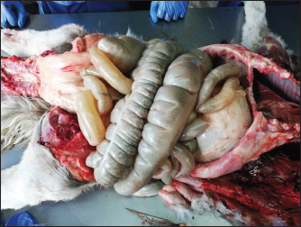
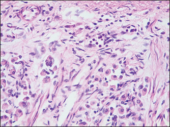
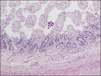
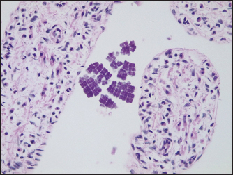
AbstractBackground: Sarcina spp. is a Gram-positive, coccoid microorganism that forms tetrads or octets, and is observed with a characteristic "bundle" arrangement. The most recognized species are Sarcina ventriculi and Sarcina maxima. It has been described as part of the normal microbiota in horses and cats, but it has also been linked to abomasal bloat in goats, lambs, and calves, although its causality has not been proven yet. Case Description: This work presents the case of a 3-months-old female horse that died of generalized tympanism. Macroscopic findings showed mild cyanosis and abundant gas in the lumen of the stomach, and small and large bowel. Microscopically, high numbers of Gram-positive microorganisms compatible with Sarcina spp. in the gastric lumen and on the surface of the small and large bowel were observed, along with mild inflammation. Conclusion: The severe tympanism was the only relevant lesion observed and could explain the death of the animal. Although it is not possible to determine a relationship between these lesions and Sarcina spp., it is interesting to highlight that the high amount of these bacteria could be associated with gas production and tympanism. It is important to continue investigating the role of Sarcina spp. in horses, and its possible link with tympanism. Keywords: Horses, Sarcina-like, Tympanism. IntroductionSarcina spp. is a Gram-positive, coccoid microorganism, generally found in tetrads or octets, and its arrangement has a typical appearance in packets or bundles. It was first described in 1842 by John Goodsir (Goodsir and Wilson, 1842). These bacteria are immotile, anaerobic, or aerotolerant. They have a fermentative metabolism, are spore-forming, and have the ability to grow at an extremely acidic pH (less than 1). The most recognized species within this genus are Sarcina ventriculi and Sarcina maxima (Canale-Parola, 1970). It has been related to cases of abomasal bloat and gastritis in some animal species such as goats, lambs, and calves (DeBey et al., 1996; Edwards et al., 2008), although causality has not been confirmed yet. Likewise, the presence of this agent has been identified in dogs, humans, cats, and horses. In the latter, it has been found in a peritoneal cavity aspirate after stomach rupture (Mocsy, 1954), but its pathogenic role is unknown since it has also been found as part of the normal microbiota (Perkins et al., 2012; Costa et al., 2015). The aim of this work is to describe a case of generalized tympanism of the digestive tract in a horse from Costa Rica, and its possible association with Sarcina-like microorganisms. Case DetailsA 3-months-old female American miniature horse was taken for a necropsy to the Veterinary Pathology Laboratory of the San Francisco de Asis School of Veterinary Medicine and Surgery (San José, Costa Rica). The animal was found dead sometime after being fed and presented no clinical signs prior to death. No changes in the diet were reported, and after its death, a necropsy was performed. At necropsy, a mild degree of autolysis was observed. Macroscopically the apparent mucous membranes were pale, except for the ocular conjunctiva, which presented cyanosis. The lungs were enlarged with the impression of the ribs on the surface. Both lungs presented a dark coloration with depressed areas. Also, a slight amount of fluid was observed in the pericardium, as well as reddish spots on the inner face of both ventricles. On the other hand, severe dilatation was found throughout the gastrointestinal tract, with abundant gas in the lumen of the stomach, and small and large bowel (Fig. 1). The intestinal contents had a liquid consistency, except for the jejunum and ileum, where the consistency was mucous. The color of the liquid was yellowish-green. The contents of the rectum also had a liquid consistency. Samples of all organs were taken for histopathology and fixed in 10% formalin. The fixed samples were processed using routine histopathological techniques to prepare paraffin-embedded blocks. Sections (4 μm thick) were cut and stained with hematoxylin and eosin and Gram stains. In the endocardium, as well as in the associated myocardium, multiple severe hemorrhages were observed. The blood vessels of the lungs were dilated and filled with erythrocytes. In some areas, there was thickening of the interstitium, with mild to moderate numbers of lymphocytes, while the BALT was slightly hyperplastic. In some sectors, edema was observed in both alveoli and bronchioles, with the presence of some areas of atelectasis. In the stomach, a moderate dilatation of the submucosal lymphatic vessels was observed, with the presence of a multifocal inflammatory infiltrate composed of lymphocytes, plasmacytes, and some neutrophils (Fig. 2). In the gastric lumen, basophilic structures of approximately 2,1-3 µm in individual diameter forming bundles were observed, with morphology suggestive of Sarcina spp. The size was obtained using Image J software by measuring 20 individual bacteria. In the small intestine, the villi were slightly atrophied. In the mucosa, some parasitic structures compatible with Strongyloides spp. were observed. In one sector, on the intestinal surface, basophilic structures forming bundles compatible with Sarcina spp. were observed (Figs. 3 and 4). The lamina propria as well as the submucosa showed edema with an inflammatory infiltrate with a moderate amount of lymphocytes and plasmacytes, and to lesser extent neutrophils, macrophages, and eosinophils, as well as a marked hyperplasia of the lymphoid tissue. Finally, a moderate to severe eosinophilic and lymphocytic infiltrate was observed in the lamina propria of the large intestine. The lymphoid tissue of the submucosa showed severe hyperplasia, with a large number of apoptotic bodies. In one sector, on the surface of the organ, basophilic structures forming bundles were observed, compatible with Sarcina spp. When Gram staining was performed, these microorganisms were observed as Gram-positive bacteria (Fig. 5). In the liver, there were some small inflammatory foci composed of a few neutrophils and some lymphocytes and macrophages. No alterations in the nervous plexuses of the gastrointestinal tract or other relevant histological alterations were observed in the rest of the organs examined. Finally, it was concluded that the death was produced as a consequence of a multiorganic failure associated with a severe tympanism of the digestive tract.
Fig. 1. Intestinal tract: severe dilatation caused by accumulation of gas. DiscussionSarcina spp. is a bacterial genus whose morphology is coccoid, Gram-positive, approximately 1.8–3 µm in individual diameter, these are generally found forming tetrads or octets, adopting the characteristic arrangement of "packets". They are usually flattened in the areas of contact with adjacent cells, and their division occurs in three perpendicular planes. Their ability to form spores has been reported, they are immotile, anaerobic, or aerotolerant. Their metabolism is fermentative, and they use carbohydrates as substrate. They can also grow in environments with extremely acidic pH, close to 1 (Garrity et al., 2006). Sarcina ventriculi cells are surrounded by a fibrous layer of cellulose that is typically 150–200 nm thick. This material functions as an intercellular cement that holds the cells together forming the characteristic bundles (Canale-Parola, 1970).
Fig. 2. Stomach: mild inflammation in the lamina propria with presence of neutrophils, lymphocytes, and plasmacytes (H&E, 400×).
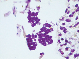
Fig. 3. Small intestine: presence of coccoid structures forming bundles compatible with Sarcina spp (H&E, 100×).
Fig. 4. Small intestine: presence of coccoid structures forming bundles compatible with Sarcina spp (H&E, 400×). In humans, the growth of S. ventriculi in the stomach has been associated as a result of some pathological conditions that delay gastric emptying, such as pyloric ulcers or stenosis (Canale-Parola, 1970; Lam-Himlin et al., 2011; Propst et al., 2020). In this context, the acid pH of the stomach and the presence of carbohydrates and other nutrients favor the growth of S. ventriculi. There are several reports that relate these bacteria to gastric pathologies in humans, although their pathogenic role is not entirely clear (Dumitru et al., 2020). Some authors suggest that Sarcina spp. could be an innocent bystander rather than a pathogenic organism (Ratuapli et al., 2013). Sarcina spp., commonly found in soil, is ingested with soil particles present in food. Sarcina ventriculi have been isolated from several places such as the surface of cereal seeds, soil, stomach contents of diseased humans, elephant, and human feces, and rabbits and guinea pigs’ stomach contents. On the other hand, S. maxima has been isolated from fresh wheat bran, horse manure, soil, and the outer coat of cereals such as oats, rice, rye, and wheat (Garrity et al., 2006).
Fig. 5. Detail of the bundles formed by Gram-positive coccoid microorganisms (Gram, 1,000×). This microorganism has been associated with cases of bloat in 3 to 6 weeks-old lambs and in 10 days-old calves. Macroscopic lesions included edema and emphysema of the abomasal wall, hemorrhage, and mucosal hyperemia. Microscopically, bacteria with morphology compatible with Sarcina spp. were observed (Edwards et al., 2008). These authors mention that there is an evident association between abomasal bloat and Sarcina spp. in young animals of these species since the presence of fermentable carbohydrates and the tolerance of this microorganism to low pH allow its overgrowth. Vatn et al. (2000a) evaluated 67 lambs from 2 to 5 weeks of age with an abomasal disease, which presented, in comparison with controls, abomasal bloat, hemorrhages, and ulcers. In 79% of the cases, Sarcina-like bacteria were found in sections and smears from the abomasum. Sarcina ventriculi was isolated and identified from the abomasal content of one of these animals. Sarcina-like bacteria were found in 93% of lambs with abomasal bloat, suggesting an association between gas accumulation in the abomasum and the presence of Sarcina-like bacteria. In regard to horses, the role of Sarcina spp. in abdominal bloat is still debatable, as it has been found to be part of the normal microbiota (Perkins et al., 2012; Costa et al., 2015). Vatn et al. (2000b) mention in their work that they have found higher numbers of Sarcina-like bacteria in horses with acute gastric distention compared to controls. These authors emphasize that it is not known if the different sizes of the packets of these bacteria are related to different metabolic levels, or if they are different species. They also emphasize that, although these bacteria have been identified as part of the normal microbiota in this species, it has not been found in canines, lambs, or goats, therefore this could suggest certain differences between animal species (Vatn et al., 2000b). On the other hand, this microorganism has also been related to a case of gastric dilatation, vomiting, and chronic enteritis in a cat (Im et al., 2017). Although its presence is described as part of the normal small intestinal microflora in this species, therefore its potential pathogenic effect is also unknown (Ritchie et al., 2008). Owens et al. (2021) mention the finding of macroscopic and microscopic lesions in chimpanzees from Sierra Leone affected with "Epizootic neurologic and gastroenteric syndrome", which are congruent with the most severe forms of Sarcina infection in animals and humans. It should be noted that this group has studied the agent found in these animals, and they describe some differences between this isolate and others previously described, therefore they conclude that the genus Sarcina could comprise a wide variety of species ranging from commensal to pathogenic microorganisms. It is important to mention that Sarcina spp. is one of the few microorganisms that can be identified by their morphology, using light microscopy and classic Hematoxylin/Eosin staining. But it should be clarified that Micrococcus spp. can also occur in tetrads or packets, therefore, it is necessary to distinguish both microorganisms by some of their differences such as size (Micrococcus spp. is smaller than Sarcina spp.), and the tendency of Micrococcus spp. to form more compact clusters (Al Rasheed and Senseng, 2016). In this case, the size and characteristics of the clusters are more suggestive of Sarcina spp. In any case, it would be important to appeal to appropriate culture methodologies in order to isolate these agents. Both macroscopic and microscopic findings found in the case of this work evidence certain similarities with those described in the previously mentioned works, in which these bacteria are linked to rupture and abomasal bloat, as well as in human cases. It should be noted that no cardiac lesions were observed that could explain the sudden death of this animal, and the inflammatory lesions observed in the liver and lungs were mild and nonspecific, so they would not have had a relevant role in the death of the animal. The parasitic lesions were also mild and probably were not related to the death of the animal, since this horse did not show diarrhea or weight loss. The only relevant change observed was the severe digestive tympanism that could explain the death by producing respiratory failure. Many of the hemorrhagic and edematous alterations observed may be agonic (King et al., 2007). Considering that this microorganism is fastidious, and its isolation is usually difficult, it would be interesting to perform molecular techniques in order to identify these bacteria and to be able to link its presence with both macroscopic and microscopic findings. In view of the scarce information related to this microorganism, its presence as part of the normal microbiota and the differences between species, its possible role as a causal agent of abomasal bloat in goats, lambs, and calves, and the findings mentioned in this work, more studies are needed to relate its possible pathogenic role in other domestic species such as horses and cats, and if the increase in the amount of Sarcina spp. could be related to the presence of tympanism in horses. AcknowledgmentsThe authors would like to thank Escuela de Medicina y Cirugía Veterinaria San Francisco de Asís for their support. Conflict of interestThe authors declare that there is no conflict of interest. Authors contributionsLB: conceptualization, data collection, image capturing and editing, image analysis, and writing of the article. RO and PP: macroscopic and histopathological diagnosis. JH and FR: macroscopic diagnosis, reading, and revision of the manuscript. PC: clinical examination and diagnosis. DZ: histopathological techniques and stains. Critical revision of the manuscript was performed by all the authors. All authors read and approved the final manuscript. ReferencesAl Rasheed, M.R. and Senseng, C.G. 2016. Sarcina ventriculi: review of the literature. Arch. Pathol. Lab. Med. 140(12), 1441–1445. Canale-Parola, E. 1970. Biology of the sugar-fermenting Sarcinae. Bacteriol. Rev. 34(1), 82–97. Costa, M.C., Silva, G., Ramos, R.V., Staempfli, H.R., Arroyo, L.G., Kim, P. and Weese, J.S. 2015. Characterization and comparison of the bacterial microbiota in different gastrointestinal tract compartments in horses. Vet. J. 205(1), 74–80. DeBey, B.M., Blanchard, P.C. and Durfee, P.T. 1996. Abomasal bloat associated with Sarcina-like bacteria in goat kids. J. Am. Vet. Med. Assoc. 209(8), 1468–1469. Dumitru, A., Aliuş, C., Nica, A.E., Antoniac, I., Gheorghiță, D. and Grădinaru, S. 2020. Fatal outcome of gastric perforation due to infection with Sarcina spp. Case Rep. 19, e00711. Edwards, G.T., Woodger, N.G., Barlow, A.M., Bell, S.J., Harwood, D.G., Otter, A. and Wight, A. R. 2008. Sarcina-like bacteria associated with bloat in young lambs and calves. Vet. Rec. 163(13), 391–393. Garrity, G., Staley, J.T., Boone, D.R., De Vos, P., Goodfellow, M. and Rainey, F.A. 2006. Bergey’s manual of systematic bacteriology. New york, NY: Springer Goodsir, J. and Wilson, G. 1842. History of a case in which a fluid periodically ejected from the stomach contained vegetable organisms of an undescribed form. Edinb. Med. Surg. J. 57(151), 430–443. Im, J.Y., Sokol, S. and Duhamel, G.E. 2017. Gastric dilatation associated with gastric colonization with Sarcina-like bacteria in a cat with chronic enteritis. J. Am. Anim. Hosp. Assoc. 53(6), 321–325. King, J.M., Roth-Johnson, L. and Newson, M.E. 2007. The necropsy book. Charles Louis Davis, DVM Foundation, Ithaca, New York, USA. Lam-Himlin, D., Tsiatis, A.C., Montgomery, E., Pai, R.K., Brown, J.A., Razavi, M., Lamps, L., Eshleman, J.R., Bhagavan, B. and Anders, R.A. 2011. Sarcina organisms in the gastrointestinal tract: a clinicopathologic and molecular study. Am. J. Surg. Pathol. 35(11), 1700–1705. Mocsy, J. 1954. Krankheiten des Magens und des Darmes. (Diseases of the stomach and intestines). In Spezielle Pathologie und Therapie der Haustiere, 10th ed. Eds., Hutyra Marek. Jena, Germany: VEB Gustav Fisher Verlag, pp: 91. Owens, L.A., Colitti, B., Hirji, I., Pizarro, A., Jaffe, J.E., Moittié, S., Bishop-Lilly, K.A., Estrella, L.A., Voegtly, L.J., Kuhn, J.H., Suen, G., Deblois, C.L., Dunn, C.D., Juan-Sallés, C. and Goldberg, T.L. 2021. A Sarcina bacterium linked to lethal disease in sanctuary chimpanzees in Sierra Leone. Nat. Commun. 12(1), 763. Perkins, G.A., den Bakker, H.C., Burton, A.J., Erb, H.N., McDonough, S.P., McDonough, P.L., Parker, J., Rosenthal, R.L., Wiedmann, M., Dowd, S.E. and Simpson, K.W. 2012. Equine stomachs harbor an abundant and diverse mucosal microbiota. Appl. Environ. Microbiol. 78(8), 2522–2532. Propst, R., Denham, L., Deisch, J.K., Kalra, T., Zaheer, S., Silva, K. and Magaki, S. 2020. Sarcina organisms in the upper gastrointestinal tract: a report of 3 cases with varying presentations. Int. J. Surg. Pathol. 28(2), 206–209. Ratuapli, S.K., Lam-Himlin, D.M. and Heigh, R.I. 2013. Sarcina ventriculi of the stomach: a case report. World. J. Gastroenterol. 19(14), 2282–2285. Ritchie, L.E., Steiner, J.M. and Suchodolski, J.S. 2008. Assessment of microbial diversity along the feline intestinal tract using 16S rRNA gene analysis. FEMS. Microbiol. Ecol. 66(3), 590–598. Vatn, S., Tranulis, M.A. and Hofshagen, M. 2000a. Sarcina -like bacteria, Clostridium fallax and Clostridium sordellii in lambs with abomasal bloat, haemorrhage and ulcers. J. Comp. Pathol. 122(2–3), 193–200. Vatn, S., Gunnes, G., Nybø, K. and Juul, H.M. 2000b. Possible involvement of Sarcina ventriculi in canine and equine acute gastric dilatation. Acta. Vet. Scand. 41(3), 333–337. | ||
| How to Cite this Article |
| Pubmed Style Bass LG, Peña-Álvarez P, Hidalgo-dittel JD, Robles-herrera FD, Cappella-flores PM, Zúñiga-cortés DS, Olivares RWI. Generalized tympanism in a horse and its possible association with Sarcina-like microorganism: A case report. Open Vet J. 2022; 12(6): 859-863. doi:10.5455/OVJ.2022.v12.i6.11 Web Style Bass LG, Peña-Álvarez P, Hidalgo-dittel JD, Robles-herrera FD, Cappella-flores PM, Zúñiga-cortés DS, Olivares RWI. Generalized tympanism in a horse and its possible association with Sarcina-like microorganism: A case report. https://www.openveterinaryjournal.com/?mno=67439 [Access: July 10, 2025]. doi:10.5455/OVJ.2022.v12.i6.11 AMA (American Medical Association) Style Bass LG, Peña-Álvarez P, Hidalgo-dittel JD, Robles-herrera FD, Cappella-flores PM, Zúñiga-cortés DS, Olivares RWI. Generalized tympanism in a horse and its possible association with Sarcina-like microorganism: A case report. Open Vet J. 2022; 12(6): 859-863. doi:10.5455/OVJ.2022.v12.i6.11 Vancouver/ICMJE Style Bass LG, Peña-Álvarez P, Hidalgo-dittel JD, Robles-herrera FD, Cappella-flores PM, Zúñiga-cortés DS, Olivares RWI. Generalized tympanism in a horse and its possible association with Sarcina-like microorganism: A case report. Open Vet J. (2022), [cited July 10, 2025]; 12(6): 859-863. doi:10.5455/OVJ.2022.v12.i6.11 Harvard Style Bass, L. G., Peña-Álvarez, . P., Hidalgo-dittel, . J. D., Robles-herrera, . F. D., Cappella-flores, . P. M., Zúñiga-cortés, . D. S. & Olivares, . R. W. I. (2022) Generalized tympanism in a horse and its possible association with Sarcina-like microorganism: A case report. Open Vet J, 12 (6), 859-863. doi:10.5455/OVJ.2022.v12.i6.11 Turabian Style Bass, Laura Gabriela, Paula Peña-Álvarez, Juan Diego Hidalgo-dittel, Fernando Daniel Robles-herrera, Paula María Cappella-flores, Diego Saúl Zúñiga-cortés, and Roberto Walter Israel Olivares. 2022. Generalized tympanism in a horse and its possible association with Sarcina-like microorganism: A case report. Open Veterinary Journal, 12 (6), 859-863. doi:10.5455/OVJ.2022.v12.i6.11 Chicago Style Bass, Laura Gabriela, Paula Peña-Álvarez, Juan Diego Hidalgo-dittel, Fernando Daniel Robles-herrera, Paula María Cappella-flores, Diego Saúl Zúñiga-cortés, and Roberto Walter Israel Olivares. "Generalized tympanism in a horse and its possible association with Sarcina-like microorganism: A case report." Open Veterinary Journal 12 (2022), 859-863. doi:10.5455/OVJ.2022.v12.i6.11 MLA (The Modern Language Association) Style Bass, Laura Gabriela, Paula Peña-Álvarez, Juan Diego Hidalgo-dittel, Fernando Daniel Robles-herrera, Paula María Cappella-flores, Diego Saúl Zúñiga-cortés, and Roberto Walter Israel Olivares. "Generalized tympanism in a horse and its possible association with Sarcina-like microorganism: A case report." Open Veterinary Journal 12.6 (2022), 859-863. Print. doi:10.5455/OVJ.2022.v12.i6.11 APA (American Psychological Association) Style Bass, L. G., Peña-Álvarez, . P., Hidalgo-dittel, . J. D., Robles-herrera, . F. D., Cappella-flores, . P. M., Zúñiga-cortés, . D. S. & Olivares, . R. W. I. (2022) Generalized tympanism in a horse and its possible association with Sarcina-like microorganism: A case report. Open Veterinary Journal, 12 (6), 859-863. doi:10.5455/OVJ.2022.v12.i6.11 |

