


| Case Report | ||
Open Vet J. 2022; 12(6): 851-854 Open Veterinary Journal, (2022), Vol. 12(6): 851–854 Case Report Superficial digital flexor tendon luxation in a Golden RetrieverMitsuhiro Isaka*, Taichi Okayama, Minori Yamaguchi and Wataru KonnoDepartment of Small Animal Clinical Sciences, Laboratory of Companion Animal Surgery,School of Veterinary Medicine, Rakuno Gakuen University, Hokkaido, Japan Submitted: 20/06/2022 Accepted: 11/10/2022 Published: 15/11/2022 *Corresponding Author: Mitsuhiro Isaka. Department of Small Animal Clinical Sciences, Laboratory of Companion Animal Surgery,School of Veterinary Medicine, Rakuno Gakuen University, Hokkaido, Japan. Email: m-isaka [at] rakuno.ac.jp © 2022 Open Veterinary Journal
AbstractBackground: Luxation of the superficial digital flexor (SDF) tendon involves the dislocation of the tendon from the groove of the calcaneal tuberosity. Although it is common in Shetland sheepdogs and collies presenting with acute, severe, and non-weight-bearing lameness, it is a rare orthopedic disease in Golden Retrievers. Case Description: A 13-month-old neutered male Golden Retriever (32.2 kg) with acute right hind lameness and pain, was diagnosed with SDF luxation based on physiological, radiographic, and ultrasound examinations. The surgical procedure used was capsulorrhaphy of the tarsal joint and fixation with Kirschner wire. Postoperatively, the tarsal joint was stretched and externally fixed to prevent tension. The patient showed good gait without pain after pin removal on postoperative day 88. Conclusion: In clinical practice, the possibility of this condition should be considered in Golden Retrievers presenting with lameness. Keywords: Golden Retriever, Superficial digital flexor, Surgical procedures, Postoperative complications, Calcaneus. IntroductionThe superficial digital flexor (SDF) tendon extends distal to the calcaneus, gliding over a bursa and the calcaneal tuber, as it is supported by the retinaculum on either side. Traumatic injury resulting in rupture of the retinaculum, with medial or lateral displacement of the tendon, has been reported in dogs and a single cat (Mauterer et al., 1993; Reinke and Mughannam, 1993; Hoscheit, 1994; McNicholas et al., 2000). A hereditary basis has been established for luxation in Shetland sheepdogs, and luxation is almost always lateral and caused by varying degrees of flattening of the lateral aspect of the calcaneal tuber (Bernard, 1977; Solanti et al., 2002; Jury, 2021). Depending on the inciting cause, clinical signs can include acute or chronic intermittent pelvic limb lameness and swelling around the calcaneal tuber (Mauterer et al., 1993; McNicholas et al., 2000; Solanti et al., 2002). In some cases, the tendon can be luxated and reduced manually, with the tarsus held in extension (Mauterer et al., 1993; McNicholas et al., 2000). Non-surgical management is ineffective in this condition (Mauterer et al., 1993; McNicholas et al., 2000; Solanti et al., 2002). Prognosis is good to excellent following surgical treatment directed at repairing the torn retinaculum, the imbrication of the redundant retinaculum, and, in some cases, deepening the groove between the medial and lateral processes of the calcaneal tuber; such correction requires postoperative external fixation for 4 to 8 weeks (Mauterer et al., 1993; McNicholas et al., 2000; Solanti et al., 2002; Jury, 2021). There are few reports in the literature on the dislocation of the flexor tendon of the shallow digit. Here, we report the luxation of the SDF tendon in a Golden Retriever. Case DetailsA 13-month-old neutered male Golden Retriever weighing 32.2 kg presented at our hospital with crepitus and swelling of the right tarsal joint. The dog had injured his right hind leg in a dog run in June 2021 and, since then, had adopted a stooped posture, with frequent rests while walking. Swelling of the caudal side of the right calcaneus and mobility of the Achilles tendon attachment were observed at the veterinary hospital at the Rakuno Gakuen University Animal Medical Center a month later. On examination at our hospital, a Monroe walk was observed, with a Grade I lameness score of the right hind limb. On orthopedic examination, swelling, and warmth of the right tarsal joint were observed, as well as crepitus and pain during extension and flexion. Lateral mobility of the Achilles tendon attachment was observed when the affected limb was flexed. Radiographic examination showed increased soft tissue opacity around the caudal aspect of the right calcaneus (Fig. 1). Ultrasonography revealed a hyperechoic structure and fluid retention around the right Achilles tendon (Fig. 2). Based on these observations, we diagnosed lateral luxation of the SDF. Presurgical medication consisted of continuous infusion of butorphanol (0.2 mg/kg intravenously), medetomidine (2 μg/kg intravenously), and lidocaine (1 mg/kg intravenously). In addition, robenacoxib (2 mg/kg) was administered subcutaneously for analgesia. Ropivacaine (3 mg/kg) was administered for the sciatic nerve block. Intravenous propofol (6 mg/kg) was administered for the induction of anesthesia, and anesthesia was maintained with oxygen-sevoflurane. The overall general anesthesia time was 91 minutes, and the operation time was 68 minutes. The collected joint fluid was serous, and postoperative Diff-Quick staining showed a large number of macrophages, suggesting chronic inflammation (Fig. 3A). Medial capsulorrhaphy of the tarsal joint, arthrodesis, in which the shallow digital flexor tendon was sutured with 2–0 polydioxanone, and fixation with a 1.5-mm Kirschner wire were performed (Figs. 3B and 4A). Postoperatively, the tarsal joint was stretched to prevent tension on the tendon. A cast made of Castrolite Alpha® (ALCARE Corporation, Tokyo, Japan) was applied without the cranial aspect; and the Robert Jones bandage technique was used for stabilization (Fig. 4B). Postoperative medication consisted of intravenous cefazolin (20 mg/kg), subcutaneous sodium carbazochrome sulfonate (0.5 mg/kg), and tranexamic acid (10 mg/kg) twice daily for 3 days. Cefazolin (20 mg/kg twice daily) was also administered until suture removal. The Robert Jones bandage was removed on postoperative day 28. Finally, we removed the pin on postoperative day 88 under general anesthesia using the same methods as previously described. The patient walked well without pain and lameness 12 days after the removal of the pin.
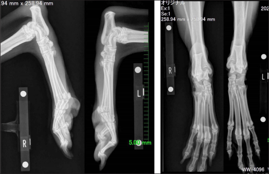
Fig. 1. Radiographs showing increased soft tissue opacity around the caudal aspect of the right calcaneus.
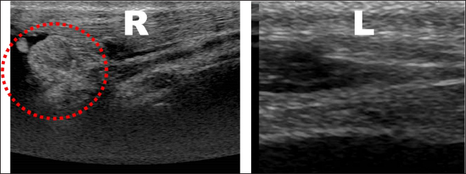
Fig. 2. Ultrasonography showing a bumpy hyperechoic structure and fluid retention around the right Achilles tendon. DiscussionIn this case, the cast was removed on the 18th postoperative day, and the fixation was changed to that using aluminum collaterals and the Robert Jones bandage technique. The purpose of this gradual loosening of the fixation was to encourage muscle use and acclimatization. Bandaging also prevents skin complications caused by licking of the surgical site by the dog on account of the discomfort of surgical pins. In previous reports, bandages were applied without the use of cast or aluminum collaterals, which were not intended to support the SDF tendon. In fact, as reported by Jury (2021), re-dislocations occurred in 3 of 23 cases (13%), requiring additional surgery (Jury, 2021). However, an earlier study reported a case, in which an Orthoplast (Johnson & Johnson Limited, Montreal, Quebec, Canada) was used and no re-dislocation occurred (McNicholas et al., 2000). Houlton and Dyce (1993) reported that polypropylene mesh could be used to correct a failed repair of the SDF tendon in three dogs (Houlton and Dyce, 1993). Therefore, while postoperative external fixation is effective, it is necessary to inform not only veterinarians but also animal owners of the need for careful postoperative management because this fixation may lead to complications, other than those related to dislocation. Although this study reports a case of SDF luxation in a Golden Retriever, previous studies have reported similar findings in Shetland sheepdogs, collies, and terriers (Solanti et al. 2002; Jury, 2021). In Shetland sheepdogs, an autosomal recessive form of inheritance of the defect that results in the flattening of the calcaneal ridge has been shown. The incidence of the disease among breeds is still unknown and needs further investigation in Golden Retrievers; however, in clinical practice, it is important to consider that the disease may occur not only in breeds that are prone to the disease—such as the collie breeds discussed in previous papers—but also in other breeds. In conclusion, surgical repair of dislocations of the flexor digitorum brevis tendon is expected to restore the tendon to its pre-injury level in many cases and the use of the wire insertion technique is very effective. However, there exist only a few reports on this subject in the literature, and detailed information is still lacking. Further research is needed to determine the incidence of such luxation among breeds, the possibility of re-dislocation, and the genetic basis of this condition. In addition, small animal practitioners should be aware of this disease if the patient presents with rear lameness.
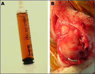
Fig. 3. (A): Gross finding of synovial fluid. (B): Medial capsulorrhaphy of the right tarsal joint and pin fixation for arthrodesis.
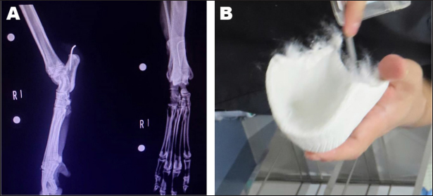
Fig. 4. (A): Postoperative radiograph of the subject. (B): The U-shaped castration. AcknowledgmentsWe would like to thank Editage (www.editage.com) for English language editing. Author contributionsDr. Isaka was the first surgeon; Dr. Okayama, Dr. Yamaguchi, and Dr. Konno were assistants for surgery. All authors wrote, revised, and agreed on the final version of the manuscript. FundingThis research did not receive any specific grant from funding agencies in the public, commercial, or not-for-profit sectors. Data availability statementThe data presented in this study are available on request from the corresponding author. Conflict of interestThe authors declare that there is no conflict of interest. ReferencesBernard, M.A. 1977. Superficial digital flexor tendon injury in the dog. Can. Vet. J. 18, 105–107. Hoscheit, L.P. 1994. Luxation of the tendon of the superficial digital flexor muscle in two dogs. Can. Vet. J. 35, 120–121. Houlton, J.E.F. and Dyce, J. 1993. The use of polypropylene mesh for revision of failed repair of superficial digital flexor tendon in three dogs. Vet. Comp. Orthop. Traumatol. 6, 129–130. Jury, A. 2021. Surgical repair for luxation of the superficial digital flexor tendon using a temporary restraining pin. J. Small. Anim. Pract. 62, 272–278. Mauterer, J.V.Jr., Prata, R.G., Carberry, C.A. and Schrader, S.C. 1993. Displacement of the tendon of the superficial digital flexor muscle in dogs: 10 cases (1983-1991). J. Am. Vet. Med. Assoc. 203, 1162–1165. McNicholas, W.T.Jr., Wilkens, B.E. and Barstad, R.D. 2000. Luxation of the superficial digital flexor tendon in a cat. J. Am. Anim. Hosp. Assoc. 36, 174–176. Reinke, J.D. and Mughannam, A.J. 1994. Lateral luxation of the superficial digital flexor tendon in 12 dogs. J. Small. Anim. Pract. 29(4), 303–309. Solanti, S., Laitinen, O. and Atroshi, F. 2002. Hereditary and clinical characteristics of lateral luxation of the superficial digital flexor tendon in Shetland sheepdogs. Vet. Ther. 3, 97–103. | ||
| How to Cite this Article |
| Pubmed Style Isaka M, Okayama T, Yamaguchi M, Konno W. Superficial digital flexor tendon luxation in a golden retriever. Open Vet J. 2022; 12(6): 851-854. doi:10.5455/OVJ.2022.v12.i6.9 Web Style Isaka M, Okayama T, Yamaguchi M, Konno W. Superficial digital flexor tendon luxation in a golden retriever. https://www.openveterinaryjournal.com/?mno=63340 [Access: July 15, 2025]. doi:10.5455/OVJ.2022.v12.i6.9 AMA (American Medical Association) Style Isaka M, Okayama T, Yamaguchi M, Konno W. Superficial digital flexor tendon luxation in a golden retriever. Open Vet J. 2022; 12(6): 851-854. doi:10.5455/OVJ.2022.v12.i6.9 Vancouver/ICMJE Style Isaka M, Okayama T, Yamaguchi M, Konno W. Superficial digital flexor tendon luxation in a golden retriever. Open Vet J. (2022), [cited July 15, 2025]; 12(6): 851-854. doi:10.5455/OVJ.2022.v12.i6.9 Harvard Style Isaka, M., Okayama, . T., Yamaguchi, . M. & Konno, . W. (2022) Superficial digital flexor tendon luxation in a golden retriever. Open Vet J, 12 (6), 851-854. doi:10.5455/OVJ.2022.v12.i6.9 Turabian Style Isaka, Mitsuhhiro, Taichi Okayama, Minori Yamaguchi, and Wataru Konno. 2022. Superficial digital flexor tendon luxation in a golden retriever. Open Veterinary Journal, 12 (6), 851-854. doi:10.5455/OVJ.2022.v12.i6.9 Chicago Style Isaka, Mitsuhhiro, Taichi Okayama, Minori Yamaguchi, and Wataru Konno. "Superficial digital flexor tendon luxation in a golden retriever." Open Veterinary Journal 12 (2022), 851-854. doi:10.5455/OVJ.2022.v12.i6.9 MLA (The Modern Language Association) Style Isaka, Mitsuhhiro, Taichi Okayama, Minori Yamaguchi, and Wataru Konno. "Superficial digital flexor tendon luxation in a golden retriever." Open Veterinary Journal 12.6 (2022), 851-854. Print. doi:10.5455/OVJ.2022.v12.i6.9 APA (American Psychological Association) Style Isaka, M., Okayama, . T., Yamaguchi, . M. & Konno, . W. (2022) Superficial digital flexor tendon luxation in a golden retriever. Open Veterinary Journal, 12 (6), 851-854. doi:10.5455/OVJ.2022.v12.i6.9 |

