


| Case Report | ||
Open Vet J. 2022; 12(1): 1-4 Open Veterinary Journal, (2022), Vol. 12(1): 01–04 Case Report Modified medial patellofemoral ligament reconstruction for severe bilateral congenital patellar luxation in a dogMitsuhiro Isaka*, Daiki Kokubo and Hiroshi UenoDepartment of Small Animal Clinical Sciences, School of Veterinary Medicine, Rakuno Gakuen University, 582 Bunkyodai Midorimachi, Ebetsu, Hokkaido 069-8501, Japan *Corresponding Author: Mitsuhiro Isaka. Department of Small Animal Clinical Sciences, School of Veterinary Medicine, Rakuno Gakuen University, Ebetsu, Japan. Tel. & Fax: +81-11-388-4774. Email: m-isaka [at] rakuno.ac.jp Submitted: 04/08/2021 Accepted: 01/12/2021 Published: 01/01/2022 © 2022 Open Veterinary Journal
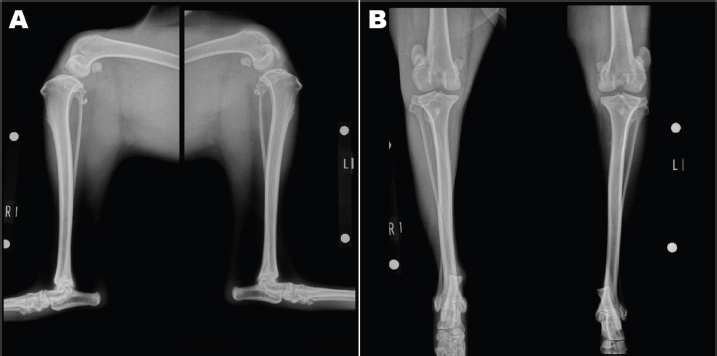
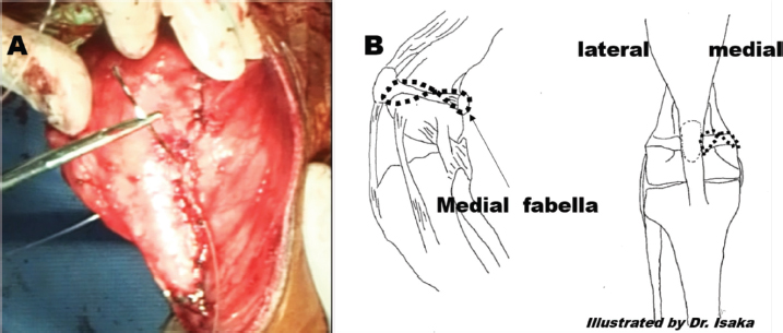
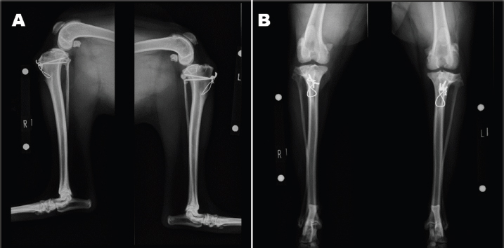
AbstractBackground: Lateral patellar luxation (LPL) is commonly diagnosed in large or giant breed dogs. In the surgical outcome for canine LPL, approximately half of the patients had complications, including reoperation and relaxation. Medial patellofemoral ligament (MPFL) reconstruction has been widely used for human repetitive patellar luxation. This case report describes that modified MPFL reconstruction with other surgical techniques might be effective for severe canine LPL repair. Case Description: An 11-month-old intact male Siberian Husky was referred to us with the main complication reported as bilateral hind lameness and LPL, diagnosed by a family doctor. Physiological examination showed bilateral patellar luxation (grade 4). We performed general surgical techniques with modified MPFL. The patient showed good prognosis. Conclusion: This report suggests that modified MPFL with general surgical techniques for LPL might be an effective surgical technique in dogs. Keywords: Dog, Lateral patellar luxation, Modified medial patellofemoral ligament. IntroductionLateral patellar luxation (LPL) is commonly diagnosed in large or giant breed dogs (Hayes et al., 1994; Harasen, 2006). In one report, which retrospectively evaluated 65 dogs of all breeds with LPL, medium and large breed dogs were more affected, and less than 10% of all dogs were small breed dogs (Kalff et al., 2014). Although there are many surgical techniques, including lowering or medialization of the tibial tubercle, division of the lateral retinaculum, plication of the medial retinaculum, lowering of the vastus medialis, and trochleoplasty (Harasen, 2006; Schneider et al., 2016), the surgical success rate is very low in canine LPLs, especially in large breeds (Kalff et al., 2014, Shaver et al., 2014). The medial patellofemoral ligament (MPFL) is a ligament that runs from the femur (thigh bone) to the inside of the patella (plate) and is responsible for keeping the patella from slipping outward. It is the most important ligament in the medial support mechanism of the patella, playing more than 50% of its role (Chahla et al., 2019). Thus, injuries to the MPFL are caused when the patella is dislocated outward. Consequently, MPFL reconstruction has been widely used for repetitive patellar luxation in humans because this technique has a low incidence of recurrent instability, postoperative apprehension, and reoperations (Schneider et al., 2016; Aframian et al., 2017). Collectively, the surgical reconstruction of the MPFL might be effective for canine patellar luxation. However, there are no reports of MPFL reconstruction for canine LPL. This case report describes the use of modified MPFL reconstruction techniques with general surgical techniques in a large breed with bilateral congenital LPL. Case DetailsAn 11-month-old intact male Siberian Husky was referred to us with the main complication reported as bilateral hind lameness and LPL, diagnosed by a family doctor. His posture showed an X-linked knee. Physiological examination showed bilateral patellar luxation with grade 4 and negative for cranial drawer sign and tibial compression test. The radiograph showed severe bilateral LPL without changes in the hip joint (Figs. 1A-B). His quadriceps (Q) angle showed right=43.37°and left=32.15°. Diazepam 0.1 mg/kg, ketamine 0.5 mg/kg, and fentanyl 5 μg/kg (intravenous), atropine 0.05 mg/kg (intramuscular), and meloxicam 0.2 mg/kg (subcutaneous) were administered as the premedication. After an induction of propofol 6 mg/kg, ropivacaine 0.22 mg/kg was used for epidural anesthesia. Under general anesthesia, the patient was immobilized in the supine position. Surgical techniques included bilateral block trochleoplasty, tibial tuberosity with tension band wiring using Kirschner wire, modified capsules (release of the lateral capsule and suturing of vastus medialis, plication), and the modified MPFL. The modified MPFL was used with the Cruciate Needle (medium, 40 lb) 40# suture (monofilament nylon) single strand (SECUROS, MA), suturing from the lateral aspect of the patella periosteum to the medial sesamoid bone (Figs. 2A-B). The postoperative radiograph is shown in Figures 3A-B. The patient has shown no luxation over 1 year after the operation.
Fig. 1. Preoperative radiograph. Severe bilateral LPL was detected.
Fig. 2. Modified MPFL reconstruction. (A): Modified MPFL reconstruction in operation. (B): Illustration of the stifle joint with modified MPFL in anterior–posterior and lateral views. DiscussionRegarding surgical outcomes for canine LPLs, approximately half of the patients had complications, including reoperation and relaxation, which has 12.5 times the odds of relaxation in a single operation for bilateral surgical repair (Shaver et al., 2014). Moreover, in one retrospective study of canine LPL, the complication rate was 37.9% (22/58). In particular, the revision surgery was approximately 11.5% (Kalff et al., 2014), although using various surgical techniques. In general, anatomical and biomechanical studies have shown that MPFL is the primary stabilizer of the patella between full extension and 30° flexion in humans (Chouteau, 2016). Injuries to the MPFL are often associated with dislocation of the patella, and are caused as follows: dysplasia of femoral condyle, increase in Q angle, dysfunction of the vastus medialis muscle, and overstrained external tissues. If the muscles and ligaments on the outside of the patella are strained, and a force is applied to pull the patella outward, making it more likely to dislocate, leading to X-linked knee (Mizuno et al., 2001; Schneider et al., 2016; Aframian et al., 2017; Chahla et al., 2019). We did not detect dysplasia of the femoral condyle. Although the median Q angle was 8.7° (range: 2.7°–14.8°) in large breed dogs (Pinna and Romagnoli, 2017), the patient’s Q angle, in our case, was 43.37° on the right and 32.15° on the left, leading to X-linked knee. Thus, the application of MPFL might be an adequate surgical technique for the patient. Although in our case, we did not apply the device of MPFL used in human medicine (Tucker et al., 2018), a 40-medium suture was employed. In this case, the patient did not show lameness, implant failure, pain, and re-luxation after surgery. This suture might be effective for modified MPFL reconstruction in canine LPLs. In conclusion, modified MPFL reconstruction might be considered for LPL repair if the standard techniques are ineffective. However, in the future, the authors will only need the MPFL technique for canine LPL repair in clinical cases.
Fig. 3. Postoperative radiograph. AcknowledgmentThe authors would like to thank Editage (www.editage.com) for English language editing. Conflict of interestThe authors declare that there is no conflict of interest. Authors’ contributionsDr. Isaka performed the surgery and wrote the article and the corresponding author, Dr. Daiki Kokubo, performed the surgery with Dr. Isaka. Dr. Uen revised the final article. ReferencesAframian, A., Smith, T.O., Tennent, T.D., Cobb, J.P. and Hing, C.B. 2017. Origin and insertion of the medial patellofemoral ligament: a systematic review of anatomy. Knee Surg. Sports Traumatol. Arthrosc. 25(12), 3755–3772. Chahla, J., Smigielski, R., LaParade, R.F. and Fulkerson, J.P. 2019. An updated overview of the anatomy and function of the proximal medial patellar restraints (medial patellofemoral ligament and the medial quadriceps tendon femoral ligament. Sports Med. Arthrosc. Rev. 27(4), 136–142. Chouteau, J. 2016. Surgical reconstruction of the medial patellofemoral ligament. Orthop. Traumatol. Surg. Res. 102(Suppl. 1), S189–S194. Harasen, G. 2006. Patellar luxation: pathogenesis and surgical correction. Can. Vet. J. 47(10), 1037–1039. Hayes, A.G., Boudrieau, R.J. and Hungerford, L.L. 1994. Frequency and distribution of medial and lateral patellar luxation in dogs: 124 cases (1982-1992). J. Am. Vet. Med. Assoc. 205(5), 716–720. Kalff, S., Butterworth, S.J., Miller, A., Keeley, B., Baines, S. and McKee, W.M. 2014. Lateral patellar luxation in dogs: a retrospective study of 65 dogs. Vet. Comp. Orthop. Traumatol. 27(2), 130–134. Mizuno, Y., Kugagai, M., Mattesshich, S.M., Elias, J.J., Cosgarea, A.J. and Chao, E.Y. 2001. Q-angle influences tibiofemoral and patellofemoral kinematics. J. Orthop. Res. 19(5), 834–840. Pinna, S. and Romagnoli, N. 2017. Radiographic measurement of the quadriceps angle in dogs. PLoS One 12(10), e0185833. Schneider, D.K., Grawe, B., Magnussen, R.A., Ceasar, A., Parikh, S.N., Wall, E.J., Colosimo, A.J., Kaeding, C.C. and Myer, G.D. 2016. Outcomes after isolated medial patellofemoral ligament reconstruction for the treatment of recurrent lateral patellar dislocations: a systematic review and meta-analysis. Am. J. Sports. Med. 44(11), 2993–3005. Shaver, S.L., Mayhew, K.N., Sutton, J.S., Mayhew, P.D., Runge, J.J., Brown, D.C. and Kass, P.H. 2014. Complications after corrective surgery for lateral patellar luxation in dogs: 36 cases (2000-2011). J. Am. Vet. Med. Assoc. 244(4), 444–448. Tucker, A., McMahon, S., McArdle, B., Rutherford, B. and Acton, D. 2018. Synthetic versus autologous reconstruction (Syn-VAR) of the medial patellofemoral ligament study protocol for a randomized controlled trial. Trials 19(1), 268. | ||
| How to Cite this Article |
| Pubmed Style Isaka M, Kokubo D, Ueno H. Modified medial patellofemoral ligament reconstruction for severe bilateral congenital patellar luxation in a dog. Open Vet J. 2022; 12(1): 1-4. doi:10.5455/OVJ.2022.v12.i1.1 Web Style Isaka M, Kokubo D, Ueno H. Modified medial patellofemoral ligament reconstruction for severe bilateral congenital patellar luxation in a dog. https://www.openveterinaryjournal.com/?mno=106253 [Access: July 11, 2025]. doi:10.5455/OVJ.2022.v12.i1.1 AMA (American Medical Association) Style Isaka M, Kokubo D, Ueno H. Modified medial patellofemoral ligament reconstruction for severe bilateral congenital patellar luxation in a dog. Open Vet J. 2022; 12(1): 1-4. doi:10.5455/OVJ.2022.v12.i1.1 Vancouver/ICMJE Style Isaka M, Kokubo D, Ueno H. Modified medial patellofemoral ligament reconstruction for severe bilateral congenital patellar luxation in a dog. Open Vet J. (2022), [cited July 11, 2025]; 12(1): 1-4. doi:10.5455/OVJ.2022.v12.i1.1 Harvard Style Isaka, M., Kokubo, . D. & Ueno, . H. (2022) Modified medial patellofemoral ligament reconstruction for severe bilateral congenital patellar luxation in a dog. Open Vet J, 12 (1), 1-4. doi:10.5455/OVJ.2022.v12.i1.1 Turabian Style Isaka, Mitsuhiro, Daiki Kokubo, and Hiroshi Ueno. 2022. Modified medial patellofemoral ligament reconstruction for severe bilateral congenital patellar luxation in a dog. Open Veterinary Journal, 12 (1), 1-4. doi:10.5455/OVJ.2022.v12.i1.1 Chicago Style Isaka, Mitsuhiro, Daiki Kokubo, and Hiroshi Ueno. "Modified medial patellofemoral ligament reconstruction for severe bilateral congenital patellar luxation in a dog." Open Veterinary Journal 12 (2022), 1-4. doi:10.5455/OVJ.2022.v12.i1.1 MLA (The Modern Language Association) Style Isaka, Mitsuhiro, Daiki Kokubo, and Hiroshi Ueno. "Modified medial patellofemoral ligament reconstruction for severe bilateral congenital patellar luxation in a dog." Open Veterinary Journal 12.1 (2022), 1-4. Print. doi:10.5455/OVJ.2022.v12.i1.1 APA (American Psychological Association) Style Isaka, M., Kokubo, . D. & Ueno, . H. (2022) Modified medial patellofemoral ligament reconstruction for severe bilateral congenital patellar luxation in a dog. Open Veterinary Journal, 12 (1), 1-4. doi:10.5455/OVJ.2022.v12.i1.1 |

