


| Case Report | ||
Open Vet. J.. 2025; 15(1): 471-477 Open Veterinary Journal, (2025), Vol. 15(1): 471-477 Case Report Rehabilitation of chronic pain and enhanced healing in feline femoral surgery: A case report on laser therapyDženita Hadžijunuzović Alagić , Kenan Baltić , Sabina Kolašinac and Nejra Hadžimusić*Department of Clinical Veterinary Sciences, Veterinary Faculty, University of Sarajevo, Sarajevo, Bosnia and Herzegovina *Corresponding Author: Nejra Hadžimusić. Department for Clinical Sciences, Veterinary Faculty, University of Sarajevo, Sarajevo, Bosnia and Herzegovina. Email: nejra.hadzimusic [at] vfs.unsa.ba Submitted: 23/10/2024 Accepted: 25/12/2024 Published: 31/01/2025 © 2025 Open Veterinary Journal
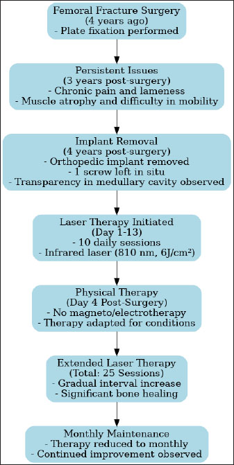
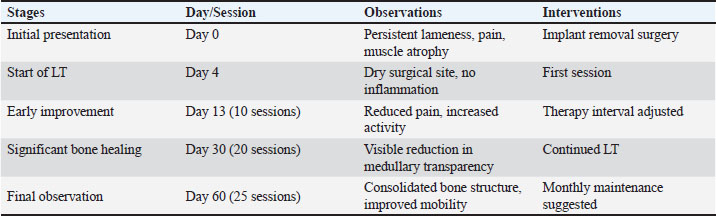
AbstractBackground: Chronic pain and delayed bone healing are significant challenges in veterinary rehabilitation following orthopedic surgeries in cats. These issues necessitate advanced modalities for effective management. Laser therapy (LT), also known as photobiomodulation, has shown promise in promoting tissue repair and alleviating pain but remains underused in feline cases. This case report aims to evaluate the efficacy of LT in managing chronic pain and enhancing bone healing in a cat following femoral surgery complicated by orthopedic implant failure. Case Description: A 7-year-old cat presented with persistent lameness and discomfort 3 years post-femoral fracture surgery. Despite initial surgical stabilization, chronic pain and restricted mobility persisted, leading to implant removal. After surgery, the cat underwent 25 sessions of LT using an 810-nm infrared semiconductor laser at a dosage of 6 J. Treatment sessions were conducted over 2 months, beginning 4 days post-surgery. Clinical and radiographic evaluations were performed to assess therapeutic outcomes. The cat demonstrated marked improvement in weight-bearing and mobility, with reduced signs of pain. Radiographic imaging revealed significant bone remodeling and consolidation. The owner reported enhanced activity levels and overall quality of life for the cat. These findings suggest that LT effectively facilitated bone healing and chronic pain relief in this case. Conclusion: LT proved to be a valuable component of multimodal rehabilitation in this feline patient, addressing both chronic pain and delayed bone healing. This case underscores the importance of integrating advanced therapeutic modalities into veterinary practice, particularly for complex orthopedic cases. Further research is recommended to validate these findings in larger, controlled studies. Keywords: Laser therapy, Femoral fracture, Chronic pain. IntroductionThe rise in cat ownership globally has led to a greater need for veterinary care for cats. Conditions such as illness, injury, and surgery can cause acute or chronic pain, necessitating effective pain management to ensure optimal feline welfare, including physical health and emotional well-being. Femoral fractures are frequently seen in cats, representing 20%–26% of all fractures (Rubinos and Meeson, 2022). Surgical repair of diaphyseal femoral fractures should not be delayed, as this can lead to quadriceps contracture due to muscle contusion or laceration (Scott et al., 2022). These fractures require surgical stabilization, typically achieved through methods, such as open reduction and internal fixation or external skeletal fixation (Malik-Tabassum et al., 2020). Orthopedic implant failure is characterized by either the failure of the bone–implant interface, leading to loosening and instability, or mechanical failures such as bending or breakage of the implant (Johnson, 2016). This failure can result from mechanical factors related to load on the construct, biological factors associated with bone healing, or a combination of both (Menghini et al., 2023). Plastic deformation or breakage of implants occurs due to cyclical loading that induces implant fatigue or, less commonly, from an acute high-load event. Larger cyclic-bending loads reduce the fatigue life of the implant (Aikawa et al., 2018). The incidence of implant failure ranges from approximately 5%–19%, depending on the affected bone, fracture configuration, and fixation method (Menghini et al., 2023). Given these challenges, this case underscores the importance of alternative modalities, such as laser therapy (LT), in addressing persistent postoperative complications in feline orthopedic cases. Physical therapy and rehabilitation are an expanding area within veterinary medicine. Unlike dogs, feline patients are less commonly represented in veterinary physical therapy and rehabilitation (Drum et al., 2015). A primary objective of rehabilitation is to achieve a return to normal or near-normal functioning (Brunke, 2017). Light therapy, also known as photobiomodulation, refers to various methods that involve irradiating tissue with different types of light to promote healing. This category includes techniques, such as laser treatment, light-emitting diode therapy, and ultraviolet and infrared light. The exact mechanism of light therapy remains unclear. Light particles, known as photons, are directed into tissues. One theory suggests that photons are absorbed by mitochondrial chromophores in cells, resulting in the photodissociation of inhibitory nitric oxide from cytochrome C oxidase (Millis and Bergh, 2023). This process enhances electron transport, enzyme activity, and Adenosine triphosphate (ATP) production, all of which are crucial for cell proliferation and tissue repair. LT may also aid in muscle recovery (Brunke, 2017). Case DetailsA 7-year-old cat presented at the veterinary surgery clinic, due to persistent lameness and pain on palpation of the left hind limb, which had persisted for over 3 years. The owner noted that the cat had severe difficulty getting onto the kitchen counter while simultaneously showing no issues with appropriately using the litter box. The cat exhibited decreased activity levels, spending more time resting and experiencing difficulty jumping up or down. The cat had undergone orthopedic surgery 4 years ago, during which a plate was placed due to a femoral fracture (Fig. 2). Despite the surgery, the patient continued to show signs of discomfort, necessitating further diagnostics and therapy. During the examination at the radiology clinic, the cat showed signs of lameness and pain. The clinical examination revealed muscle atrophy of the left leg and pain on extension and flexion. Radiographic imaging of the pelvis and left femur revealed changes associated with the implant. Removal of the implant was recommended and subsequently performed. Figure 3 shows the radiographs of the cat’s left femur post-plate removal, showing 1 screw remaining in situ. Increased transparency in the medullary cavity indicates the need for LT to enhance healing and promote tissue regeneration. Therapy was initiated using the Medio Light Amplification by Stimulated Emission of Radiation (LASER) combi device (Bender GmbH&Co), which employs an infrared semiconductor laser (810 nm) with a continuous emission mode and a dosage of 6 J. The dosage was determined based on established guidelines, with 2–4 J/cm2 recommended by the World Association of Laser Therapy for muscle pain (Hochman, 2018). The rationale for the chosen dosage of 6 J was based on these parameters (2 J/cm2), considering the patient’s condition and treatment goals. The duration was set automatically based on the device’s recommendation, with a treatment time of 5 minutes. Over the first 13 days, a total of 10 treatments were administered (one session per day with breaks on weekends), after which the treatment intervals were gradually reduced. The first physical therapy session was conducted 4 days post-surgery, while the sutures were still present. The surgical site appeared dry and showed no signs of inflammation. Due to the presence of the metal screw, magnetotherapy was contraindicated. Additionally, electrotherapy was not feasible due to the patient’s lack of cooperation. The patient has undergone a total of 25 LT sessions (Fig. 1).
Fig. 1. Timeline of interventions and treatments in feline femoral surgery rehabilitation. The owner reported continuous improvement, noting that the cat is increasingly bearing weight on the left hind limb, showing fewer signs of pain, and displaying an improved disposition. Follow-up radiographs revealed improvement in the condition of the femur post-therapy (Fig. 4). There is a noticeable improvement in bone structure, indicating enhanced healing and remodeling processes. After 2 months of intensive therapy and visible clinical improvement, the intervals between treatments were further extended. The radiograph shows the bone structure to be more consolidated, indicating effective recovery and remodeling (Fig. 5). Overall, the imaging suggests a positive outcome following treatment. In agreement with the owner, the treatments have continued on a monthly basis, and the cat continues to show progress. To better illustrate the progression of the cat’s recovery, Table 1 summarizes the key clinical observations and treatment stages throughout the LT rehabilitation process. DiscussionBone regeneration is typically an efficient physiological process; however, the presence of bone defects can significantly hinder this process. Additionally, factors such as pathological fractures, infections, inadequate blood supply, or systemic diseases can further complicate healing (Berni et al., 2023). Orthopedic implant failure is characterized by either a failure at the bone–implant interface, leading to loosening and instability of the fracture, or mechanical issues, such as bending or breakage of the implant. This failure can result from mechanical factors related to load on the construct, biological factors affecting bone healing, or a combination of both (Menghini et al., 2023). The research conducted by Menghini et al. (2023) found that the mechanical failure of implants raises the likelihood of delayed or abnormal fracture healing. Bone regeneration typically involves a combination of intramembranous and endochondral ossification, where progenitor cells differentiate into chondroblasts and osteoblasts. Over several weeks, the fibrocartilaginous callus is progressively replaced by a more organized, calcified, and stable callus. This callus is then remodeled over the following months, adapting to stress forces, until it closely resembles normal bone.
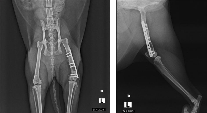
Fig. 2. The radiographs of a cat’s left femur show (a) a ventrodorsal and (b) a mediolateral view following fracture repair using a 6-hole plate. The fracture is not visible, indicating satisfactory healing. Bone transparency is observed in the medullary cavity of the femur, further enhancing visualization of the internal bone structure. The plate is properly aligned, with no signs of complications.
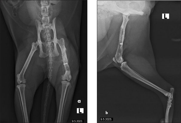
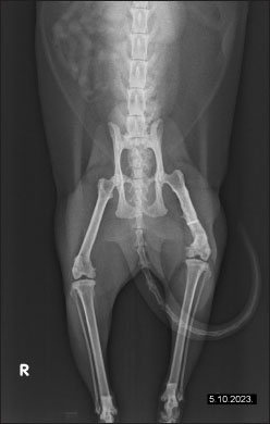
Fig. 3. Radiographs of a cat’s left femur showing (a) a ventrodorsal and (b) a mediolateral view post-plate removal, with one screw remaining in situ. Increased medullary cavity transparency highlights the need for LT to support bone healing and tissue regeneration.
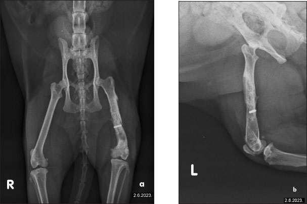
Fig. 4. The radiographs of a cat’s left femur show (a) a ventrodorsal and (b) a mediolateral view demonstrating the femur after 1 month of LT, demonstrating reduced medullary transparency and early signs of bone consolidation. Various clinical approaches have been explored to stimulate and accelerate bone healing, including physical stimulation, chemical treatments, and photostimulation. Photobiomodulation, the scientific term for LT, describes a photochemical process where light interacts with cells, triggering biochemical changes. The term is preferred as it reflects that some cellular processes are upregulated while others are downregulated. Numerous studies have examined photobiomodulation, many focusing on its cellular effects in vitro. Research highlights include increased angiogenesis, neurite extension, ion channel normalization, and cellular membrane stabilization. Moreover, LT promotes osteogenesis; both in vitro and in vivo studies demonstrate enhanced osteoblast proliferation, survival, and metabolism. The exact mechanisms behind photobiomodulation remain debated. Likely, multiple mechanisms exist depending on the target and cell type. The most recognized is the cytochrome c pathway in mitochondria, where cytochrome c, acting as a photoreceptor, absorbs light (500–1,100 nm). This excitation breaks nitric oxide bonds, enabling oxygen bonding and optimizing cytochrome c oxidase production. This enzyme is crucial for ATP formation, essential for cellular energy. The resulting ATP boosts secondary biological effects such as pain reduction, inflammation control, and tissue healing. LT also stimulates alkaline phosphatase activity and osteopontin, a protein crucial for osteoclast attachment to the mineralized bone matrix. Clinically, this leads to improved calcification and accelerated fracture healing.
Fig. 5. Radiographic evidence of consolidated bone structure and reduced medullary transparency following 25 sessions of LT. Assis et al. (2015) assessed the impact of LT on biochemical markers and the morphology of skeletal muscle in rats following an endurance training protocol. A study conducted by Barbosa et al. (2014) examined bone healing in rats with femur osteotomies, comparing no treatment, 660 nm laser, and 830 nm laser. Laser-treated groups showed faster bone development, with the 830 nm group achieving the best results by day 21. In addition to its impact on bone defect regeneration, LT plays a significant role in managing chronic pain. Chronic pain persists beyond the expected healing period for a specific injury or may remain due to incomplete healing. While it serves no biological purpose, it significantly impacts the quality of life. Traditional pain management strategies in veterinary medicine, such as non-steroidal anti-inflammatory drugs or opioids, can sometimes be ineffective (Alorfi, 2023). In such cases, LT can be integrated into a comprehensive multimodal treatment plan for animals, offering a complementary approach to improve pain relief. In human medicine, cold LT has been shown to alleviate pain and improve physical disability associated with knee osteoarthritis (OA), even delaying the need for knee replacement surgery and enhancing physical activity (Angelova and Ilieva, 2016; Youssef et al., 2016). Other studies on OA in former professional athletes have also demonstrated significant pain reduction and increased activity following just a few treatment sessions (Elvir-Lazo et al., 2020). However, there are fewer documented cases in veterinary medicine. A total of 50 studies on laser treatment for the musculoskeletal system were reported, including 28 involving dogs and none involving cats. In the treatment of pain with laser, three studies were regarding dogs, one was regarding cats, and six studies were on horses (Millis and Bergh, 2023). Within veterinary literature, photobiomodulation therapy has been reported to expedite the time to ambulation in dogs undergoing surgery for myelopathy caused by disc herniation (Draper et al., 2012), enhance peak vertical force following cranial cruciate disease surgical repair (Rogatko et al., 2017), and assist in the healing of sterile granulomatous pododermatitis (Perego et al., 2016). However, it has also been found ineffective in accelerating wound healing in canine patients (Kurach et al., 2015) and improving recovery-related variables when used alone or as part of a rehabilitation protocol for surgically treated intervertebral disc disease (Bennaim et al., 2017). Table 1. Progression of clinical observations and treatment stages during LT rehabilitation.
In our case, LT not only contributed to the healing of the bone defect but also facilitated a faster overall recovery. This was evident in the patient’s improved condition, as noted by the owner, who observed a significant change in the animal’s behavior. For the first time since the femoral fracture—over 4 years prior—the animal eagerly engages in movement and play. This marked improvement highlights the effectiveness of LT as a crucial component of the rehabilitation process, both for bone regeneration and long-term pain management. The main limitation of this report is the small sample size (one patient), which limits the generalizability of the results. Additionally, the lack of a control group makes it difficult to determine the specific effects of LT compared to other treatments or a placebo. Quantitative measurements, such as lameness scoring or weight-bearing metrics, were not included, which would have strengthened the objective assessment of the therapy’s effectiveness. Future research should focus on larger, randomized controlled trials with a broader population of feline patients to better assess the effectiveness of LT compared to conventional treatments or placebo. Incorporating quantitative metrics such as lameness scoring, gait analysis, and weight-bearing measurements would improve the objectivity and reliability of the findings. Additionally, exploring the molecular mechanisms behind LT, particularly its role in osteogenesis and cellular repair, could provide deeper insights into how this treatment promotes bone healing and pain management. Long-term follow-up studies are also necessary to evaluate the durability of the therapeutic benefits observed in the short term. These studies could help determine whether the improvements seen in the immediate aftermath of treatment are sustained over time. ConclusionThis case report highlights the transformative role of LT not only in enhancing bone regeneration but also in redefining multimodal pain management strategies in feline rehabilitation. Despite a lengthy recovery period of over 4 years since the initial fracture, the implementation of LT significantly improved the patient’s mobility and overall quality of life. The positive clinical outcomes observed—such as increased weight-bearing on the affected limb and enhanced engagement in physical activity—demonstrates LT’s potential as an integral component of veterinary rehabilitation. Furthermore, this case underscores the importance of adopting multimodal pain management strategies in veterinary practice, particularly when traditional methods are insufficient. Overall, LT represents a valuable tool for both facilitating bone regeneration and alleviating chronic pain, contributing to better patient outcomes in veterinary medicine. AcknowledgmentsThe authors would like to thank all the staff of the Radiology Clinic at the Veterinary Faculty, Department of Clinical Veterinary Sciences, for their cooperation and kind support throughout this work. Conflict of interestThe authors declare that they have no conflict of interest. FundingThis study was not supported by any sponsor or funder. Authors’ contributionsDŽHA and NH were responsible for the conceptualization and drafting of the manuscript. KB and SK contributed to the methodology. DŽHA and NH reviewed the manuscript. All authors approved the final version of the manuscript. Data availabilityAll data generated or analyzed during this work are included in the manuscript. ReferencesAikawa, T., Miyazaki, Y., Shimatsu, T., Iizuka, K. and Nishimura, M. 2018. Clinical outcomes and complications after open reduction and internal fixation utilizing conventional plates in 65 distal radial and ulnar fractures of miniature-and toy-breed dogs. Vet. Comp. Orthop. Traumatol. 31, 214–217. Alorfi, N.M. 2023. Pharmacological methods of pain management: narrative review of medication used. Int. J. Gen. Med. 16, 3247–3256. Angelova, A. and Ilieva, E.M. 2016. Effectiveness of high intensity laser therapy for reduction of pain in knee osteoarthritis. Pain. Res. Manag. 1, 9163618. Assis, L., Yamashita, F., Magri, A.M., Fernandes, K.R., Yamauchi, L. and Renno, A.C. 2015. Effect of low-level laser therapy (808 nm) on skeletal muscle after endurance exercise training in rats. Braz. J. Phys. Ther. 19, 457–465. Barbosa, D., Villaverde, A.G.J.B., LoschiavoArisawa, E.Â. and Souza, R.A.D. 2014. Laser therapy in bone repair in rats: analysis of bone optical density. Acta Ortop. Bras. 22, 71–74. Bennaim, M., Porato, M., Jarleton, A., Hamon, M., Carroll, J.D., Gommeren, K. and Balligand, M. 2017. Preliminary evaluation of the effects of photobiomodulation therapy and physical rehabilitation on early postoperative recovery of dogs undergoing hemilaminectomy for treatment of thoracolumbar intervertebral disk disease. Am. J. Vet. Res. 78, 195–206. Berni, M., Brancato, A.M., Torriani, C., Bina, V., Annunziata, S., Cornella, E. and Pasta, G. 2023. The role of low-level laser therapy in bone healing: systematic review. Int. J. Mol. Sci. 24(8), 7094. Brunke, M.W. 2017. Laser therapy and injury rehabilitation. In Laser therapy in veterinary medicine: photobiomodulation. Eds., Riegel, R.J. and Godbold, J.C. Hoboken, NJ: John Wiley & Sons, Inc., pp: 252–266. Draper, W.E., Schubert, T.A., Clemmons, R.M. and Miles, S.A. 2012. Low-level laser therapy reduces time to ambulation in dogs after hemilaminectomy: a preliminary study. J. Small Anim. Pract. 53, 465–469. Drum, M.G., Bockstahler, B., Levine, D. and Marcellin-Little, D.J. 2015. Feline rehabilitation. Vet. Clin. North Am. Small Anim. Pract. 45, 185–201. Elvir-Lazo, O.L., White, P.F., Yumul, R. and Eng, H.C. 2020. Management strategies for the treatment and prevention of postoperative/postdischarge nausea and vomiting: an updated review. F1000Res 9, F1000 Faculty Rev-983. Hochman, L. 2018. Photobiomodulation therapy in veterinary medicine: a review. Top. Companion Anim. Med. 33, 83–88. Johnson, A. 2016. Implant failure. In Complications in small animal surgery. Eds., Griffon, D. and Hamaide, A. Hoboken, NJ: Wiley. pp: 641–648. Kurach, L.M., Stanley, B.J., Gazzola, K.M., Fritz, M.C., Steficek, B.A., Hauptman, J.G. and Seymour, K.J. 2015. The effect of low-level laser therapy on the healing of open wounds in dogs. Vet. Surg. 44, 988–996. Malik-Tabassum, K., Pillai, K., Hussain, Y., Bleibleh, S., Babu, S., Giannoudis, P.V. and Tosounidis, T.H. 2020. Post-operative outcomes of open reduction and internal fixation versus circular external fixation in treatment of tibial plafond fractures: a systematic review and meta-analysis. Injury 51, 1448–1456. Menghini, T.L., Shriwise, G. and Muir, P. 2023. Fracture healing in 37 dogs and cats with implant failure after surgery (2013–2018). Animals 13, 1549. Millis, D.A. and Bergh, A. 2023. A systematic literature review of complementary and alternative veterinary medicine: laser therapy. Animals 13, 667. Perego, R., Proverbio, D., Zuccaro, A. and Spada, E. 2016. Low-level laser therapy: case-control study in dogs with sterile pyogranulomatous pododermatitis. Vet. World. 9, 882. Rogatko, C.P., Baltzer, W.I. and Tennant, R. 2017. Preoperative low level laser therapy in dogs undergoing tibial plateau levelling osteotomy: a blinded, prospective, randomized clinical trial. Vet. Comp. Orthop. Traumatol. 30, 46–53. Rubinos, C. and Meeson, R.L. 2022. Traumatic physeal fractures in cats: a review of 36 cases (2010–2020). J. Feline Med. Surg. 24, 98–106. Scott, H., Marti, J. and Witte, P. 2022. Fractures of the femur. In Feline orthopaedics. Eds., Scott, H., Marti, J. and Witte, P. Boca Raton, FL: CRC Press, pp: 168–181. Youssef, M., Ashkar, S., Hamade, E., Gutknecht, N., Lampert, F. and Mir, M. 2008. The effect of low-level laser therapy during orthodontic movement: a preliminary study. Lasers Med. Sci. 23, 27–33. | ||
| How to Cite this Article |
| Pubmed Style Alagić DH, Baltić K, Kolašinac S, Hadžimusić N. Rehabilitation of chronic pain and enhanced healing in feline femoral surgery: A case report on laser therapy. Open Vet. J.. 2025; 15(1): 471-477. doi:10.5455/OVJ.2025.v15.i1.43 Web Style Alagić DH, Baltić K, Kolašinac S, Hadžimusić N. Rehabilitation of chronic pain and enhanced healing in feline femoral surgery: A case report on laser therapy. https://www.openveterinaryjournal.com/?mno=225781 [Access: January 25, 2026]. doi:10.5455/OVJ.2025.v15.i1.43 AMA (American Medical Association) Style Alagić DH, Baltić K, Kolašinac S, Hadžimusić N. Rehabilitation of chronic pain and enhanced healing in feline femoral surgery: A case report on laser therapy. Open Vet. J.. 2025; 15(1): 471-477. doi:10.5455/OVJ.2025.v15.i1.43 Vancouver/ICMJE Style Alagić DH, Baltić K, Kolašinac S, Hadžimusić N. Rehabilitation of chronic pain and enhanced healing in feline femoral surgery: A case report on laser therapy. Open Vet. J.. (2025), [cited January 25, 2026]; 15(1): 471-477. doi:10.5455/OVJ.2025.v15.i1.43 Harvard Style Alagić, D. H., Baltić, . K., Kolašinac, . S. & Hadžimusić, . N. (2025) Rehabilitation of chronic pain and enhanced healing in feline femoral surgery: A case report on laser therapy. Open Vet. J., 15 (1), 471-477. doi:10.5455/OVJ.2025.v15.i1.43 Turabian Style Alagić, Dženita Hadžijunuzović, Kenan Baltić, Sabina Kolašinac, and Nejra Hadžimusić. 2025. Rehabilitation of chronic pain and enhanced healing in feline femoral surgery: A case report on laser therapy. Open Veterinary Journal, 15 (1), 471-477. doi:10.5455/OVJ.2025.v15.i1.43 Chicago Style Alagić, Dženita Hadžijunuzović, Kenan Baltić, Sabina Kolašinac, and Nejra Hadžimusić. "Rehabilitation of chronic pain and enhanced healing in feline femoral surgery: A case report on laser therapy." Open Veterinary Journal 15 (2025), 471-477. doi:10.5455/OVJ.2025.v15.i1.43 MLA (The Modern Language Association) Style Alagić, Dženita Hadžijunuzović, Kenan Baltić, Sabina Kolašinac, and Nejra Hadžimusić. "Rehabilitation of chronic pain and enhanced healing in feline femoral surgery: A case report on laser therapy." Open Veterinary Journal 15.1 (2025), 471-477. Print. doi:10.5455/OVJ.2025.v15.i1.43 APA (American Psychological Association) Style Alagić, D. H., Baltić, . K., Kolašinac, . S. & Hadžimusić, . N. (2025) Rehabilitation of chronic pain and enhanced healing in feline femoral surgery: A case report on laser therapy. Open Veterinary Journal, 15 (1), 471-477. doi:10.5455/OVJ.2025.v15.i1.43 |

