


| Research Article | ||
Open Vet. J.. 2025; 15(1): 261-269 Open Veterinary Journal, (2025), Vol. 15(1): 261-269 Research Article Evaluation of factors affecting the physiological levels of copper and iron in sheep and cattle in some areas of Diyala Governorate, IraqAl-Zubaidi Raad Mahmood Hussein1*, Marah Salim Hameed2, Waleed Ibrahim Jalil1 and Ali Ibrahim Ali AL-Ezzy31Department of Medicine, College of Veterinary Medicine, University of Diyala, Baqubah, Iraq 2Department of Physiology, College of Veterinary Medicine, University of Diyala, Baqubah, Iraq 3Department of Pathology, College of Veterinary Medicine, University of Diyala, Baqubah, Iraq *Corresponding Author: Al-Zubaidi Raad Mahmood Hussein. Department of Medicine, College of Veterinary Medicine, University of Diyala, Baqubah, Iraq. Email: raad.m [at] uodiyala.edu.iq Submitted: 13/09/2024 Accepted: 10/12/2024 Published: 31/01/2025 © 2025 Open Veterinary Journal
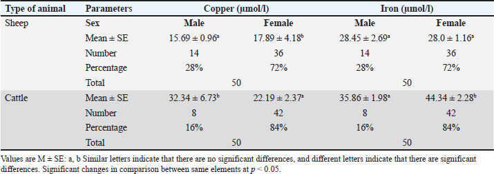
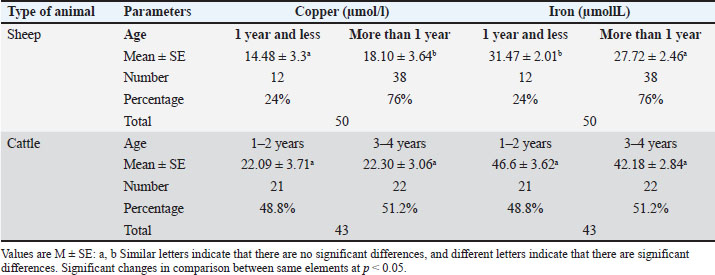
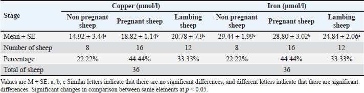
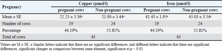
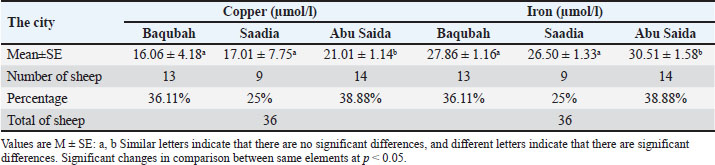
AbstractBackground: Trace minerals are important components of many physiological functions, including growth, development, and the immune response. Aim: This study aimed to evaluate copper and iron status in sheep and cattle in some areas of Diyala governorate, Iraq. Methods: One hundred blood samples were collected, 50 from sheep and 50 from cattle in order to measure the levels of copper and iron in the serum. Results: In sheep, the serum level of copper significantly increased among females compared to males, while among cattle, the serum level of copper significantly decreased in females compared with males. The level of iron showed a significant increase in females compared with males. Serum levels of copper were increased with the age of more than 1 year accordingly compared with sheep at the first year of life or less than a year. Among cattle, age plays no significant role in the level of copper. The levels of copper and iron were significantly increased among pregnant ewes compared with non-pregnant. The levels of copper were significantly increased in lambing compared with pregnant and non-pregnant ewes in the value of copper. The level of iron was significantly decreased in lambing sheep compared with pregnant and non-pregnant sheep. The levels of copper and iron showed no significant changes between pregnant and non-pregnant cows. Sheep from Abu Saida showed significant increase in serum levels of copper and iron compared with those from Baqubah and Saadia cities. Cows with good body scores have a significant increase in serum levels of copper and iron compared with those with medium and poor body scores. There was no linear correlation between iron and copper levels in serum and the studied parameters for cows and sheep. Conclusion: The levels of copper and iron in the body of sheep and cattle are affected by age, gender, pregnancy, and soil composition. Keywords: Copper, Iron, Trace minerals, Sheep, Cattle. IntroductionThe body needs trace minerals at extremely minute levels, but these elements are crucial for many different biochemical activities. Sheep need the following trace minerals: zinc (Zn), molybdenum (Mo), manganese (Mn), copper (Cu), iodide (I), iron (Fe), and cobalt (Co). It is commonly recognized that virtually all of the body’s metabolic functions depend on trace minerals to function normally. They are necessary to preserve the health and production of animals since they are a component of many enzymes and regulate a vast array of biological processes. Adequate trace mineral levels along with optimal nutrition ensure that the body performs its essential functions, the most crucial of which are physiological, structural, catalytic, and regulatory. Previous studies stated that the most frequent reason to evaluate the trace mineral status of the animals is when their performance falls short of expectations (Suttle, 2010; Swecker and Van Saun, 2023). The assessment of trace minerals is carried out in farm animals to ascertain if dietary deficiencies are common in animals or not, which have a direct effect on production and performance. Additionally, assessments of trace minerals are essential to determine the effectiveness of dietary supplements, which reflect the good management policies in large-scale farms (Byrne and Murphy, 2022). As a co-factor in hundreds of enzymatic activities involved in the synthesis of collagen, the generation of red blood cells, the production of energy, the formation of hormones, and the defense against oxidative damage, copper is a necessary element for life (Jomova et al., 2022). Many clinical signs, such as a pale coat, poor quality sheep fleece, anemia, spontaneous fractures, poor capillary integrity, myocardial degeneration, demyelination of the spinal cord, impaired reproductive function, decreased resistance to infectious disease, diarrhea, and generalized ill health, have been linked to copper deficiency, resulting in significant economic losses (Riaz and Muhammad, 2018). The second most common mineral deficiency effect in grazing animals is hypocuprosis. Several studies on the mechanisms underlying the body’s copper activity have focused mainly on the distribution of copper in different tissues, the changes that occur in the blood after various conditions, and the interactions between copper and different enzyme systems, vitamins, and minerals (Hefnawy and El-Khaiat, 2015). Intracellular homeostasis and the appropriate balance of iron stores are therefore closely regulated. Iron is an essential nutrient and cofactor for the synthesis of hemoglobin and myoglobin as well as several cellular functions including oxygen transport, respiration, growth, gene regulation, and the proper functioning of iron-dependent enzymes (Cronin et al., 2019). Poor dietary intake of absorbable iron (or insufficient intake to meet increased demands during pregnancy) and/or iron loss from parasitic infections (e.g., hookworm) or blood loss are the causes of iron deficiency (Pena-Rosas et al., 2015). The majority of dietary iron is nonheme iron, which is found in plant-based meals such as green leafy vegetables. However, the primary source of dietary iron for mammals is hemoglobin, which is found in foods such as fish and animal meats because it is more quickly absorbed and has a higher bioavailability (Cairo et al., 2006). The aim of the current study was to assess the effect of age, sex, and pregnancy on the levels of copper and iron in sheep and cattle in certain regions of the Diyala governorate in Iraq. Materials and MethodsStudy area and study populationThis study was conducted on sheep and cows, reared in Baqubah, Saadia, and Abu Saida in Diyala province, Iraq. Study designIn order to assess the levels of copper and iron in the serum, a cross-sectional study was designed (Al-Ezzy, 2016a), involving the collection of 100 blood samples, which were randomly collected from 50 sheep and 50 cattle from various sites in Diyala Governorate. Sample collectionFive milliliters of jugular vein blood were taken according to Pugh et al. (2020) and collected in a dry, clean tube. The samples were centrifuged for 10 minutes at 3,000 rpm after being left to clot in a slanting posture at room temperature throughout the whole night (Sultan et al., 2023; Hameed and Al-Ezzy, 2024). According to Hameed et al. (2024a,b), the clear sera were thoroughly aspirated using an automated pipette and then dispensed into clear, dry, labeled Eppendorf tubes, and kept at −20°C until testing. Assessment of serum iron and copperCopper biochemical analysis was performed using the spectrophotometer (AA-5) according to Dawson et al. (1968), Ogunsanmi et al. (2002), Akhtar et al. (2009), using a special copper kit. The working solution (working reagent) is prepared according to the recipe attached to the kit. Equal amounts of reagent A are mixed with reagent B. This reagent remains for 20 days at room temperature. When stored in the refrigerator before work, reagent A may precipitate in the form of grains. In this case, it must be placed in a water bath at a temperature of −2,530°C for 5 minutes. This will make it clear and suitable for mixing. After preparing the working reagent, we prepare three tubes to mix the sample in. After mixing, we wait 10 minutes; then, the samples are read by the device under a wavelength of 580 nm. The reaction color remains constant for 30 minutes. We apply the following equation: Amount of copper (CU)=Wavelength of the model/Wavelength of the standard x×3.15 Estimating the amount of iron in the serumThe spectrophotometer was used according to the method indicated by Doumas and Biggs (1972) using a special kit to examine the amount of iron in the serum. Ethical approvalThe Helsinki Declaration’s guiding principles were followed in the conduct of this investigation. A thorough explanation of the study’s objectives to each owner prior to its commencement. Complete consent forms were acquired from all owners. Before beginning work, the Department of Medicine at Diyala University in Iraq’s College of Veterinary Medicine obtained approval from an ethical review commission (AL-Ezzy et al., 2020; Al-Khalidi et al., 2020a,b; Al-Zuhairi et al., 2020a,b; Humadi et al., 2020; Hameed et al., 2020a,b; Hameed and Al-Ezzy, 2024; Hameed et al., 2024a) Statistical analysisData were expressed as (mean ± SE) (Al-Ezzy, 2016b,c). T-test was used for data analysis (Al-Ezzy et al., 2016; Hameed et al ., 2024b). The Pearson correlation coefficient is used to measure the linear correlation between parameters (Fajer et al., 2023a,b). The SPSS is used for the analysis of variables with significant levels (p < 0.05) (Hameed and Al-Ezzy, 2019; Hameed et al., 2020b) ResultsThe results of the current study showed the blood serum value of copper and iron (14 male 28% and 36 female 72%). A significant increase in serum copper in females compared with the serum value of copper of males, while the result of iron showed no significant changes between sex. As shown in Table 1, in cattle, the serum values of copper and iron (8 males 16% and 42 females 84%) significantly decreased in serum copper in females compared with the serum value of copper of males, while the result of iron showed a significant increase in serum iron in females compared with the serum value of iron of males (Table 1). The results of blood serum value for copper and iron [12 (24%) ≤ 1 year and 38 (76%) ≥ 1 year] showed a significant increase in ages of more than 1 year compared with sheep in 1 and less year, but the result of iron showed a significant decrease in ages of more than 1 year compared with sheep in 1 and less year. However, the blood serum value of copper and iron [21 cows (1–2 year) 48.8% and 22 cows (3–4 year) 51.2%] showed no significant changes between ages in the examined cows (Table 2). Among non-pregnant sheep (8/36; 22.22%), pregnant sheep (16/36; 44.44%), and lambing sheep (12/36; 33.33%), there was a significant increase in the serum levels of copper among pregnant compared with non-pregnant sheep. On the other hand, there was a significant increase in serum copper among lambing sheep compared with pregnant and non-pregnant sheep. There was a significant decrease in serum iron levels among lambing sheep compared with pregnant and non-pregnant sheep. There were no significant changes in serum iron levels between pregnant and non-pregnant sheep (Table 3). The results of blood serum value for copper and iron (19 pregnant cows 44.19%, 24 non-pregnant cows 55.81%) showed no significant changes between pregnant and non-pregnant cows (Table 4). The results of blood serum value for copper and iron (13) sheep (36.11%) from Baqubah, 9 sheep (25%) from Saadia, and 14 sheep (38.88%) from Abu Saida city showed a significant increase in serum copper and iron in sheep from Abu Saida compared with Baqubah and Saadia cities, but no significant changes between sheep from Baqubah and Saadia cities (Table 5). Table 1. Value of serum copper and iron according to sex of sheep and cattle.
Table 2. Value of serum copper and iron according to age of sheep and cattle.
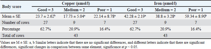
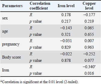
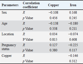
The results of blood serum value for copper and iron (27), good body score (62.7%), (9) medium body score (20.9%), and (7) poor body score (16.4%) showed a significant increase in good body score compared with medium and poor body scores in value of copper, while in iron significant decrease in good and medium body scores compared with poor body score as shown in Table 6. As shown in Table 7, the serum level of copper and iron was not correlated with age, sex, body score, and pregnancy status among studied cows. As shown in Table 8, the serum level of copper and iron was not correlated with age, sex, location, and pregnancy status among studied sheep. A negative correlation was reported between iron and copper levels in sheep. DiscussionBecause blood is less intrusive to collect than liver and blood measurements are highly connected with nutritional status of specific trace elements, blood measures are commonly utilized in evaluation (Herdt and Hoff, 2011). Mineral concentrations in whole blood frequently fluctuate gradually. There is a correlation between the amount of bioavailable copper in bovine diets and the concentration of copper in their livers (Ensley, 2020). Physiological requirements influence the concentration of copper in the liver in addition to bioavailable copper from food. Genetics has a role in copper metabolism, and breeds of sheep differ significantly in the amounts of copper in their plasma and liver (López-Alonso and Miranda, 2020). When compared to males, females’ serum levels of copper increase substantially. For feeding purposes, the animal’s mineral status should be taken into consideration when preparing the final diets, as diets and feedstuffs lacking in trace mineral requirements can have detrimental effects on reproduction functions in both males and females of both species (Biru, 2021). According to Hussein’s (2012) study, the levels of copper and iron in goats from the Iraqi region are unaffected by age or gender. The present study’s findings on low levels of copper in men might perhaps be attributed to the dietary differences between males and females, particularly in cases when iron intake is elevated (Constable et al., 2017). However, Khwedim (2013) noted that the copper content of the Iraqi soil is low. In cattle, the results indicated that the blood serum values of iron and copper significantly increased the serum iron value in females when compared to males, while the blood serum value of copper significantly decreased in females when compared to the serum value of males. The animal breeds have great role and directly affect the efficacy of copper metabolism (Sefdeen, 2017). The source of the high iron intake in ruminants surpassing several hundred micrograms was the increase in soil iron brought on by pollution, which also caused an increase in iron (Yildiz Küçük and Gökçek, 2024). Table 3. Value of serum copper and iron in non-pregnant, pregnant and lambing sheep.
Table 4. Value of serum copper and iron in pregnant and non-pregnant cows.
Table 5. Value of serum copper and iron of female sheep in the Baqubah, Saadia and Abu Saida city.
Table 6. Value of serum copper and iron according to body score of cows.
Table 7. Correlation between sex, age, pregnancy, body score and serum copper and iron levels among cows.
Table 8. Correlation between sex, age, pregnancy status, location and serum copper and iron levels among sheep.
Cattle and sheep that were fed iron compounds or 10% dry matter from soils high in iron consistently saw an increase in iron levels and a decrease in copper in their livers (Sousa et al., 2012). Conversely, in sheep and goats, the impact of iron on copper metabolism was noted. When iron was given at doses of 247 and 827 mg/kg dry matter, the greatest levels of iron and lowest levels of copper were seen in all tissues, including lung tissue, in grazing sheep (Grace and Lee, 1990; Mohammed et al., 2016). According to Suttle (2022), there are notable variations in the concentrations of copper in the plasma and liver of different sheep breeds. These variations may be due to genetic influences on copper metabolism (Adeniyi et al., 2024). The results of blood serum copper values significantly increased in ages greater than 1 year when compared to sheep younger than 1 year. These results disagree with those of Suttle (2010). Although all of the animals in this study are of post-weaning age, Mohammed et al. (2016) stated that “copper absorption was much higher in milk supplied to pre-ruminant lambs (75%–90%) than in post-weaned lambs and adult ewes (8%–9%).” However, breed-specific variations exist in sheep’s copper absorption efficiency (Autukaite et al., 2021). Using meals containing 12 to 20 mg Cu/kg dry matter, a study by Woolliams et al. (1982) found that Texel lambs store more than twice as much iron in their livers than pure black-faced lambs. The blood serum values for iron and copper in the cattle did not significantly alter between the ages of the cows that were investigated. When chelated amino acid complexes were used instead of inorganic feed sources, copper absorption was greater (Pal et al., 2010). While less than 5% of copper may be absorbed via herbage, sheep, and cattle can receive 60%–80% of it from milk (Suttle, 2010). Due to species variations in Cu availability, the availability in cattle was calculated to be around 3.1%, but the projected value using equations based on sheep data was 4.0% (Suttle, 2010). Ruminants seldom experience iron deficiency, with the exception of young ruminants (Joerling and Doll, 2019) and calves given milk. Up to the age of six, cows’ liver and spleen have higher concentrations of iron (Nogara et al., 2024). When comparing pregnant and non-pregnant sheep, the serum copper value significantly increased in the former group and significantly decreased in the latter. A significant increase was found in lambing sheep’s serum copper value when compared to pregnant and non-pregnant sheep. In addition to dietary considerations, there are several additional factors that influence plasma copper concentrations. According to Rodríguez et al. (2021), cows’ serum has more copper during the estrus period on day 21 than nulliparous calves and less copper on the day of parturition in meat cows. According to Shoushtari et al. (2015), the plasma copper level peaked five weeks prior to birth. Moreover, infections raise serum copper levels (Besold et al., 2016). The study’s findings support the assertions made by Kapper et al. (2024) that there is a connection between an animal’s mineral status and its reproductive cycle phases. For instance, during pregnancy, both the mother and the fetus experience fast development and cell differentiation as well as extremely sensitive dietary imbalances (Caton et al., 2020). For instance, during pregnancy, both the mother and the fetus experience fast development and cell differentiation as well as extremely sensitive dietary imbalances (Caton et al. 2020).The neurohormonal connection and the intricate way biomineral molecules work are to blame (Zatta and Frank 2007). Since sheep milk is high in iron, low iron in mother ewes plasma might be the result of increased iron excretion from milk (Molik et al., 2008). When comparing the blood serum values of sheep from Abu Saida with those from Baqubah and Saadia Cities, the results showed a considerable increase in both copper and iron. These findings are corroborated by other studies that looked at the concentration of minerals and how they relate to one another in the soil, feed, and serum of sheep and goats in Mexico. These studies found that there are mineral imbalances, with the excess of phosphorus and iron in the sheep and copper deficiencies linked to an excess of iron and copper in the soil and forages (Domínguez-Vara and Huerta-Bravo, 2008). It is possible that there is a difference in the diets followed by the breeders and what is available from the pastures, which led to these differences between different regions of the current study. In another context, Qudoos et al. (2023) indicated that there is another difficult factor represented in the recommendations made to animals in the fattening field system. These recommendations stated that grazing animals meet all their needs of minerals due to the high concentration of trace elements in pastures and what is available in soil and water. However, the amount of trace elements in fodder varies depending on the kind of soil, fertilization methods, species of forage, age of the plants, composition of the pasture’s plants, climate, and season (Mirzaei, 2012). A study by Hussein (2012) reported that the average serum levels of copper and iron for healthy goats were 21.66 and 29.24 µmol/l, respectively. When compared to goats that seemed to be in good health, the anemic goats’ average values showed a significant reduction. Thus, one of the factors that may have contributed to the variation in copper and iron contents across the study’s many locations is the management and health conditions. According to the studies by Sousa et al. (2012) and Mohammed et al. (2016), feeding sheep with different iron compounds or supplementing soil with iron at a rate of 10% of its dry matter results in high iron levels and a corresponding decrease in copper levels in the animal’s body, particularly in the liver. Older cattle on farms have an adequate amount of iron; the primary issue is an excess of dietary iron (Suttle, 2010). These findings may help to explain the current study’s findings. Serum values for iron and copper (27/43 , 62.7%, with good body scores; 9/43, (20.9%), with medium body scores, and 7/43, 16.4% with poor body scores) showed a significant decrease in the good and medium body scores compared to the poor body scores, while the good and medium body scores significantly increased in the value of iron. These results are consistent with the findings of Nawito et al. (2015), who reported that an assessment of the nutritional status of animal herds revealed that the mean body condition score was below mean levels, but differed markedly between sheep and goats or lambs. The findings of the current study are in accordance with Constable et al. (2017), who stated that “the clinical manifestations of copper deficiency in cattle occur yearly in approximately 0.9% of cattle herds in the United Kingdom.” The primary cause of the largest loss is thought to be the animals’ inability to develop copper deficiency, which impairs tissue oxidation and interferes with intermediate metabolism, leading to status or growth failure. Conversely, when more copper was given to the cattle, the physical state of the cows improved compared to the cows with little copper in the same field circumstances. In the current study, there was no linear correlation between serum levels of copper and iron with age, sex, body score, and pregnancy status among studied cows. There was no linear correlation between serum level of copper and iron with age, sex, location, and pregnancy status among studied sheep. No previous study reported such correlation between iron, copper, and other parameters of the current study in sheep and cow. The absence of such linear correlation might attribute to the fact that to estimate the linear correlation among studied parameters, a large number of samples required, and study groups equally distributed regarding parameters under investigation, such as age, sex, location, and pregnancy status. ConclusionAlthough the serum levels of copper and iron in the body of sheep and cattle are affected by age, sex, pregnancy, and soil composition, there is no linear correlation between serum level of copper and iron with age, sex, body score, and pregnancy status among studied cows as well as age, sex, location, and pregnancy status among studied sheep. AcknowledgmentsThe authors deeply appreciated for the efforts of Technicians at the College of Veterinary Medicine, University of Diyala. Conflict of interestThe authors disclosed that there is no conflict of interest FundingThe authors disclosed that they share the total cost of current research project. Authors’ contributionsAuthors disclosed that they were equally contribute in planning, methodology, data collection and analysis, and writing of manuscript. Data availabilityAll the required data were included with in the text. ReferencesAdeniyi, O., Lenstra, J., Mastrangelo, S. and Lühken, G. 2024. Genome-wide comparative analyses for selection signatures indicate candidate genes for between-breed variability in copper accretion in sheep. Animal 18(10), 101329. Akhtar, M., Farooq, A. and Mushtaq, M. 2009. Serum concentrations of copper, iron, zinc and selenium in cyclic and anoestrus Nili-Ravi buffaloes kept under farm conditions. Pak. Vet. J. 29(1), 47–48. Al-Ezzy, A.I.A. 2016a. Evaluation of endoscopy based H. Pylori diagnostic techniques in Iraqi patients with upper gastrointestinal disorders. Indian. J. Sci. Technol. 9(22), 1–10. Al-Ezzy, A.I.A. 2016b. Immunomodulatory effect of H. Pylori CagA genotype and gastric hormones on gastric versus inflammatory cells fas gene expression in Iraqi patients with gastroduodenal disorders. OAMJMS 4(3), 364–373. Al-Ezzy, A.I.A. 2016c. In situ nick end labeling as a molecular immunopathological indicator for the severity of DNA fragmentationand gastroduodenal tissue damage among H. Pylori Cag A positive patients. Ind. J. Sci. Tech. 9, 78512; doi:10.17485/ijst/2016/v9i2/78512 Al-Ezzy, A.I.A., Al-Khalidi, A.A.H. and Hameed, M.S. 2020. Evaluation of C-reactive protein in Iraqi children presented with acute enteropathogenic Escherichia coli associated diarrhea with special emphasis to age and gender. GMJ 31(2), 143–148. Al-Ezzy, A.I.A., Hameed, M.S., Jalil, W.I. and Mohamad, W.M. 2016. Pathophysiological effects of vitamin C and E-selenium combination on lipid profile and serum glucose of experimentally induced sodium nitrate intoxication in mice. Res. J. Pharm. Biol. Chem. Sci. 7(2), 958–964. Al-Khalidi, A.A.H., Hameed, M.S. and Al-Ezzy, A.I.A. 2020a. Effects of saccharomyces cerevisiae as probiotic on blood indices, humoral immunity and performance of Isa brown laying hens in Diyala Province-Iraq. Biochem. Cell. Arch. 20(1), 2727–2733. Al-Khalidi, A.A.H., Hameed, M.S. and Al-Ezzy, A.I.A. 2020b. Effect of drinking water quality on physiological blood parameters and performance of laying hens in Diyala province-Iraq. Biochem. Cell. Arch. 20(1), 2649. Al-Zuhairi, A.H., Al-Khafaji, M.N., Al-Khafaji Nazar Jabbar and Al-Ezzy, A.I.A. 2020a. Comparative treatment of awassi ewes naturally infested with psoroptes ovis, in Diyala Province, Iraq. Biochem. Cell. Arch. 20(1), 2621–2628. Al-Zuhairi, A.H., Jalil, W.I., Hameed, M.S. and Al-Ezzy, A.I.A. 2020b. Hepatoprotective effect of aqueous—methanol extract of Cordia dichotoma in experimental paracetamol induced hepatitis in rabbits. Biochem. Cell. Arch. 20(1), 2575–2579. Autukaite, J., Juozaitiene, V., Antanaitis, R., Poskiene, I., Baumgartner, W., Zilinskas, H. and Zilaitis, V. 2021. The impact of breed, testing time and metabolic profile on the variation of copper concentration in sheep blood serum. Ind. J. Animal Res. 55(7), 767–773. Besold, A.N., Culbertson, E.M. and Culotta, V.C. 2016. The Yin and Yang of copper during infection. JBIC J. Biol. Inorg. Chem. 21, 137–144. Biru, B. 2021. Mineral and digestive responses to dietary challenges in the gastro-intestinal system of ruminants. Ghent, Belgium: Ghent University. Byrne, L. and Murphy, R.A. 2022. Relative bioavailability of trace minerals in production animal nutrition: a review. Animals 12(15), 1981. Cairo, G., Bernuzzi, F. and Recalcati, S. 2006. A precious metal: Iron, an essential nutrient for all cells. Genes Nutr. 1(1), 25–39. Caton, J.S., Crouse, M.S., Mclean, K.J., Dahlen, C.R., Ward, A.K., Cushman, R.A., Grazul-Bilska, A.T., Neville, B.W., Borowicz, P.P. and Reynolds, L.P. 2020. Maternal periconceptual nutrition, early pregnancy, and developmental outcomes in beef cattle. J. Anim. Sci. 98(12), skaa358. Constable, P.D., Hinchcliff, K.W., Done, S.H. and Gruenberg, W. 2017. A textbook of the diseases of cattle, horses, sheep, pigs, and goats. Vet. Med. 11, 412–418. Cronin, S. J., Woolf, C. J., Weiss, G. and Penninger, J. M. 2019. The role of iron regulation in immunometabolism and immune-related disease. Front. Mol. Biosci. 6, 116. Dawson, J., Ellis, D. and Newton-John, H. 1968. Direct estimation of copper in serum and urine by atomic absorption spectroscopy. Clin. Chim. Acta. 21(1), 33–42. Domínguez-Vara, I.A. and Huerta-Bravo, M. 2008. Mineral concentration and interrelationship in soil, forage and blood serum of sheep during two seasons in the Toluca Valley, Mexico. Agrociencia 42(2), 173–183. Doumas, B. and Biggs, H. 1972. Standard methods of clinical chemistry. New York, NY: Academic Press. Vol. 7, p. 175. Ensley, S. 2020. Evaluating mineral status in ruminant livestock. Vet. Clin. North Am. Food Anim. Pract. 36(3), 525–546. Fajer, Z.B., Al-Ezzy, A.I.A. and Al-Zuhairi, A.H. 2023a. Molecular prevalence of MecA and Blaz Genes with phenotypic analysis of antibiotic sensitivity pattern for S. aureus isolated from dermal lesions of sheep breeders in Diyala governorate–Iraq. DJM 25(1), 12–26. Fajer, Z.B., Al-Ezzy, A.I.A. and Al-Zuhairi, A.H. 2023b. Sociodemographic risk factors for dermal infections with methi-cillin sensitive and methicillin resistant Staphylococcus aureus among sheep breeders in Diyala Governorate, Iraq. DJM 24(1), 66–84. Grace, N. and Lee, J. 1990. Effect of increasing Fe intake on the Fe and Cu content of tissues in grazing sheep. In Proceedings of the New Zealand Society of Animal Production, New Zealand. Hameed, M.S. and Al-Ezzy, A.I.A. 2019. Evaluation of possible stress factors affecting physiological level of gamma interferon during first six months of life in healthy calves. Adv. Anim. Vet. Sci. 7(5), 370–377. Hameed, M.S. and Al-Ezzy, A.I.A. 2024. Evaluation of antioxidant, nephroprotective and immunomodulatory activity of vitamins C and E sodium selenite in mice intoxicated with sodium nitrate. Adv. Anim. Vet. Sci. 12(6), 1018–1027. Hameed, M.S., Al-Ezzy, A.I.A., Jalil, W.I. and Al-Khalidi, A.a.H. 2020a. Physiological protective effects of ascorbic acid versus Dl-A-tocopheryl acetate–sodium selenite combination in mice under experimental sodium nitrate intoxication. Biochem. Cell. Arch. 20(1), 2593–2601. Hameed, M.S., Al-Ezzy, A.I.A., Jalil, W.I. and Khalidi, A.A.H.A. 2020b. Impact of stress factors on physiological level of interleukin 10 in healthy calves in Diyala Province –Iraq. IJPR 12 (suppl.2), 2050–3056. Hameed, M.S., Al-Zubaidi, R.M.H. and Al-Ezzy, A.I.A. 2024a. Effectiveness of aqueous versus alcoholic extracts of Melia azedarach in amelioration of lipid profile, liver enzymes and innate inflammatory indices for white new Zealand Rabbits. Adv. Animal Vet. Sci. 12(7), 1256–1265. Hameed, M.S., Hasson, S.J., Mahmood, M.A. and Al-Ezzy, A.I.A. 2024b. Physiological effect of multivitamins supplementation on hematological parameters, lipid profile, hepato-renal function of ross 308 broilers. Assiut Vet. Med. J. 70(183), 585–595. Hefnawy, A. and El-Khaiat, H. 2015. The importance of copper and the effects of its deficiency and toxicity in animal health. Int. J. Livestock Res. 5(12), 1–20. Herdt, T.H. and Hoff, B. 2011. The use of blood analysis to evaluate trace mineral status in ruminant livestock. Vet. Clin. North Am. Food Anim. Pract. 27(2), 255–283. Humadi, A.A., Al-Kaisei, B.I., Humadai, T.J. and Al-Ezzy, A.I.A. 2020. Toxicopathological, cytogenetic effects of acetothioamide on female albino mice reproductive system. OAMJMS 7, 858. Hussein, R.M. 2012. Study of some biochemical values changes associated with morphological anemia in local breed goats. Iraqi J. Vet. Med. 36(0A), 77–85. Joerling, J. and Doll, K. 2019. Monitoring of iron deficiency in calves by determination of serum ferritin in comparison with serum iron: a preliminary study. Open Vet. J. 9(2), 177–184. Jomova, K., Makova, M., Alomar, S.Y., Alwasel, S.H., Nepovimova, E., Kuca, K., Rhodes, C.J. and Valko, M. 2022. Essential metals in health and disease. Chem-Biol. Interact. 367, 110173. Kapper, C., Oppelt, P., Ganhör, C., Gyunesh, A.A., Arbeithuber, B., Stelzl, P. and Rezk-Füreder, M. 2024. Minerals and the menstrual cycle: impacts on ovulation and endometrial health. Nutrients 16(7), 1008. Khwedim, K. 2013. Study of distribution of some trace elements contents in the soil of Basra city using Geographic Information System (GIS). J. Babylon Univ. Pure Appl. Sci. 21(2), 479–509. López-Alonso, M. and Miranda, M. 2020. Copper supplementation, a challenge in cattle. Animals 10(10), 1890. Mirzaei, F. 2012. Minerals profile of forages for grazing ruminants in Pakistan. Open J. Anim. Sci. 2(3), 133–141. Mohammed, A., Osman, N.E.H.I.E.D. and Youssef, F.G. 2016. Review on copper’s functional roles, copper X mineral interactions affecting absorption, tissue storage, and Cu deficiency swayback of small ruminants. ARC J. Anim. Vet. Sci. 2(2), 1–14. Molik, E., Murawski, M., Bonczar, G. and Wierzchoś, E. 2008. Effect of genotype on yield and chemical composition of sheep milk. Ani. Sci. Papers Rep. 26(3), 211–218. Nawito, M.F., Mahmoud, K.G.M., Kandiel, M.M.M., Ahmed, Y.F. and Sosa, A.S.A. 2015. Effect of reproductive status on body condition score, progesterone concentration and trace minerals in sheep and goats reared in South Sinai, Egypt. African J. Biotech. 14(43), 3001–3005. Nogara, K.F., Tavares, Q.G., Palmeira, M., Ribeiro, C.H.M. and Zopollatto, M. 2024. Effect of metals (Hg, Cd, Pb, Zn, Cr and Cu) on ruminants. Toxicology of essential and xenobiotic metals. Boca Raton, FL: CRC Press, pp: 59–78. Ogunsanmi, A., Ozegbe, P., Ogunjobi, O., Taiwo, V. and Adu, J. 2002. Haematology, plasma biochemistry and whole blood minerals of the captive adult African grasscutter (Thryonomys swinderianus, Temminck). Trop. Vet. 20(1), 27–35. Pal, D., Gowda, N., Prasad, C., Amarnath, R., Bharadwaj, U., Babu, G.S. and Sampath, K. 2010. Effect of copper-and zinc-methionine supplementation on bioavailability, mineral status and tissue concentrations of copper and zinc in ewes. J. Trace Elements. Med. Biol. 24(2), 89–94. Pena-Rosas, J., De-Regil, L., Garcia-Casal, M. and Dowswell, T. 2015. Daily oral iron supplementation during pregnancy. Cochrane Database Syst. Rev. 2015(7), CD004736. Pugh, D.G., Baird, N.N., Edmondson, M. and Passler, T. 2020. Sheep, goat, and cervid medicine-E-Book. Edinburgh, UK: Elsevier Health Sciences. Qudoos, A., Tahir, U., Ahmad, S., Tariq, M., Imran, M., Nadeem, R., Noreen, S., Luqman, N., Maqbool, M. and Jan, I. 2023. Trace mineral concentration of forages in connection with sheep dietary requirements. S. Afr. J. Anim. Sci. 53(3), 361–368. Riaz, M. and Muhammad, G. 2018. Copper deficiency in ruminants in Pakistan. MSM 2(1), 18–21. Rodríguez, A.M., Valiente, S.L., Mattioli, G. and Maresca, S. 2021. Effects of inorganic copper injection in beef cows at late gestation on fetal and postnatal growth, hematology and immune function of their progeny. Res. Vet. Sci. 139, 11–17. Sefdeen, S.M. 2017. Effect of dietary iron on copper metabolism in sheep. Newport, UK: Harper Adams University. Shoushtari, S.M.A., Rezaie, S.A., Khaki, A., Belbasi, A. and Tahmasebian, H. 2015. Copper and zinc concentrations in uterine fluid and blood serum during the estrous cycle and pre-pubertal phase in water buffaloes. Urmia, Iran: Urmia University. Sousa, I.K.F.D., Hamad Minervino, A.H., Sousa, R.D.S., Chaves, D.F., Soares, H.S., Barros, I.D.O., Araújo, C.a.S.C.D., Junior, R.A.B. and Ortolani, E.L. 2012. Copper deficiency in sheep with high liver iron accumulation. Vet. Med. Int. 2012(1), 207950. Sultan, A.A., Hameed, M.S., Humadi, A.A., Al-Kaisei, B.I. and Al-Ezzy, A.I.A. 2023. Protective role of chlorophyllin against thyroid adenoma induced by polychlorinated biphenyls:(pathological and hormonal assay). In AIP Conference Proceedings, Melville, NY: AIP Publishing. Suttle, N. 2010. Mineral nutrition of livestock, 4th ed. Wallingford, UK: CABI. Suttle, N. 2022. Mineral nutrition of livestock. London, UK: Cabi GB. Swecker, W.S. and Van Saun, R.J. 2023. Vitamins and trace minerals in ruminants, an issue of veterinary clinics of North America: food animal practice, E-Book. Edinburgh, UK: Elsevier Health Sciences. Woolliams, J., Suttle, N., Wiener, G., Field, A. and Woolliams, C. 1982. The effect of breed of sire on the accumulation of copper in lambs, with particular reference to copper toxicity. Anim. Sci. 35(3), 299–307. Yildiz Küçük, N. and Gökçek, R. 2024. Determination of some minerals and heavy metals in raw cow’s milk. AKUJSE 24(4), 839–846. Zatta, P. and Frank, A. 2007. Copper deficiency and neurological disorders in man and animals. Brain Res. Rev. 54(1), 19–33. | ||
| How to Cite this Article |
| Pubmed Style Hussein ARM, Hameed MS, Jalil WI, Al-ezzy AIA. Evaluation of factors affecting the physiological levels of copper and iron in sheep and cattle in some areas of Diyala Governorate, Iraq. Open Vet. J.. 2025; 15(1): 261-269. doi:10.5455/OVJ.2025.v15.i1.24 Web Style Hussein ARM, Hameed MS, Jalil WI, Al-ezzy AIA. Evaluation of factors affecting the physiological levels of copper and iron in sheep and cattle in some areas of Diyala Governorate, Iraq. https://www.openveterinaryjournal.com/?mno=220334 [Access: January 25, 2026]. doi:10.5455/OVJ.2025.v15.i1.24 AMA (American Medical Association) Style Hussein ARM, Hameed MS, Jalil WI, Al-ezzy AIA. Evaluation of factors affecting the physiological levels of copper and iron in sheep and cattle in some areas of Diyala Governorate, Iraq. Open Vet. J.. 2025; 15(1): 261-269. doi:10.5455/OVJ.2025.v15.i1.24 Vancouver/ICMJE Style Hussein ARM, Hameed MS, Jalil WI, Al-ezzy AIA. Evaluation of factors affecting the physiological levels of copper and iron in sheep and cattle in some areas of Diyala Governorate, Iraq. Open Vet. J.. (2025), [cited January 25, 2026]; 15(1): 261-269. doi:10.5455/OVJ.2025.v15.i1.24 Harvard Style Hussein, A. R. M., Hameed, . M. S., Jalil, . W. I. & Al-ezzy, . A. I. A. (2025) Evaluation of factors affecting the physiological levels of copper and iron in sheep and cattle in some areas of Diyala Governorate, Iraq. Open Vet. J., 15 (1), 261-269. doi:10.5455/OVJ.2025.v15.i1.24 Turabian Style Hussein, Al-zubaidi Raad Mahmood, Marah Salim Hameed, Waleed Ibrahim Jalil, and Ali Ibrahim Ali Al-ezzy. 2025. Evaluation of factors affecting the physiological levels of copper and iron in sheep and cattle in some areas of Diyala Governorate, Iraq. Open Veterinary Journal, 15 (1), 261-269. doi:10.5455/OVJ.2025.v15.i1.24 Chicago Style Hussein, Al-zubaidi Raad Mahmood, Marah Salim Hameed, Waleed Ibrahim Jalil, and Ali Ibrahim Ali Al-ezzy. "Evaluation of factors affecting the physiological levels of copper and iron in sheep and cattle in some areas of Diyala Governorate, Iraq." Open Veterinary Journal 15 (2025), 261-269. doi:10.5455/OVJ.2025.v15.i1.24 MLA (The Modern Language Association) Style Hussein, Al-zubaidi Raad Mahmood, Marah Salim Hameed, Waleed Ibrahim Jalil, and Ali Ibrahim Ali Al-ezzy. "Evaluation of factors affecting the physiological levels of copper and iron in sheep and cattle in some areas of Diyala Governorate, Iraq." Open Veterinary Journal 15.1 (2025), 261-269. Print. doi:10.5455/OVJ.2025.v15.i1.24 APA (American Psychological Association) Style Hussein, A. R. M., Hameed, . M. S., Jalil, . W. I. & Al-ezzy, . A. I. A. (2025) Evaluation of factors affecting the physiological levels of copper and iron in sheep and cattle in some areas of Diyala Governorate, Iraq. Open Veterinary Journal, 15 (1), 261-269. doi:10.5455/OVJ.2025.v15.i1.24 |

