Open Veterinary Journal, (2021), Vol. 11(3): 441–446
Case Report
10.5455/OVJ.2021.v11.i3.17
Bilateral cataracts extraction by lens aspiration and foldable intraocular lens implantation in a black kite (Milvus migrans)
Natthanet Sritrakoon1 , Kaset Sutasha2, Chaiyan Kasorndorkbua3, Winyu Karntip1, Noppasin Jindawattana1 and Aree Thayananuphat4*
1Ophthalmology Unit, Kasetsart University Veterinary Teaching Hospital, Bangkok, 10900, Thailand
2Exotic Clinic, Kasetsart University Veterinary Teaching Hospital, Bangkok, 10900, Thailand
3Department of Pathology, Faculty of Veterinary Medicine, Kasetsart University, Bangkok, and Raptor Rehabilitation Unit, Kasetsart University Veterinary Teaching Hospital, Kamphaengsaen, Nakhon Pathom, Thailand
4Department of Companion Animal Clinical Sciences, Faculty of Veterinary Medicine, Kasetsart University, Bangkok, Thailand
*Corresponding Author: Aree Thayananuphat. Department of Companion Animal Clinical Sciences, Faculty of Veterinary Medicine, Kasetsart University, Bangkok, Thailand. Email: areethaya [at] gmail.com
Submitted: 05/07/2021 Accepted: 11/08/2021 Published: 25/08/2021
© 2021 Open Veterinary Journal
This is an Open Access article distributed under the terms of the Creative Commons Attribution-Non Commercial-No Derivatives License (http://creativecommons.org/licenses/by-nc-nd/4.0/), which permits non-commercial re-use, distribution, and reproduction in any medium, provided the original work is properly cited, and is not altered, transformed, or built upon in any way.
Abstract
Background: Cataract is a common disease in the lenses of birds. Cataract surgery is the recommended treatment for birds with visual impairment. Nevertheless, reports of the results of cataract surgery in birds are limited.
Case Description: An unknown-aged male black kite (Milvus migrans) was presented to Kasetsart University Veterinary Teaching Hospital to evaluate and treat bilateral ocular cloudiness. Bilateral hypermature cataracts were diagnosed. A-scan biometry was carried out to calculate the intraocular lens’ (IOL) diopter power. Bilateral cataract extraction by lens aspiration with +17.5 diopter foldable IOL implantation was successfully completed. Two months after surgery, the black kite navigated well and could fly and catch his food. Retinoscopy at 4 months postoperative revealed −0.5 diopters OD, whereas OS could not be assessed due to a poor reflection. Follow-up at 3 years after surgery, the black kite’s vision remained favorable.
Conclusion: Phacoemulsification and IOL implantation were successful in this black kite because the black kite’s vision was noticeably improved, and the black kite was able to navigate well. To the authors’ knowledge, this is the first report of bilateral cataract surgery with IOL implantation using a calculated dioptric power (+17.5 D) IOL in a black kite.
Keywords: Biometry, Black kite, Cataract, Intraocular lens (IOL), Milvus migrans.
Introduction
Cataracts are caused by the opacity of the lens and can occur in many species, including birds (Keymer, 1977; Williams, 2008). Causes of cataracts in birds can be congenital, secondary to trauma, electrical injury, nutritional deficiencies, infectious, neoplasia, genetics, aging, and other ocular problems such as uveitis and retinal degeneration (Keller, 1992; Bayon et al., 2007; Dees and MacLaren, 2012). Inherited cataract has been described in canaries (Bayon et al., 2007). In free-living raptors, cataracts frequently occur due to trauma (Murphy et al., 1982; Buyukmihci et al., 1988).
Cataracts can cause blindness in birds. Birds with cataracts lose their ability to navigate the environment or find food resulting in death from emaciation, trauma, or predation (Keymer, 1977). Surgical treatments can cure cataracts. Cataract surgery has been reported in a few birds (Kern et al., 1984; Keller, 1992; Carter et al., 2007; Rainwater et al., 2015; Sigmund et al., 2019). After cataract surgery without intraocular lens (IOL) implantation, the aphakic eye was far-sighted or hyperopic (Brooks, 1997). In addition, the images would be blurry (Brook, 1997). Thus, cataract surgery with IOLs implantation is recommended to restore emmetropia after cataract removal. A-scan biometry should be used to calculate the strength of an IOL. To date, ocular biometry has been reported in only a few birds (Kuhn et al., 2015).
This is the first report of a successful intraocular foldable lens implantation following cataract surgery incorporating the calculation of the refractive power of the IOL using ultrasonographic ocular biometry before cataract surgery in a black kite (Milvus migrans).
Case Details
History and ophthalmic examination
An unknown-aged male black kite (M. migrans) was captured and presented to Kasetsart University Veterinary Teaching Hospital to evaluate ocular cloudiness oculus uterque (OU) (both eyes) for an unknown duration. The bird was unable to navigate for food and was reluctant to move.
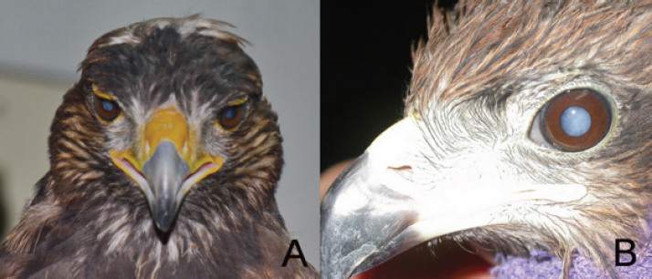
Fig. 1. Hypermature cataract in an unknown age male black kite: (A) OU and (B) OS. Other anterior parts of both eyes were normal.
The general physical examination was normal. The visual examination revealed that the bird showed blindness, determined by negative menace and absence of visual tracking response. The dazzle reflex was positive OU. The direct pupillary light reflex was brisk and completed OU. Consensual pupillary light reflex was not carried out due to complete optic nerve fiber decussation at the optic chiasm in birds (Kern et al., 1984; Bayon et al., 2007). Ocular examination was carried out using a slit-lamp biomicroscope (SL-15 portable slit lamp, Kowa Company, Ltd., Nagoya, Japan). The cornea, anterior chamber, and iris were normal. A hypermature cataract was observed OU (Fig. 1). The fundus could not be assessed due to the substantial opacity of the lenses. A clinical diagnosis of cataract OU was made based on the appearance of bilateral opacity of the lenses obstructing photo-translumination to the fundus. No other eye problem, such as lens-induced uveitis, was detected.
Ocular biometry
For complete ocular examination, anesthesia was carried out by masking with 5% isoflurane and maintaining 2% isoflurane (Terrell, Minrad Inc., PA). A-scan biometry (E-Z Scan AB 5500+, Sonomed, NY) with an immersion technique was carried out to evaluate the axial length (Fig. 2). The average axial lengths were 21.85 mm oculus dexter (OD) (right eye) and 21.70 mm oculus sinister (OS) (left eye). The corneal curvature was measured using keratometry (ARK-30, Nidex Co. Ltd., Gamagori Aichi, Japan). The average corneal refractive power was 51.75 D (K1, vertical axis) and 54.25 D (K2, horizontal axis). IOL power was calculated using the SPK-2 formula. The average IOL power was +17.5 D. B-scan ultrasonography (E-Z Scan AB 5500+, Sonomed, NY) revealed hyperechogenicity of lens OU with no posterior segment abnormalities. The complete blood count and serum biochemistry profile were within referent limits.
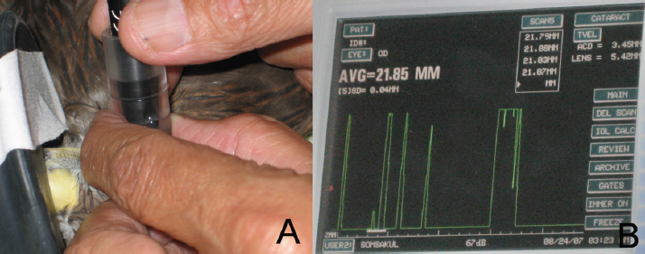
Fig. 2. (A) Emersion technique of A-scan ultrasonography carried out in the black kite under general anesthesia. (B) Screen showing A-scan ultrasonography in the black kite OD.
Currently, there is no effective medical therapy to resolve the advanced lesion of cataracts discovered in animals and human beings. Therefore, bilateral cataract surgery and IOL implantation were planned to improve eye condition and return vision. Topical 1% prednisolone acetate eye suspension (Inf-oph, Seng Thai Company, Bangkok, Thailand) was applied OU thrice daily for 3 days preoperatively.
Surgical management
Anesthesia induction with 5% isoflurane and maintained with 2% isoflurane (Terrell, Piramal Critical Care, Inc., PA) were delivered via a face mask. The periocular area was prepared for sterile surgery by gentle cleansing with 1:50 povidone:iodine solution diluted with normal saline. Subsequently, the animal was positioned lateral recumbency on a sandbag. The operative area was draped with an antimicrobial incise drape (Ioban, 3M Health Care, MN). Then, 5-0 silk stay sutures were placed to facilitate both upper and lower eyelid retraction. A 3-mm-long corneal incision near the limbus was made at the 12 o’clock position with a 3.0 mm angled Slit Knife (SharpointTM, Surgical Specialties Corporation, Tijuana, CP, Mexico). The neuromuscular blocker, 0.2 mg of atracurium 10 mg/ml (Tracrium, GlaxoSmithKline Manufacturing SPA, Parma, Italy), was injected into the anterior chamber to achieve pupil dilation. The anterior chamber was maintained by a viscoelastic agent containing 4% sodium chondroitin sulfate and 3% sodium hyaluronate (Viscoat, Alcon Surgical Inc., TX) to protect the corneal endothelium. A curvilinear capsulorhexis was made using Vannas scissors. Utrata capsulorhexis forceps were used to remove the torn anterior lens capsule. Due to the softness of the cataractous lenses in birds, an irrigation and aspiration technique was chosen instead of phacoemulsification. Aspiration of the soft cataractous cortical and nuclear materials using an I/A probe of a phacoemulsification machine (AMO Prestige, Allergan, CA) resulted in the successful removal of all cataractous materials. The fluid used during the procedure was a mixture of balanced salt solution (Ocusol, A.N.B. Laboratories Co., Ltd., Bangkok, Thailand) mixed with 2 ml of 1 mg/ml epinephrine bitartrate (Adrenaline, Atlantic Laboratories Corporation Ltd., Samut Prakan, Thailand) and 0.1 ml (25,000 IU/5 ml) of heparin sodium (Nuparin, Troikaa Pharmaceuticals Ltd., Gujarat, India) per 500 ml of fluid. The posterior lens capsule was polished to reduce the chance of postoperative posterior lens capsular opacity. The anterior chamber and capsular bag were refilled with the viscoelastic agent. A + 17.5 D acrylic foldable single-piece lens with 13.0 mm overall length and 6.0 mm optic diameter (AcrySof model SA60AT, Alcon Laboratories, Fort Worth, TX) was inserted via a lens inserter. The viscoelastic agent was removed from the capsular bag and the anterior chamber while the balanced salt solution was flushed to reinflate the anterior chamber. The corneal incision was closed with a 10-0 nylon suture in a simple interrupted pattern OU. The same procedure was carried out in the contralateral eye. A subconjunctival injection was administered with 2 mg of gentamicin sulphate (Gentamicin Sulphate, T.P. Drug Laboratories, Bangkok, Thailand). One suture of a temporary tarsorrhaphy with 5-0 silk in a simple interrupted pattern was carried out OU to prevent self-trauma to the eyes postoperatively.
Topical application of a neomycin, polymixin-B, and gramicidin solution (Poly-oph, Seng Thai Company, Bangkok, Thailand) was administered postoperatively every 6 hours as antibacterial prophylaxis. In addition, topical 1% prednisolone acetate eye suspension (Inf-oph, Seng Thai Company, Bangkok, Thailand) was prescribed for anti-inflammation every 6 hours OU.
Postoperative outcome
Three days after surgery, tarsorrhaphy was removed. The visual tracking test was still negative. The cornea did not retain fluorescein dye OU. Mild generalized corneal edema was present and aqueous flare was observed (+0.5) in the anterior chamber OU. The frequency of topical prednisolone acetate administration was increased to six times per day OU. The next day, the corneal edema and aqueous flare had decreased OU. Four days after surgery, both eyes were open comfortably. The improved vision was observed because the black kite was able to follow a moving hand, and the menace response was positive OU. Decreased corneal edema and aqueous flare were observed OU.
One week after surgery, OU observed a minimal amount of corneal edema and aqueous flare (Fig. 3). Posterior capsular opacity was observed OD (Fig. 3A). Posterior synechia was presented at 7–5 o’clock OS (Fig. 4). Therefore, 0.1 mg of atracurium was intracamerally injected OS to induce mydriasis under general anesthesia with 2% isoflurane inhalation. This procedure was unable to break the synechia, and this was regularly observed in the later examination. The frequency of topical 1% prednisolone acetate was decreased to six times per day. Two weeks after surgery, corneal edema was resolved; however, a small amount of aqueous flare was still present. The topical antibiotic solution was discontinued.
Four weeks after surgery, no corneal edema or aqueous flare was observed OU. All medications were discontinued. The black kite was able to capture his prey when fed with live mice. Six weeks after surgery, the bird’s vision had improved, and the bird was able to follow moving objects such as cars or people outside his cage.
Two months after surgery, the bird was allowed to fly in a 1 × 1 m cage. Refraction was evaluated by streak retinoscopy 4 months after surgery. The refraction was −0.50 D OD, whereas OS could not be assessed due to a poor reflection. However, the black kite had a good response when stimulated with moving objects visible from both eyes. The vision of this black kite was judged as substantially improved and adequate for his normal life because the black kite could fly without bumping into any objects and was able to catch all running mice offered. The bird was kept in the Kasetsart University Raptor Rehabilitation Unit since it was tame. Three years after surgery, the bird could still fly without hitting objects and could precisely catch running prey.
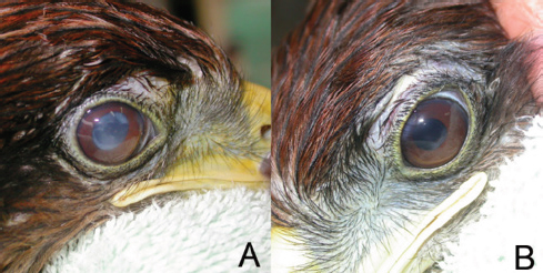
Fig. 3. (A) One week postoperatively, mild corneal edema and posterior capsular opacity were investigated OD. (B) Mild corneal edema was evident OS.
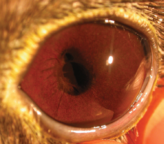
Fig. 4. Posterior synechia presented at 7–5 o’clock OS.
Discussion
Cataract is a common disease in lenses of birds (Murphy et al., 1982; Williams, 2008). Cataract surgery is recommended in birds exhibiting visual deficits (Brooks, 1997; Williams, 2008). Nevertheless, reports of cataract surgery results in birds have been limited (Kern et al., 1984; Keller, 1992; Carter et al., 2007; Rainwater et al., 2015; Sigmund et al., 2019). A successful outcome (improved postoperative visual acuity) was reported in 83.3% (10 out of 12) of birds undergoing cataract removal (5 out of 12 of those birds were alive >3 years postoperatively) (Rainwater et al., 2015). 47% of affected eyes that did not receive suitable surgical treatment were reported to develop lens-induced uveitis (Rainwater et al., 2015). Several surgical techniques to treat cataracts have been described for birds (Brooks, 1997; Ofri, 2018). Intracapsular cataract extraction is recommended only in the luxated lens because of the fusion of the ciliary process to the lens in birds (Brooks et al., 1983). Extracapsular cataract extraction has been reported in an Andean condor, a canary, and a crow (Slatter et al., 1983; Moore et al., 1985; Keller, 1992). However, this technique might cause severe uveitis, which results in a loss of all vision in the eye after cataract surgery (Keller, 1992). In general, phacoemulsification is an alternative technique for all stages of cataracts (Kern et al., 1984; Brooks, 1997; Rainwater et al., 2015; Sigmund et al., 2019). Another report showed that phacoemulsification with or without IOL implantation was successfully carried out in the following species: one great horned owl (Carter et al., 2007), one screech owl (Kern et al., 1984), two peregrine falcons (Kern et al., 1984; Sigmund et al., 2019), four bald eagles, and two red-tailed hawks (Sigmund et al., 2019). However, in one report of a falcon, the vision was limited due to posterior capsular opacification (Kern et al., 1984). Cataracts in birds are generally soft (Brooks, 1997; Sigmund et al., 2019). The aspiration and irrigation technique is an alternative procedure that provides a substantially better result in a soft cataract (Keller, 1992; Sigmund et al., 2019). The aspiration and irrigation technique is also suggested for small eyes that cannot accommodate standard ophthalmic instrumentation (Ofri, 2018). Our case report chose the aspiration and irrigation technique because both cataracts were soft despite being hypermature.
There are several particular concerns for cataract surgery in birds due to their different ocular anatomy from mammals. For example, the fusion of the ciliary process to the lens capsule is unique in birds. The excessive tension applying to the ciliary process may cause intraocular hemorrhaging during anterior capsule incision (Kern et al., 1984; Keller, 1992). Furthermore, the ciliary body and iris of the bird contain striated muscle rather than smooth muscle (Keller, 1992). Therefore, parasympathetic blocking agents such as tropicamide or atropine, which are routinely used in a mammal, cannot be used to dilate the pupil in birds (Verschueren and Lumeij, 1991). In addition, there are only smooth muscle responses to topical parasympatholytic agents; thus, intracameral injection of a neuromuscular blocking agent is required (Verschueren and Lumeij, 1991; Carter et al., 2007). Intracameral atracurium or d-tubocurarine has been reported for successful pupillary dilatation in birds (Murphy, 1987; Verschueren and Lumeij, 1991; Carter et al., 2007). On the other hand, topical d-tubocurarine has no mydriasis effect in birds (Verschueren and Lumeij, 1991). Studies using other topical curare derivatives (topical vecuronium bromide and rocuronium bromide) have been reported in pupillary dilatation in raptors without side effects (Mikaelian et al., 1994; Barsotti et al., 2010). Topical vecuronium bromide (4 mg/ml), 2 drops every 15 minutes for 2 instillations, achieved full mydriasis within 30 minutes, and was maintained for 4 hours in kestrels (Mikaelian et al., 1994). A single dose of topical rocuronium bromide (0.35 mg) achieved full mydriasis within 80 minutes and was maintained for 4 hours in tawny owls (Barsotti et al., 2010). When used at an excessive dose, the side effects of neuromuscular blocking agents are eyelid, neck, hindlimb, and respiratory muscle paralysis (Mikaelian et al., 1994).
The postoperative complications in this black kite included corneal edema, anterior uveitis, posterior synechia, and posterior capsular opacity. These complications are commonly observed postoperatively (Williams, 2008; Sigmund et al., 2019). Posterior capsular opacity and corneal scarring were the most common postoperative ocular abnormalities reported at 22.2% and 11.1%, respectively (Rainwater et al., 2015). Corneal edema is usually present 24–72 hours postoperatively (Brooks, 1997). Other serious postoperative complications could include glaucoma, persistent uveitis, persistent iridocyclitis, plasmoid aqueous, iris bombe, corneal ulceration, corneal fibrovascular infiltrates, retained lens cortex, wound leakage, vitreous presentation into the anterior chamber, retinal degeneration, retinal detachment, and infectious endophthalmitis (Brooks, 1997; Sigmund et al., 2019). None of these severe conditions were observed with the bird in the current study.
Theoretically, the postoperative aphakic eye probably results in farsightedness or hyperopia, and the visual image would be minimized and blurry. Furthermore, the loss of ocular accommodation potentially causes severe debilitation in some birds. Therefore, an IOL implantation after cataract surgery is imperative to the lifestyle of diurnal raptors such as the black kite in the current study to restore emmetropia (Kuhn et al., 2015). Prior to surgery, the appropriate IOL refractive power corresponding to a given species needs to be determined (Brooks, 1997). The IOL strength has been studied previously in other birds, especially raptors, but not the black kite. Examples of IOL strength in particular birds have been reported: +13.82 D for a great horned owl (Carter et al., 2007), +17 D for a red-tailed hawk, +18 D for a bald eagle (Sigmund et al., 2019), and more generally +16.4 to 17.4 for bald eagles (Kuhn et al., 2015). The calculation method for the optimal IOL power requires parameters derived using keratometry and biometry (Sigmund et al., 2019). The IOL power in this black kite was +17.5 D, which is similar to the human IOL. Therefore, the implantation of a human foldable IOL was selected, and the surgery was considered successful because the bird’s vision was substantially improved, as displayed through successful flight navigation. To the authors’ knowledge, this is the first report of bilateral cataract surgery with IOL implantation using a calculated dioptric power (+ 17.5 D) IOL in a black kite.
Acknowledgment
The authors appreciate Dr. Manthanee Pairachavet for the technical assistance.
Conflict of interest
The authors declare that there is no conflict of interest.
References
Barsotti, G., Briganti, A., Spratte, J.R., Ceccherelli, R. and Breghi, G. 2010. Mydriatic effect of topically applied rocuronium bromide in tawny owls (Strix aluco): comparison between two protocols. Vet. Ophthalmol. 13(Suppl 1), 9–13.
Bayon, A., Almela, R.M. and Talavera, J. 2007. Avian ophthalmology. Eur. J. Companion. Anim. Pract. 17(3), 253–265.
Brooks, D.E. 1997. Avian cataracts. Semin. Avian. Exot. Pet. Med. 6(3), 131–137.
Brooks, D.E., Murphy, C.J., Quesenberry, K.E. and Walsh, M.T. 1983. Surgical correction of a luxated cataractous lens in a barred owl. J. Am. Vet. Med. Assoc. 183(11), 1298–1299.
Buyukmihci, N.C., Murphy, C.J. and Schulz, T. 1988. Developmental ocular disease of raptors. J. Wildl. Dis. 24(2), 207–213.
Carter, R.T., Murphy, C.J., Stuhr, C.M. and Diehi, K.A. 2007. Bilateral phacoemulsification and intraocular lens implantation in a great horned owl. J. Am. Vet. Med. Assoc. 230(4), 559–561.
Dees, D.D. and MacLaren, N.E. 2013. Presumptive electric cataracts in a great horned owl (Bubo virginianus). Vet. Ophthalmol. 16(1), 73–76.
Keller, C.B. 1992. Bilateral extraction of cataracts in a crow. Can. Vet. J. 33(4), 273–274.
Kern, T.J., Murphy, C.J. and Riis, R.C. 1984. Lens extraction by phacoemulsification in two raptors. J. Am. Vet. Med. Assoc. 185(11), 1403–1406.
Keymer, I.F. 1977. Cataracts in birds. Avian. Pathol. 6(4), 335–341.
Kuhn, S.E., Hendrix, D.V.H., Jones, M.P., Ward, D.A., Baine, K.H. and Franklin, S.R. 2015. Biometry, keratometry, and calculation of intraocular lens power for the bald eagle (Haliaeetus leucocephalus). Vet. Ophthalmol. 18(Suppl 1), 106–112.
Mikaelian, I., Paillet, I. and Williams, D. 1994. Comparative use of various mydriatic drugs in kestrels (Falco tinnunculus). Am. J. Vet. Res. 55(2), 270–272.
Moore, C.P., Pickett, J.P. and Beehler, B. 1985. Extracapsular extraction of senile cataract in an Andean condor. J. Am. Vet. Med. Assoc. 187(11), 1211–1213.
Murphy, C.J. 1987. Raptor ophthalmology. Compend. Cont. Educ. Pract. Vet. 9, 241–260.
Murphy, C.J., Kern, T.J., McKeever, K., McKeever, L. and MacCoy, D. 1982. Ocular lesions in free-living raptors. J. Am. Vet. Med. Assoc. 181, 1302–1304.
Ofri, R. 2018. Diseases of the lens. In Slatter’s fundamentals of veterinary ophthalmology. 6th ed. Eds., Maggs, D.J., Miller, P.E. and R. Ofri, St. Louis, MO: Elsevier, pp: 306–333.
Rainwater, K.L., Sykes, J.M. and Sapienza, J.S. 2015. Retrospective investigation of cataract management in avian species in a zoologic collection. J. Zoo. Wildl. Med. 46(4), 858–869.
Sigmund, A.B., Jones, M.P., Ward, D.A. and Hendrix, D.V.H. 2019. Long-term outcome of phacoemulsification in raptors-A retrospective study (1999–2014). Vet. Ophthalmol. 22(3), 360–367
Slatter, D.H., Bradley, J.S., Vale, B., Constable, I.J. and Cullen, L.K. 1983. Hereditary cataracts in canaries. J. Am. Vet. Med. Assoc. 183(8), 872–874.
Verschueren, C.P. and Lumeij, J.T. 1991. Mydriasis in pigeons (Columbia livia domestica) with d-tubocurarine: topical instillation versus intracameral injection. J. Vet. Pharmacol. Therap. 14(2), 206–208.
Williams, D.L. 2008. Raptor ophthalmology. In BSAVA manual of raptors, pigeons and passerine birds. Eds., Chitty, J. and M. Lierz, Gloucester, UK: British Small Animal Veterinary Association, pp: 278–283.


