


| Case Report | ||
Open Vet J. 2022; 12(5): 628-631 Open Veterinary Journal, (2022), Vol. 12(5): 628–631 Case Report A rare urachal abscess in a young bull with conservative managementJalal M. Abdelhadi1*, Aiman A. Shalgum2 and Mohamed Abushhiwa11Department of Surgery and Theriogenology, Faculty Veterinary Medicine, University of Tripoli, Tripoli, Libya 2Department of Anatomy, Histology and Embryology, Faculty Veterinary Medicine, University of Tripoli, Tripoli, Libya *Corresponding Author: Jalal M. Abdelhadi. Department of Surgery and Theriogenology, Faculty Veterinary Medicine, University of Tripoli, Tripoli, Libya. Email: abdelhadijalal [at] gmail.com Submitted: 27/04/2022 Accepted: 10/08/2022 Published: 08/09/2022 © 2022 Open Veterinary Journal
AbstractBackground: The urachus is an embryonic remnant occurring as a result of the involution of the allantoic duct and the ventral cloaca. This canal becomes progressively obliterated after birth. It uncommonly persists to different degrees after birth. Case Description: A young bull was presented with distended abdomen and clinical signs of chocking, with low-grade fever, loss of appetite, frothy mouth arched back. On the first inspection, the animal was suspected to have simple indigestion. The treatment was attempted in this stage by introducing a stomach tube but only little relief was achieved. Therefore, 5 days later, an exploratory laparotomy was done and a big balloon-like cyst structure extended ventro-latrally in the abdominal cavity was noticed. That structure was located on the floor of the abdominal cavity extending from the pelvic rim caudally to the umbilical region cranially. The structure was then incised and evacuated and a rubber tube was fixed for constant drainage for up to one month later. The bull was followed-up and made a good recovery after a month post-surgery. Conclusion: We found that urachal abscess could be treated simply via surgical evacuation of the abscess and proper drainage for some time with a very promising outcome. Additionally, this affection can be diagnosed based on history, clinical signs, and exploratory surgery, when relatively modern diagnostic techniques are not available. Keywords: Abscess, Anatomy, Bull, Embryology, Urachus. IntroductionThe embryology of the umbilicus and the developmental basis for surgical abnormalities are still not well described in ruminants. Most of the cases like umbilical hernias, abdominal wall defects, urachus abscesses, and omphalomesenteric remnants are considered umbilical abnormalities and they need urgent intervention (Abdelhadi and Oheida, 2021). A stark contrast is noticed between the physio-anatomical importance of the umbilicus during different fetal developmental stages and its function after birth. During fetal development, the umbilicus functions as vessel which allow blood flow between the placenta and the fetus (Wang and Zhao, 2010). It also has an important role in the development of the intestine and the urinary system (Basta and Lipsett, 2021). While after birth, the umbilical cord undergoes autolysis and disappears, and no evidence of functionality is shown by these connections after birth. Nevertheless, umbilical disorders, such as urachus, are rarely encountered in the domestic animal clinic (Parsons, 2009). An understanding of the anatomy and embryology of the abdominal wall and umbilicus is important for identifying and properly treating such a condition. Anatomically, urachus is located among the fascia transversalis and the peritoneum collectively (Schott et al., 2018), which is more adjacent to umbilical ligaments and the remnants of the umbilical arteries during fetal life (Barrell et al., 2020). The urachus varies in length and diameter according to the animal species (Visser et al., 2020). It is surrounded by a fascial sheath known as the umbilicovesical sheath, which extends laterally to each umbilical artery and cranially above the dorsal wall of the urinary bladder to the hypogastric arteries caudally and the pelvic diaphragm cranially, forming a self-contained space (Begg, 1930). Histologically, the urachus is composed of three layers; an inner mucosal layer, a submucosal tissue layer, and an outer smooth muscle layer, while the central lumen is irregular, beaded and packed with desquamated epithelial debris (Begg, 1930). During developmental stages of the embryo, the embryonic disk is in contact with the yolk sac anteriorly. As the embryo grows, and the differential growth of tissues leads to the folding appearance of the embryo and its ventral attachment to the yolk sac becomes narrower (Charles and Faye-Petersen, 2014). The yolk sac intracoelomic portion becomes the primitive alimentary canal (Bhatia et al., 2021) and connects with the extracoelomic portion through the vitelline duct. The allantois raises from the hindgut and grows into the body stalk. Then, the yolk stalk and the body stalk eventually fuse to become the umbilical cord (Laverty and Salisbury, 2002). Urachus is derived from the bladder, and its proximal portion is formed from the allantoic duct (Begg, 1930). Once the bladder descends into the pelvis during developmental stages, it keeps its attachment to the umbilicus by the urachus (Begg, 1930). As the urachus develops, it loses its attachment to the umbilicus (Begg, 1930). Five anatomical urachal terminations will occur based on the differential growth rates of the urachus (Nguyen and Cilento, 2010). Failure of the normal obliterative processes of the vitelline duct and the urachus leads to the abnormal formation of a patent urachus, a urachal sinus, and/or a urachal cyst (Rich et al., 1983). Case DetailsAn inbred Friesian young bull was presented at Al-Etgaan private clinic in Tripoli, Libya. The owner noticed for a week or more that the young bull is physically not active and suffering from a sharp weight loss. Other signs were also observed like loss of appetite, frothy mouth, arched back restlessness, distended abdomen, rough wrinkled coat, and the animal was laying down and getting up frequently. The clinical examination revealed a low-grade fever, pale mucous membranes, a markedly distended left flank and hypersalivation with ruminal stasis (Fig. 1). Primarily, the case was suspected to be a simple indigestion. The initial treatment was the placement of a stomach tube. The tube was passed directly through the oesophagus smoothly with simple massaging from outside. Large amount of ingesta was passed out through the tube but without a reduction in the distended flank. After 5 days of treatment, no improvement was noticed although the animal was receiving antipyretic, antacid and antibiotics. At this point, a surgical intervention was done through the left flank. An exploratory laparotomy on the standing position and under local anaesthetic infiltration was performed (Fig. 2). After an incision through the skin, subcutaneous tissues and the muscles, a big balloon-like cyst structure was protruded from the incision (Fig. 3) and its boundaries extended ventrally on the floor of the abdominal cavity from the pelvic rim caudally to umbilical region cranially and almost touching the urinary bladder. A huge amount of creamy bus flowed up strongly with very bad odour gases came out, when a small cut was made through this cyst accidentally. A complete aseptic evacuation of about four litters of bus was followed by washing the whole pocket using normal saline, hydrogen peroxide, and betadine solution. This lavage was repeated many times until a clear exudate came out. Moreover, a counter opening for drainage tube was applied for a complete drainage of the extra fluid (Fig. 2). A systemic antibiotic was also given IV; 3 gm Ceftriaxone, beside a nonsteroidal anti-inflammatory, flunixin meglumine, with a dose of 2.2 mg/kg once daily for 7 days. The animal had a good postoperative recovery after one-month follow-up and the animal is currently gaining weight and appearing clinically healthy.
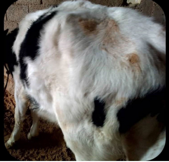
Fig. 1. A young bull suffering from urachal abscess. Loss of weight, rough coat and distended abdomen with arched back. DiscussionThe urachus, as previously mentioned, arises from the upper part of the bladder and both of them arise from the ventral part of the cloaca (Begg, 1930). There are five types of urachal abnormalities: the patent urachus; in which the entire tubular structure fails to close, the urachal sinus; in which both ends of the canal close leaving an open central portion, the urachal cyst; which drains proximally into the umbilicus, the vesicourachal diverticulum; where the distal communication to the bladder persists, and the alternating sinus: which can drain to either bladder or umbilicus (Wilson et al., 2019). The incidence of urachus in human adults is unknown but it is considered rare, while no reports are available about it in domesticated animals (Wilson et al., 2019). Manners of presentation of urachal anomalies in newly born animal differ from those understood in adult animals. In adults, the commonest form is the urachal cyst, with no drainage and the abscess being the usual mode of presentation. The septic contamination of this structure could be through the lymphatics and /or as ascending from the urinary bladder infections (Qureshi et al., 2013). Urachus cyst is usually asymptomatic until it becomes infected (Qureshi et al., 2013). The treatment of choice for urachal cyst is not always performed by complete excision as with this case. Earlier reports suggested that appropriate antibiotic therapy of infected urachus after complete drainage gave good results (Lipskar et al., 2010). In the present case, we adopted an operative procedure for complete drainage of the cyst with a good management and administration of broad-spectrum antibiotics post-surgery.
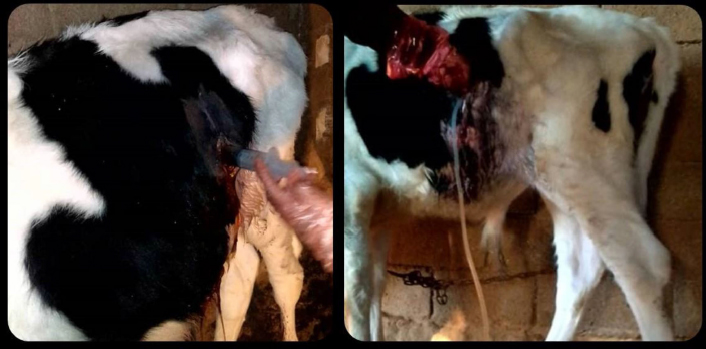
Fig. 2. The laparotomy procedure performed on the young bull for urachus evacuation and washing and abscess drainage. The site preparation and local anaesthesia (left) and the drainage tube (right).
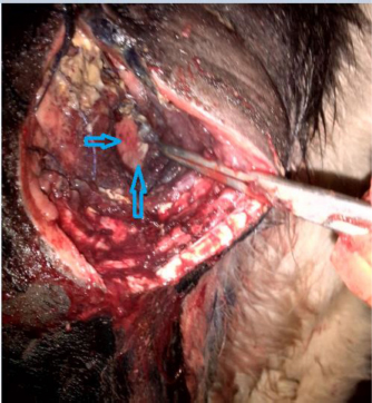
Fig. 3. Urachal abscess localization after an exploratory laparotomy. Arrows indicate the site of the urachal abscess. ConclusionThe current report documented a rare condition of urachal abscess in a young bull based on the clinical presentation and exploratory surgery. The diagnosis of such conditions requires some relatively more advanced diagnostic imaging techniques, namely, contrast radiography and CT scan. Those methods were unfortunately not available but the clinical signs and the laparotomy surgery confirmed the diagnosis. This affection was treated using simple, cost effective and relatively non-invasive surgical technique with great success. The case was followed-up for almost 2 months post-surgery until complete recovery and the bull became clinically normal. AcknowledgmentsSpecial thanks to all the staff of Anatomy, Histology and Embryology Department for their contributions in this work. ReferencesAbdelhadi, J.M. and Oheida, A.H. 2021. Herniorrhaphy in two newborn lambs with omphalocele. Open. Vet. J. 11(4), 667–671. Barrell, E.A., Burton, A.J., Arroyo, L.G., Saulez, M.N., Beasley, E.M., Schott, H.C. and Dodd, C.C. 2020. Chapter 34-diseases of the renal system. In Large animal internal medicine. Eds., Smith, B. P., Van Metre, D. C. and Pusterla N. 6th ed. St. Louis, MO: Mosby, pp: 956–1005.e1009. Basta, M. and Lipsett, B.J. 2021. Anatomy, abdomen and pelvis, umbilical cord. In: StatPearls [Internet]. Treasure Island (FL): StatPearls Publishing; PMID: 32491321. Begg, R.C. 1930. The urachus: its anatomy, histology and development. J. Anat. 64(Pt 2), 170–183. Bhatia, A., Shatanof, R.A. and Bordoni, B. 2021. Embryology, gastrointestinal. In: StatPearls [Internet]. Treasure Island (FL): StatPearls Publishing; PMID: 30725857. Charles, A. K. and Faye-Petersen, O.M. 2014. Human placental development from conception to term. In Pathobiology of human disease. Eds., McManus, L. M. and Mitchell, R.N. San Diego, CA: Academic Press, pp: 2322–2341. Laverty, P.H. and Salisbury, S.K. 2002. Surgical management of true patent urachus in a cat. J. Small. Anim. Pract. 43(5), 227–229. Lipskar, A.M., Glick, R.D., Rosen, N.G., Layliev, J., Hong, A.R., Dolgin, S.E. and Soffer, S.Z. 2010. Nonoperative management of symptomatic urachal anomalies. J. Pediatr. Surg. 45(5), 1016–1019. Nguyen, H.T. and Cilento, B.G. 2010. Bladder diverticula, urachal anomalies, and other uncommon anomalies of the bladder. In Pediatric urology. Eds., Gearhart, J. P., Rink, R.C. and Mouriquand, P.D.E. Philadelphia, PA: W.B. Saunders, pp: 416–424. Parsons, D.A. 2009. The newborn foal. In Equine breeding management and artificial insemination. Eds., Samper, J.C. Saint Louis, MO: W.B. Saunders, pp: 261-276. Qureshi, K., Maskell, D., McMillan, C. and Wijewardena, C. 2013. An infected urachal cyst presenting as an acute abdomen-a case report. Int. J. Surg. Case Rep. 4(7), 633–635. Rich, R.H., Hardy, B.E. and Filler, R.M. 1983. Surgery for anomalies of the urachus. J. Pediatr. Surg. 18(4), 370–372. Schott, H.C., Waldridge, B. and Bayly, W.M. 2018. Disorders of the urinary system. In Equine internal medicine. Eds., Reed, S.M., Bayly, W.M. and Sellon, D.C. 4th ed. Philadelphia, PA: W.B. Saunders, pp: 888–990. Visser, J., Kummeling, A., van Nugteren, M.A., Grinwis, G.C.M. and Brocks, B.A.W. 2020. Resection of urachal anomalies in dogs with recurrent lower urinary tract disease. Vet. Surg. 49(1), 214–221. Wang, Y. and Zhao, S. 2010. Vascular biology of the placenta. San Rafael, CA: Morgan and Claypool Life Sciences. PMID: 21452443. Wilson, A.L., Gandhi, J., Seyam, O., Rahmani, B., Patel, S., Joshi, G. and Khan, S.A. 2019. Urachal anomalies: a review of pathological conditions, diagnosis, and management. Transl. Res. Anat. 16, 100041. | ||
| How to Cite this Article |
| Pubmed Style Abdelhadi JM, Shalgum AA, Abushhiwa MH. A rare urachal abscess in a young bull with conservative management. Open Vet J. 2022; 12(5): 628-631. doi:10.5455/OVJ.2022.v12.i5.6 Web Style Abdelhadi JM, Shalgum AA, Abushhiwa MH. A rare urachal abscess in a young bull with conservative management. https://www.openveterinaryjournal.com/?mno=8787 [Access: April 19, 2024]. doi:10.5455/OVJ.2022.v12.i5.6 AMA (American Medical Association) Style Abdelhadi JM, Shalgum AA, Abushhiwa MH. A rare urachal abscess in a young bull with conservative management. Open Vet J. 2022; 12(5): 628-631. doi:10.5455/OVJ.2022.v12.i5.6 Vancouver/ICMJE Style Abdelhadi JM, Shalgum AA, Abushhiwa MH. A rare urachal abscess in a young bull with conservative management. Open Vet J. (2022), [cited April 19, 2024]; 12(5): 628-631. doi:10.5455/OVJ.2022.v12.i5.6 Harvard Style Abdelhadi, J. M., Shalgum, . A. A. & Abushhiwa, . M. H. (2022) A rare urachal abscess in a young bull with conservative management. Open Vet J, 12 (5), 628-631. doi:10.5455/OVJ.2022.v12.i5.6 Turabian Style Abdelhadi, Jalal Mohamed, Aiman A: Shalgum, and Mohamed H. Abushhiwa. 2022. A rare urachal abscess in a young bull with conservative management. Open Veterinary Journal, 12 (5), 628-631. doi:10.5455/OVJ.2022.v12.i5.6 Chicago Style Abdelhadi, Jalal Mohamed, Aiman A: Shalgum, and Mohamed H. Abushhiwa. "A rare urachal abscess in a young bull with conservative management." Open Veterinary Journal 12 (2022), 628-631. doi:10.5455/OVJ.2022.v12.i5.6 MLA (The Modern Language Association) Style Abdelhadi, Jalal Mohamed, Aiman A: Shalgum, and Mohamed H. Abushhiwa. "A rare urachal abscess in a young bull with conservative management." Open Veterinary Journal 12.5 (2022), 628-631. Print. doi:10.5455/OVJ.2022.v12.i5.6 APA (American Psychological Association) Style Abdelhadi, J. M., Shalgum, . A. A. & Abushhiwa, . M. H. (2022) A rare urachal abscess in a young bull with conservative management. Open Veterinary Journal, 12 (5), 628-631. doi:10.5455/OVJ.2022.v12.i5.6 |

