Open Veterinary Journal, (2021), Vol. 11(3): 468–470
Short Communication
10.5455/OVJ.2021.v11.i3.20
Findings suggestive of coronary microvascular dysfunction in cats with myocardial ischemia
Guillermo Belerenian1, Pablo Alejandro Donati2,3, Cristian Daniel Rodriguez2, Victor Castillo1, Juan Manuel Guevara2, Claudia Pucheta1, Sergio Ferraris4 and Roberto Walter Israel Olivares5*
1Luis Pasteur Zoonosis Institute,, Buenos Aires, Argentina
2Veterinaria UCIcoop, Buenos Aires, Argentina
3Universidad de Buenos Aires, Facultad de Ciencias Veterinarias, Cátedra de Anestesiología y Algiología, Buenos Aires, Argentina
4Research and Experimental Models Center, Maimonides University, Buenos Aires, Argentina
5Servicio de Patología Diagnóstica LAPAVET-ESFA. Cátedra de Patología e Histología, Escuela de Medicina y Cirugía Veterinaria San Francisco de Asís, Universidad Veritas, San José, Costa Rica
*Corresponding Author: Roberto Walter Israel Olivares. Servicio de Patología Diagnóstica LAPAVET-ESFA. Cátedra de Patología e Histología, Escuela de Medicina y Cirugía Veterinaria San Francisco de Asís, Universidad Veritas, San José, Costa Rica. Email: rolivares [at] veterinariaveritas.ac.cr
Submitted: 13/04/2021 Accepted: 14/08/2021 Published: 04/09/2021
© 2021 Open Veterinary Journal
This is an Open Access article distributed under the terms of the Creative Commons Attribution-Non Commercial-No Derivatives License (http://creativecommons.org/licenses/by-nc-nd/4.0/), which permits non-commercial re-use, distribution, and reproduction in any medium, provided the original work is properly cited, and is not altered, transformed, or built upon in any way.
Abstract
Background: Myocardial infarction (MI) is an important cause of death and disability among humans worldwide. Few studies have reported the occurrence of MI in small animals as well. Reports in human medicine indicate that up to 30% of patients with clinical signs compatible with myocardial ischemia suggestive of coronary disease exhibit normal epicardial arteries at angiography. These symptoms have been associated with a syndrome characterized by alterations in cardiac microvasculature, known as coronary microvascular dysfunction (CMD).
Aim: This study aimed to describe the necropsy findings and clinical-pathological characterization (when available) of cats with histopathological findings suggesting CMD.
Methods: Necropsy records of cats presenting histopathological diagnosis compatible with acute and/or chronic MI, with normal epicardial arteries and microvascular disorders were evaluated.
Results: Twenty animals met the inclusion criteria. Eight cats (40%) exhibited findings compatible with mild hypertrophic cardiomyopathy (HCM) without left atrial enlargement, one (5%) presented restrictive cardiomyopathy, and another one (5%) had lesions consistent with histiocytoid cardiomyopathy. The remaining cats (50%) showed alterations compatible with severe HCM with left atrial enlargement. In all cases, epicardial arteries were normal (without obstruction). All the evaluated hearts exhibited myocardial multifocal fibrosis along with replacement of cardiomyocytes by adipose tissue and blood vessels with hyperplasia and hypertrophy of the muscular layer with protrusion of the nuclei of the endothelial cells.
Conclusion: These findings suggest the presence of microvascular dysplasia of the coronary arteries. Further studies are necessary to confirm and clinically characterize these results.
Keywords: Coronary microcirculation, microvascular dysfunction, feline ischemia.
Introduction
Myocardial infarction (MI) is an important cause of death and disability in humans worldwide (Thygesen et al., 2007). Few studies have reported the occurrence of MI in small animals. In a previous post-mortem examination study, the presence of MI was described in 32 dogs and 5 cats (Driehuys et al., 1998). All cats showed lesions compatible with left ventricular hypertrophy (LVH). Previous studies indicate that cats with end-stage hypertrophic cardiomyopathy (HCM) might exhibit thinning of the myocardial walls (Cesta et al., 2005). This phenomenon might be associated with the presence of chronic myocardial ischemia. Reports in human medicine indicate that up to 30% of patients with clinical signs compatible with myocardial ischemia suggestive of coronary disease exhibit normal epicardial arteries at angiography (Crea and Lanza, 2004; Camici and Crea, 2007). These lesions have been associated with a syndrome characterized by alterations in cardiac microvasculature, known as coronary microvascular dysfunction (CMD) (Camici and Crea, 2007). To the authors’ best knowledge, CMD has not been previously described in cats. This work aimed to describe the necropsy findings along with clinical-pathological characterization (when available) of cats with histopathological findings suggesting CMD.
Material and Methods
Of a total of 480 necropsies of cats performed in the Luis Pasteur Zoonosis Institute between January 2016 and January 2020, those cats presenting the following inclusion criteria were selected:
- Histopathological diagnosis compatible with acute and/or chronic MI.
- Normal epicardial arteries (without obstruction).
- Histological evidence of microvascular disorders.
The epicardial arteries were initially rinsed with a saline solution searching for obstructive lesions. Subsequently, they were inspected in detail by performing longitudinal sections along the major axis of the vessel.
Fragments of the left ventricle, right ventricle, and septum were taken for histopathology and placed in 10% formalin at room temperature. The formalin fixed samples were processed with the routine technique to make paraffin-embedded blocks. 4 μm thick serial sections were cut and stained with hematoxylin-eosin and Masson’s trichrome. Serial sections of the epicardial vessels were made to rule out lesions by microscopic evaluation.
Animals were considered to have HCM when the thickness of the free wall of the ventricle and/or the septum measured at the midventricular level from a section performed perpendicular to the long axis of the left ventricle was greater than 6 mm (Häggström et al., 2015). Restrictive cardiomyopathy (RCM) was defined as the necropsy finding of enlargement of the left atrium in the presence of a non-severely hypertrophied left ventricle along with some of the following factors:
1) patchy to diffuse interstitial myocardial fibrosis; or 2) severe endomyocardial scar (which may appear as a distinct “tubular” lesion bridging the interventricular septum and the left ventricular free wall); or 3) diffuse endomyocardial scarring resulting in a decrease or obliteration of the left ventricular cavity) (Chetboul et al., 2019).
Histiocytoid cardiomyopathy (HC) was defined by the presence of subendocardial, epicardial, or valvular yellow-tan nodules, histologically characterized by the presence of Purkinje-like cells (Finsterer, 2008; Belerenian et al., 2018).
Ethical approval
Not applicable. No experimental animals were used for this study.
Results and Discussion
A total of 20 cats that met the inclusion criteria were selected. All the studied cats were of undefined breed. Of them, 17 were males (85%), and 3 (15%) were females, ranging between 1 and 11 years. 19 of the 20 animals were referred to the anatomic pathology service of the institution due to sudden death. The only patient that did not undergo sudden death died due to congestive cardiac failure. Eight cats (40%) exhibited findings compatible with mild HCM without left atrial enlargement, one (5%) presented RCM, and another one (5%) had findings consistent with HC (5%). The remaining cats (50%) showed alterations compatible with severe HCM (with left atrial enlargement).
In all the evaluated hearts, multifocal zones of fibrosis with few macrophages and lymphocytes were observed in the myocardium, which often adopted a perivascular distribution. In some cases, in a multifocal way and affecting the myocardium in-depth, areas of replacement of cardiomyocytes by adipose tissue were observed. Adipose tissue replacement was observed mainly on the free wall of the left ventricle.
Many blood vessels located in the myocardium showed moderate hyperplasia and hypertrophy of the muscular layer, with protrusion of the nuclei of the endothelial cells toward the light (myointimal hyperplasia) (Fig. 1). Myointimal hyperplasia was observed in the edges hypertrophied regions, on the free wall of the left ventricle, and in the case of the cat with HC, in the areas where the presence of Purkinje-like cells was observed.
Some animals had histopathological alterations suggesting acute MI (areas of coagulative necrosis, with loss of striations, intracytoplasmic calcium deposition, increased acidophilia, and maintenance of tissular structure). In contrast, others evidenced signs of chronicity (areas of fibrosis, angiogenesis, mononuclear inflammation) (Fig. 2).
One of the cats with LVH showed areas of transmural hemorrhagic infarction. This animal, in addition, exhibited findings suggesting acute and chronic damage, as described in the previous sentence, distributed in a patchy pattern.
In this study, the detected necropsy findings in cat hearts suggest the presence of a CMD with similar characteristics to those described in human medicine. CMD is considered as one of the potential causes of heart failure in human patients with preserved ejection fraction (Pries et al., 2015).
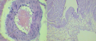
Fig. 1. Presence of marked myointimal hyperplasia (left) of an intromyocardiac coronary arteriole. Fatty transmural replacement of the free wall of the left ventricle (right). Both sections were stained with hematoxylin-eosin.
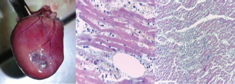
Fig. 2. Heart of a cat with a thinned area of the free wall of the left ventricle due to subendocardial infarction (left). Histopathological alterations compatible with acute myocardium infarction with polymorphonuclear infiltrations (middle). Changes suggesting chronic myocardium infarction with fibrosis areas distributed in a patchy pattern (right).
Ischemia affecting large vessels has different characteristics from that in the microvascular network (Pries et al., 2015). In the pathology of epicardial vessels, an overall dysfunction of the myocardium affected region occurs (Pries et al., 2015). By contrast, in microvascular dysfunction, flow heterogeneity increases, causing both hypoperfusion and hyperperfusion regions (Sakr et al., 2004; Pries et al., 2015).
It should be considered that the extramural compression forces of hypertrophy of the left ventricle may alter the microcirculation in patients with HCM (Maron and Spirito, 1998). In these patients, the presence of more severe vascular remodeling is often observed in the subendocardial regions (Maron et al., 1979; Maron and Spirito, 1998). However, some of the studied cats had a histopathological diagnosis of ischemia in the absence of severe HCM. On the other hand, arterioles with myointimal hyperplasia were more frequently associated with the fibrosis areas, suggesting a causal relationship. Furthermore, in the present study, cats with mild HCM evidenced a lower degree of myofibrillar disorganization, and a higher degree of myointimal hyperplasia than previously described in cats at end-stage HCM (Cesta et al., 2005). These findings, along with the absence of obstruction of epicardial arteries, suggest that the lesions described in this work would not correspond to an end-stage HCM but would probably have a microvascular origin. In addition, the transmural fatty replacement observed in the left ventricle free wall would not be attributed to end-stage HCM. The observed histological alterations would also not be the consequence of an extension to the left ventricle of an arrhythmogenic right ventricular dysplasia, since no cat presented the alterations characteristic of this syndrome in the right ventricle (Fox et al., 2000).
In conclusion, the findings of the present work suggest the possibility that cats may suffer from CDM, similar to humans. Further studies are necessary to better characterize this syndrome.
Conflict of interest
The Authors declares that there is no conflict of interest.
References
Belerenian, G., Castillo, V., Iachini, R., Pucheta, C., Donati, P.A., Guevara, J.M., Ferraris, S. and Mucha, C. 2018. Cardiomiopatía histiocitoide asociada a fibrosis endomiocárdica en un felino siamés de 2 años. Clincardiovet 9, 10–16.
Camici, P.G. and Crea, F. 2007. Coronary microvascular dysfunction. N. Engl. J. Med. 356, 830–840.
Cesta, M.F., Baty, C.J., Keene, B.W., Smoak, I.W. and Malarkey, D.E. 2005. Pathology of end-stage remodeling in a family of cats with hypertrophic cardiomyopathy. Vet. Pathol. 42, 458–467.
Chetboul, V., Passavin, P., Trehiou-Sechi, E., Gouni, V., Poissonnier, C., Pouchelon, J.L. and Desquilbet, L. 2019. Clinical, epidemiological and echocardiographic features and prognostic factors in cats with restrictive cardiomyopathy: a retrospective study of 92 cases (2001-2015). J. Vet. Intern. Med. 33, 1222–1231.
Crea, F. and Lanza, G.A. 2004. Angina pectoris and normal coronary arteries: cardiac syndrome X. Heart 90, 457–463.
Driehuys, S., Van Winkle, T.J., Sammarco, C.D. and Drobatz, K.J. 1998. Myocardial infarction in dogs and cats: 37 cases (1985-1994). J. Am. Vet. Med. Assoc. 15, 1444–1448.
Finsterer, J. 2008. Histiocytoid cardiomyopathy: a mitochondrial disorder. Clin. Cardiol. 31, 225–227.
Fox, P.R., Maron, B.J., Basso, C., Liu, S.K. and Thiene, G. 2000. Spontaneously occurring arrhythmogenic right ventricular cardiomyopathy in the domestic cat: a new animal model similar to the human disease. Circulation 102, 1863–1870.
Häggström, J., Luis Fuentes, V. and Wess, G. 2015. Screening for hypertrophic cardiomyopathy in cats. J. Vet. Cardiol. 17, S134–S149.
Maron, B.J., Epstein, S.E. and Roberts, W.C. 1979. Hypertrophic cardiomyopathy and transmural myocardial infarction without significant atherosclerosis of the extramural coronary arteries. Am. J. Cardiol. 43, 1086–1102.
Maron, B.J. and Spirito, P. 1998. Implications of left ventricular remodeling in hypertrophic cardiomyopathy. Am. J. Cardiol. 81, 1339–1344.
Pries, A.R., Badimon, L., Bugiardini, R., Camici, P.G., Dorobantu, M., Duncker, D.J., Escaned, J., Koller, A., Piek, J.J. and De Wit, C. 2015. Coronary vascular regulation, remodelling, and collateralization: mechanisms and clinical implications on behalf of the working group on coronary pathophysiology and microcirculation. Eur. Heart. J. 36, 3134–3146.
Sakr, Y., Dubois, M.J., De Backer, D., Creteur, J. and Vincent, J.L. 2004. Persistent-microcirculatory alterations are associated with organ failure and death in patients with septic shock. Crit. Care. Med. 32, 1825–1831.
Thygesen, K., Alpert, J.S. and White, H.D. 2007. Universal definition of myocardial infarction. J. Am. Coll. Cardiol. 50, 2173–2195.


