


| Case Report | ||
Open Vet J. 2021; 11(3): 422-430 Open Veterinary Journal, (2021), Vol. 11(3): 422–430 Case Report Dystocia and cesarean section in a free-ranging ocelot (Leopardus pardalis) after traumatic spinal cord injury resulting from dog (Canis familiaris) attackEduardo Alfonso Díaz1,2*, Carolina Sáenz1, Gilberto Segnini3, Andrés Villagómez3, Ramiro F. Díaz2,4 and Rebecca Zug51Hospital de Fauna Silvestre TUERI, Instituto iBIOTROP, Universidad San Francisco de Quito USFQ, Quito, Ecuador 2Escuela de Medicina Veterinaria, Universidad San Francisco de Quito USFQ, Quito, Ecuador 3Hospital Docente de Especialidades Veterinarias, Universidad San Francisco de Quito USFQ, Quito, Ecuador 4Instituto de Investigaciones en Biomedicina, Universidad San Francisco de Quito USFQ, Quito, Ecuador 5Colegio de Ciencias Biológicas y Ambientales, Universidad San Francisco de Quito USFQ, Quito, Ecuador *Corresponding Author: Eduardo Alfonso Díaz. Escuela de Medicina Veterinaria, Universidad San Francisco de Quito USFQ, Quito, Ecuador. Email: eadiaz [at] usfq.edu.ec Submitted: 29/03/2021 Accepted: 19/07/2021 Published: 16/08/2021 © 2021 Open Veterinary Journal
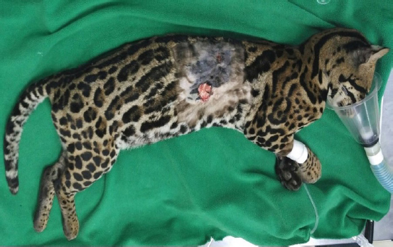
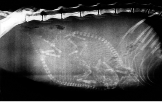
AbstractBackground: Ex situ breeding programs are essential to establish genetic resource banks and produce offspring to strengthen the in situ conservation of endangered species. However, many programs fail to maintain viable ex situ populations due to reproductive problems, including dystocia in pregnant females. Dystocia encompasses different emergency obstetric situations for the lives of dams and fetuses that require urgent intervention. This condition has been studied in domesticated species but published records in wildlife, specifically in felines species, are scarce. Case Description: An adult female ocelot (Leopardus pardalis) was referred to the wildlife hospital of the Universidad San Francisco de Quito after being attacked by dogs (Canis familiaris). Neurological tests revealed traumatic spinal cord injury at a thoracolumbar level. Complementary tests (laboratory exams, radiographs, and ultrasound) revealed a full-term pregnancy, failure in the labor progress, and critical fetal stress. A cesarean section was performed, and the newborns received resuscitation care after assessing their viability using the Apgar score system. The neonate with the lowest Apgar score died within the first hour after birth, while the second one showed an increase in Apgar score after resuscitation care and survived the procedure. Conclusion: We provide new obstetric data that could be relevant to save the lives of dams and newborns in related cases for ocelots and other species of wild felids. Furthermore, this study confirms the adverse effects that domestic dogs have on wildlife species. Keywords: Afferent innervation, Apgar score, Neonatal resuscitation, Obstetric anesthesia, Wild felid. IntroductionThe ocelot (Leopardus pardalis) is a medium-sized neotropical feline distributed from the southern United States to northern Argentina and Chile (Sunquist and Sunquist, 2002). The species is one of the most abundant in its entire distribution range, playing a crucial role in the dynamics of the ecosystems it inhabits (Rocha et al., 2016). The International Union for Conservation of Nature (IUCN) classifies the species as least concern, but the current population trend is decreasing (Paviolo et al., 2015), and in some areas such as Ecuador, ocelots are classified as near threatened due to illegal hunting and habitat loss (Espinosa et al., 2011). Domestic dogs are considered one of the causes of the decline in threatened wildlife species globally (Doherty et al., 2017). Specifically, the presence of domestic dogs (Canis familiaris) negatively affects the distribution of ocelots (Paschoal et al., 2018), including the death of specimens by direct attacks (Haines et al., 2005). However, its real impact is not currently recognized in Ecuador (Zapata-Ríos and Branch, 2018). Traumatic spinal cord injury (SCI) is a devastating disease resulting in changes, temporary or permanent, in motor, sensory, or autonomic function (Webb et al., 2010; Eminaga et al., 2011). As has been reported in humans, pregnant women with SCI are at significant risk of dystocia due to failure to progress in labor and have higher rates of cesarean sections than the general population (Cross et al., 1992; Sterling et al., 2013). Although dystocia caused by SCI has been reported in domestic cats (Özdemir Salci et al., 2020), information on wildlife veterinary medicine is scarce. Dystocia is an emergency that requires urgent diagnosis and timely intervention since improper management can compromise both fetal and maternal survival (Pretzer, 2008; Traas, 2008a). Surgical intervention, namely cesarean section, is indicated if medical therapy fails and expulsion of all fetuses through the birth canal is not possible without delay (Purohit and Gaur, 2004; Reichler and Michel, 2009). This procedure has been performed routinely in domestic cats (Ekstrand and Linde-Forsberg, 1994; Dejneka et al., 2015), but few scientific publications have reported cases of dystocia and consecutive cesarean sections in wild felids (Batista-Arteaga et al., 2011; Khan et al., 2011; Alves et al., 2018). The reproductive management of ex situ wildlife populations can assist in retaining existing genetic diversity for the future maintenance of free-ranging populations, especially for threatened species. The Felidae Taxon Advisory Group identifies feline species housed at accredited institutions. It uses a registry of captive animals (e.g., Studbook) to manage ex situ breeding programs and avoid inbreeding. There are 15 feline breeding programs included in the IUCN Red List of Threatened Species (Wildt et al., 2010). As such, zoos and other ex situ wildlife institutions play an important role in species conservation. However, many of these institutions fail to meet demographic and genetic goals to ensure long-term viability due to reproductive problems, including dystocia in pregnant females (Penfold et al., 2014). The obstetric data obtained from this study could be relevant in related cases for ocelots and other threatened feline species included in ex situ breeding programs. Consequently, the present report aims to describe for the first time dystocia and cesarean section performed on an ocelot with traumatic spinal cord injury after a dog attack. Case DetailsAn adult female ocelot was remitted from Northwest of Ecuador to the Universidad San Francisco de Quito (USFQ) wildlife hospital for evaluation after being attacked by domestic dogs; firefighters rescued the specimen after residents reported the attack. Upon admission, the patient was in lateral recumbency, and chemical restriction was not necessarily due to its depressed state of consciousness. Physical examination revealed two fresh thoracic lacerations, one dorsal approximately 1 cm2, and the other on the right flank approximately 3 cm2, about 5 cm apart (Fig. 1), poor body condition (7.5 kg), dehydration (10 %), dry and pale mucous membranes, delayed capillary refill time (> 3 seconds), hypoglycemia (52.2 mg/dl), hypothermia (34.2°C), and bradypnea (13 breath/minutes). Blood samples were collected from the cephalic vein to analyze complete blood cell and serum biochemistry panel; significative findings included normocytic, normochromic anemia (hematocrit 10 l/l, MCV 51 fl, MCHC 350 g/l), leukocytosis (white blood cells 23.60 109/l, band neutrophils 0.71 109/l, segmented neutrophils 16.52 109/l, lymphocytes 4.48 109/l, monocytes 1.65 109/l, eosinophils 0.24 109/l), and hypoalbuminemia 22.10 g/l (ocelots reference values in Widmer et al., 2016). Blood samples were tested for feline leukemia virus, feline immunodeficiency virus, and blood parasites; all tests were negative. Initial treatment consisted of oxygen supplementation by face mask (FiO2 60%), fluid resuscitation with tempered lactated ringer’s solution (50 ml/kg/h IV) plus 50% dextrose bolus (250 mg/kg IV), analgesia (meloxicam: 0.2 mg/kg IV q 24 hours) and antibiotic therapy (ampicillin sulbactam: 25 mg/kg IV q12 plus ceftriaxone: 30 mg/kg IV q 12 hours). Following the initial assessment, a complete neurological examination was performed. The ocelot showed a perception of superficial and deep pain in all four limbs. Normal spinal reflexes were observed in both forelimbs and normal withdrawal reflex but patellar hyperreflexia in both hindlimbs. At the time of examination, the bladder was distended, and the perianal reflex was present, although diminished. These findings localize the lesion in the thoracolumbar (T3-L3) region (Garosi, 2009). Since SCI was suspected, to avoid excessive manipulation and exacerbation of the patient's lesion, latero-lateral and ventro-dorsal radiographic (Medical X-Ray, Radiography System, model sharp Ray LWX-20p) views of the spine were obtained with the ocelot in the left lateral decubitus. The radiographs did not show any injuries at the thoracolumbar level, but the presence of two fetuses was evidenced (Fig. 2).
Fig. 1. Ocelot after clipping and skin disinfection upon admission. Two open, fresh lacerations are evident at the thoracic level, one dorsal and the other on the right flank of the patient.
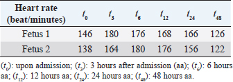
Fig. 2. Latero-lateral radiography of the ocelot. The image shows the presence of two fetuses in an advanced stage of development. An ultrasound (SonoScape, Portable Digital Color Doppler Ultrasound System Model S6V, Micro-Curved Array, 7.5 MHz) was performed to monitor fetal development and vitality. It was found that the fetuses were fully developed due to the differentiation of intestinal layers and the existence of intestinal peristalsis (Lopate, 2018). Both fetuses showed heart rates <150 beats/minutes, indicative of fetal stress (Traas, 2008a). Furthermore, low concentrations of progesterone were detected in the blood (2.1 ng/ml), but obstetric inspection did not show any signs associated with labor onset. Three hours later, the fetuses’ heart rate increased to normal values (Table 1), so the initial treatment was maintained, and a urinary catheter was placed to allow the emptying of the bladder. In the following hours, the female progressively stabilized, but 48 hours after admission, it was decided to perform a cesarean section; the fetuses' heart rate became critical again. The female continued to show no signs of labor progress. The ocelot was premedicated with fentanyl (3 μg/kg IV) for the safe handling of the animal. Anesthesia was induced with propofol (4 mg/kg IV) to allow intubation of the patient. It was maintained with 2.5% sevoflurane in oxygen at 1 l/minutes using a partial rebreathing system (Smiths Medical SurgiVet CDS 9000, Vernon Hills IL60061, USA). The dam was positioned in dorsal recumbency, and a ventral midline approach for cesarean was performed following a modified surgical technique from that described by Fossum et al. (2013), which included a more meticulous surgical skin preparation (trichotomy of the middle plus lateral abdominal region, and antisepsis with 4% soapy chlorhexidine gluconate solution plus 2% hydroalcoholic chlorhexidine gluconate solution), and reinforcement of skin closure (continuous intradermal pattern plus simple interrupted cutaneous pattern, both with 3-0 absorbable monofilament suture). Heart rate, respiratory rate, central temperature, mean arterial pressure, arterial oxygen saturation, end-tidal carbon dioxide concentration, and arterial oxygen concentration were monitored (Model CMS6000, Contec Medical Systems Co. Ltd, China) continuously during the surgery (Table 2). Administration of intravenous fluid (Lactated Ringer’s solution: 5 ml/kg/hours) started at induction and continued throughout surgery to maintain an emergency venous access. The total anesthesia time was 35 minutes, and the surgery lasted 20 minutes. Cardiorespiratory alterations were not detected during the procedure, and anesthetic recovery was smooth and without complications. Immediately after delivery, the newborns received neonatal care consisting of clamping the umbilical cord, suctioning fluids from nose and mouth with a bulb syringe, and rubbing with warm towels. To assess neonatal viability, an Apgar score test was performed 5 minutes after delivery (Veronesi et al., 2009). Both newborns were classified as critical (Table 3) and received resuscitation care following the protocol described by Traas (2008b). The neonate with the lowest score died within the first hour, while the other, with the highest score, increased their Apgar score 2 hours after birth and survived the procedure. Postoperative therapy for the dam included analgesic (pregabalin: 5 mg/kg PO q 12 hours for 30 days), anti-inflammatory (carprofen: 2.2 mg/kg PO q 12 hours for 5 days), antibiotic (amoxicillin-clavulanic: 20 mg/kg PO q 12 hours for 10 days and cefalexin: 30 mg/kg PO q 12 hours for 10 days) and vitamins (Vit E: 400 UI PO q 24 hours for 30 days). The specimen was placed in a resting cage (95 cm length × 50 cm width) for 8 days, after which it was transferred to a larger cell (200 cm length × 65 cm width) to assess its clinical evolution. No swelling or dehiscence of the surgical wound was observed during this period, and a progressive improvement in the patient's posture and locomotion was found. Four weeks after admission, the patient was neurologically reevaluated. Superficial sedation was performed (dexmedetomidine: 4 μg/kg IM plus ketamine: 2 mg/kg IM), and the recovery of the spinal reflexes of the four extremities could be verified. The dam was discharged and transferred to a wildlife rescue center 45 days after surgery, from where it was subsequently reintroduced into the wild. The surviving kitten was hand-reared following the techniques described by Edwards and Hawes (1997) for wild felids until self-feeding was safely established. At 3 months of age, the specimen was transferred to a rescue center where it is evaluated for release into the wild or inclusion in an environmental education program. Table 1. Fetus heart rates monitored by ultrasound.
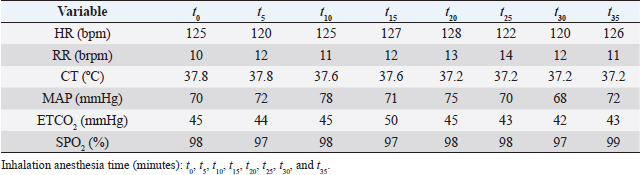
Table 2. Monitored intraoperative heart rate (HR), respiratory rate (RR), central temperature (CT), mean arterial pressure (MAP), end-tidal carbon dioxide concentration (ETCO2) and arterial oxygen concentration (SPO2).
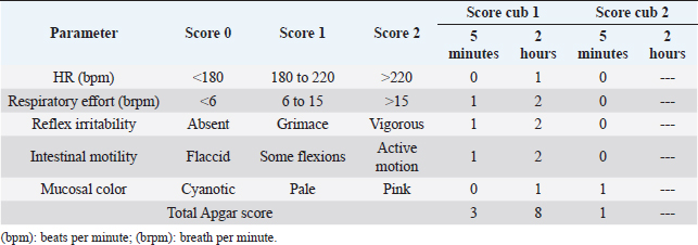
Table 3. Scores obtained in both cubs 5 minutes and 2 hours after delivery using the Apgar score system (Veronesi et al., 2009).
Ethical approvalThis study was conducted under a permit issued by Ecuador’s Ministry of Environment (019-2018-IC-FAU-DNB/MAE) and authorized by the Animal Ethics Committee of Universidad San Francisco de Quito USFQ (2018-011). DiscussionTraumatic SCI is a joint emergency presentation in feline veterinary medicine practice. In most cases, the trauma is caused by being run over or hit by a vehicle or falling from a height, but other causes, such as bite wounds, are also described. The primary injuries to the spinal cord involve T3-L3 segments thoracolumbar region (Beşalti et al., 2002; Grasmueck and Steffen, 2004; Voss and Montavon, 2004; Bruce et al., 2008; Bali et al., 2009; Gonçalves et al., 2009). Although SCI can cause devastating and irreversible damage to the nervous system, patients often present with concurrent injuries, and clinicians must perform a complete physical examination to stabilize life-threatening systemic problems before focusing on neurological examination. The basic tenants of the ABCs (airway, breathing, circulation) should always be the priority (Eminaga et al., 2011). Neurological examination should be performed after the patient is stabilized to determine the severity of the injury and the affected spinal cord segment (Park et al., 2012). The neurological exam should assess the animal's state of consciousness, awareness, body position, gait, postural reactions, muscle tone, spinal reflex, and nociception (Garosi, 2009). The confirmation in the initial neurological examinations of superficial and deep nociception in the four extremities has a good prognosis of recovery (Webb et al., 2010; Park et al., 2012). In addition, the prognosis for return of urinary bladder control is good if the perineal sensation is present on initial examination (Eminaga et al., 2011). The neurological examination must be complemented with diagnostic imaging techniques. Radiographs of the spine should be obtained as part of the initial evaluation of an acute SCI. It is essential to be aware of the possibility of multiple spinal injuries, so it is wise to acquire radiographs of the entire spine. It is generally more convenient to obtain lateral radiographs as a survey, followed by ventrodorsal views of the suspicious regions with the animal in the lateral decubitus position, to minimize excessive movement in any patient with suspected spinal trauma. If anesthesia is required, extreme care must be taken during the movement and position of the animal for radiographs due to the associated loss of muscle tone (Jeffery, 2010). In the present case, radiographs could not confirm any injuries at the thoracolumbar level and support the limited diagnostic accuracy of radiography for acute SCI compared to other advanced imaging, such as computed tomography or magnetic resonance imaging (Kinns et al., 2006). Still, its advantages include the fact that it does not require general anesthesia (Park et al., 2012). Confirmation of pregnancy in a patient with SCI presents an additional obstetric challenge by exacerbating the problems associated with the safety of the dam and fetuses. In addition, this circumstance conditions diagnostic techniques, medications, and other factors related to emergency treatments (Jain et al., 2015). Although radiographs can be used to diagnose pregnancy and confirm the number of fetuses, ultrasound is more accurate in determining fetal age and readiness for delivery (Gatel et al., 2015). It is essential to ensure that each fetus has reached, but not exceeded, its maximum gestational age before delivery to ensure the viability of the neonates. In domestic cats, differentiation of the fetal intestinal layers and intestinal peristalsis is evident in the last days of pregnancy (Lopate, 2018). However, the accuracy of predicting parturition by ultrasound measurement of fetal structures decreases toward the end of gestation. It can be even more challenging in domestic cats of different breed sizes (Keiser et al., 2017). One of the most influential, and widely used parameters to verify fetal viability is ultrasound monitoring of fetal heart rate. Fetal stress resulting from hypoxia can occur during dystocia and manifests as a decrease in heart rate (Gil et al., 2014; Lopate, 2018). A fetal heart rate less than 150 beats/minutes is considered critical fetal stress, rates of 150 to 170 indicate moderate to severe fetal stress, while a rate greater than 180 is standard (Traas, 2008a). Additionally, Keiser et al. (2017) recommend the combination of fetal ultrasound measurement with the determination of progesterone in maternal blood as a predictor of delivery since progesterone decreases towards labor until reaching 3.1 ± 1.6 ng/ml on the day of delivery in domestic cats. In our case, fetal development (intestinal layers and peristalsis), fetal stress (< 150 beats/minutes), dam’s low progesterone level (2.1 ng/ml), and the absence of signs of labor were indications of dystocia. If dystocia is confirmed, the general condition of the dam and fetal vitality will be decisive of whether therapy is indicated (Reichler and Michel, 2009). Specifically, fetal values < 150 beats/minutes indicate that a cesarean should be performed (Traas 2008a). However, the dam should be stabilized as much as possible before surgery, and any deficits can be addressed before surgery (Pascoe and Moon, 2001; Traas 2008a; Kushnir and Epstein, 2012; Robertson, 2016). Therefore, although the condition of the ocelot was not at the limit at admission, dehydration, hypothermia, bradypnea, hypoglycemia, or anemia detected in the physical evaluation could be enough to endanger the life of the dam in case of intraoperative complications. Furthermore, fetal heart rates increased in the first hours, initially deciding to stabilize the patient and perform a cesarean section when fetal stress was critical again. At the late pregnancy, fetal stimuli activate neural circuits involving primary sensory nerves and projections from the lumbosacral spinal cord into the area of the paraventricular nucleus of the female’s hypothalamus to promote uterine cervix remodeling (Puder and Papka 2005; Yellon et al., 2010). The cervix must become soft, flexible, and dilated to allow fetal expulsion from the upper vaginal cavity. However, the transection of the afferent nerves that supply the cervix prolongs or completely blocks parturition, suggesting that these nerves could be part of a neurogenic process involved in cervical changes (Higuchi et al., 1987; Burden et al., 1990; Martínez-Gómez et al., 1998; Collins et al., 2002). In domestic cats, a large proportion of afferent neurons innervate the cervix. Information from the cervix is conveyed centrally, mainly via the pudendal and pelvic nerves (Kawatani et al., 1990; Kawatani and De Groat 1991). Therefore, an SCI could interrupt the afferent connections between the uterine cervix and the hypothalamus, resulting in non-remodeling of the cervix and consequent retention of the fetuses and dystocia detected in the ocelot. The Apgar score has been widely used in human hospitals worldwide for decades as an accepted method for assessing the viability of the newborn after delivery (Finster et al., 2005; Li et al. 2013). This tool-score represents a simple and feasible method for the veterinary clinician for the effective evaluation of the newborn, which promotes a faster and more efficient response in case of need (Veronesi, 2016). Scoring is done by examining the newborn's heart rate, respiratory effort, reflex irritability, motility, and mucosal color 5 minutes after birth. Each parameter is graded from 0 to 2, and the total score determines the viability of the neonates (Veronesi et al., 2009). Low Apgar scores indicate lower viability of newborns and the need for resuscitation care: neonates in critical condition have scores from 0 to 3, moderate viability from 4 to 6, and normal neonates obtain 7 to 10 scores (Batista et al., 2014). Apgar score protocols have been developed and evaluated for different domestic species (Mila et al., 2017; Revermann et al., 2018; Bonelli et al., 2020; Flora et al., 2020), but there are no records in wild species. In our case, none of the newborns achieved an Apgar score greater than three in the first 5 minutes, so both neonates received immediate resuscitation care. The lower score neonate died within the first hour, while the one with the highest score increased its Apgar score to eight at two hours after birth. These results are in agreement with Veronesi et al. (2009), who suggest that, although a good Apgar score did not guarantee survival, newborns with higher scores have a survival advantage over those with lower scores. Additionally, early detection of unviable newborns, followed by resuscitation attempts, could improve neonatal survival, so it should also be considered in the obstetric management of wildlife newborns. The type of delivery appears to significantly influence the neonatal Apgar score (Veronesi, 2016). An important determinant of the lack of vitality in newborn animals is fetal hypoxia resulting from prolonged labor or dystocia (Mota-Rojas et al., 2018). This is corroborated by the lower Apgar scores found in neonates born by cesarean section than those born by normal vaginal delivery (Silva et al., 2009; Batista et al., 2014; Vassalo et al., 2015), and could explain the low Apgar score detected in both newborn ocelots. In addition, there is also an impact of the different anesthetic drugs submitted in cesarean sections on neonatal viability (Veronesi, 2016). The anesthesia of pregnant animals, especially during cesarean section, presents a challenge because the choice of therapy must ensure a good outcome for both the fetus and the dam. The cesarean section could be managed through a regional anesthesia approach using a spinal block to reduce risks. Still, general anesthesia is the only option for potentially dangerous animals (Kushnir and Epstein, 2012). Premedication for managing aggressive patients minimizes maternal distress and reduces the dose of induction and maintenance agents, helping to reduce the exposure of fetuses to depressing drugs. Most pregnant domestic cats respond well to opioids, and the usual doses of these drugs provide analgesia and sedation (Pascoe and Moon, 2001; Traas, 2008a; Robertson, 2016). Rapid induction with an injectable agent followed by maintenance with an inhalant agent reduces the mortality rate compared to long induction times administered by mask (Brodbelt et al., 2008). Propofol is a good choice as an initial induction agent because of the short duration of effect after a bolus and better neurological reflex scores in newborns (Luna et al., 2004; Traas, 2008a). Inhaled sevoflurane maintenance is a widely accepted technique after induction with injectable agents (Fossum et al., 2013; Robertson, 2016; De Cramer et al., 2017). However, despite anesthetic precautions, newborns are often depressed after cesarean section and require intensive care (Kushnir and Epstein, 2012). In conclusion, veterinarians still experience higher neonatal mortality rates among their patients than those observed among humans. The establishment of an assessment protocol is essential for the identification of the clinical status of the dam, the newborn, and the need for emergency intervention, but knowledge of reference values in different species is fundamental to establishing adequate treatments (Vassaloet et al., 2015; Mota-Rojas et al., 2018). As far as we know, there is almost no information in veterinary medicine on obstetric data in free-ranging wild felids. Furthermore, although there is no comparable research in the veterinary field, our study suggests that traumatic SCI in pregnant wild felines can result in an obstetric emergency that must be evaluated and treated timely. Finally, the present case report confirms the adverse effects domestic dogs have on Ecuadorian wildlife (Zapata-Ríos and Branch, 2016; Zapata-Ríos and Branch, 2018; Díaz et al., 2020). This has been a unique opportunity for researchers to document the physiological effects of predation on pregnant wildlife and highlights the need for better control of domestic dogs. AcknowledgmentsThe authors thank the Ministerio del Ambiente y Agua (MAAE), Ministerio del Interior (MI) and Unidad de Protección de Medio Ambiente (UPMA) for supporting this research. Publication of this article was funded by the Universidad San Francisco de Quito Research Publication Fund. Conflict of interestThe authors declare that there is no conflict of interest. ReferencesAlves, S.E., Joyner, P.H., Aitken-Palmer, C., Crosier, A.E. and Ware, L. 2018. Full-term pregnancy with vaginal birth following dystocia and caesarean section in two cheetahs (Acinonyx jubatus). Vet. Rec. Case. Rep. 6(2), e000582. Bali, M.S., Lang, J., Jaggy, A., Spreng, D., Doherr, M.G. and Forterre, F. 2009. Comparative study of vertebral fractures and luxations in dogs and cats. Vet. Comp. Orthop. Traumatol. 22(01), 47–53. Batista, M., Moreno, C., Vilar, J., Golding, M., Brito, C., Santana, M., and Alamo, D. 2014. Neonatal viability evaluation by Apgar score in puppies delivered by cesarean section in two brachycephalic breeds (English and French bulldog). Anim. Reprod. Sci. 146(3–4), 218–226. Batista-Arteaga, M., Santana, M., Lozano, O., Méndez, J., Quesada, O., Arbelo, M. and Espinosa, J. 2011. Medical and surgical management of a dystocia because of foetopelvic disproportion in an African lioness (Panthera leo). Reprod. Domest. Anim. 46(2), 362–365. Beşalti, O., Ozak, A. and Tong, S. 2002. Management of spinal trauma in 69 cats. Dtsch. Tierarztl. Wochenschr. 109(7), 315–320. Bonelli, F., Nocera, I., Conte, G., Panzani, D. and Sgorbini, M. 2020. Relation between Apgar scoring and physical parameters in 44 newborn Amiata donkey foals at birth. Theriogenology. 142, 310–314. Brodbelt, D.C., Pfeiffer, D.U., Young, L.E. and Wood J.L. 2008. Results of the confidential enquiry into perioperative small animal fatalities regarding risk factors for anesthetic-related death in dogs. J. Am. Vet. Med. Assoc. 233(7), 1096–1104. Bruce, C.W., Brisson, B.A. and Gyselinck, K. 2008. Spinal fracture and luxation in dogs and cats. Vet. Comp. Orthop. Traumatol. 21(03), 280–284. Burden, H.W., Price, G.T., Renegar, R.H., & Hodson, C.A. 1990. Effects of peripheral nerve lesions during pregnancy on parturition in rats. Anat. Embryol. 182(5), 499–501. Collins, J.J., Usip, S., McCarson, K.E., and Papka, R.E. 2002. Sensory nerves and neuropeptides in uterine cervical ripening. Peptides. 23(1), 167–183. Cross, L.L., Meythaler, J.M., Tuel, S.M. and Cross, A.L. 1992. Pregnancy, labor and delivery post spinal cord injury. Spinal. Cord. 30(12), 890–902. De Cramer, K.G.M., Joubert, K.E. and Nöthling, J.O. 2017. Puppy survival and vigor associated with the use of low dose medetomidine premedication, propofol induction and maintenance of anesthesia using sevoflurane gas-inhalation for cesarean section in the bitch. Theriogenology. 96, 10–15. Dejneka, G.J., Niżański, W. and Bielas, W. 2015. Cesarean section in the cat: a survey of 126 cases. Med. Weter. 71(6), 386–389. Díaz, E.A., Donoso, G., Sáenz, C., Dueñas, I. and Cabrera, F. 2020. Clinical and pathological findings in a dwarf red brocket Mazama rufina (Mammalia: cetartiodactyla: cervidae) attacked by dogs. J. Threat. Taxa. 12(13), 16885–16890. Doherty, T.S., Dickman, C.R., Glen, A.S., Newsome, T.M., Nimmo, D.G., Ritchie, E.G., Vanak, A.J. and Wirsing, A.J. 2017. The global impacts of domestic dogs on threatened vertebrates. Biol. Conserv. 210, 56–59. Edwards, M.S. and Hawes, J. 1997. An overview of small felid hand-rearing techniques and a case study for Mexican margay Leopardus wiedii glaucula at the Zoological Society of San Diego. Int. Zoo. Yearb. 35(1), 90–94. Ekstrand, C. and Linde-Forsberg, C. 1994. Dystocia in the cat: a retrospective study of 155 cases.mJ. Small. Anim. Pract. 35(9), 459–464. Eminaga, S., Palus, V. and Cherubini, G.B. 2011. Acute spinal cord injury in the cat: causes, treatment and prognosis. J. Feline. Med. Surg. 13(11), 850–862. Espinosa, S., Zapata R.G. and Tirira, D.G. 2011. Ocelote (Leopardus pardalis). In: Libro Rojo de los mamíferos del Ecuador, 2nd ed. Ed., Tirira, D.G. Fundación Mamíferos y Conservación, Pontifica Universidad Católica del Ecuador y Ministerio del Ambiente del Ecuador, Ecuador: Publicación especial sobre los mamíferos del Ecuador 8. Quito, p: 267. Finster, M., Wood, M. and Raja, S.N. 2005. The Apgar score has survived the test of time. J. Am. Soc. Anesth. 102(4), 855–857. Flora, T., Smallman, M. and Kutzler, M. 2020. Developing a modified Apgar scoring system for newborn lambs. Theriogenology. 157, 321–326. Fossum, T.W., Dewey, C.W., Horn, C.V., Johnson, A.L., MacPhail, C.M., Radlinsky, M.G., Schulz, K.S. and Willard M.D. 2013. Surgery of the reproductive and genital systems. In Small animalsSurgery. 4th ed. St. Louis, Missouri: Elsevier Mosby, pp: 780–855. Garosi, L. 2009. Neurological examination of the cat. How to get started. J. Feline. Med. Surg. 11(5), 340–348. Gatel, L., Rault, D., Chalvet-Monfray, K., Saunders, J. and Buff, S. 2015. Prediction of parturition time in queens using radiography and ultrasonography. Anat. Histol. Embryol. 44(4), 241–246. Gil, E.M.U., Garcia, D.A.A., Giannico, A.T. and Froes, T.R. 2014. Canine fetal heart rate: do accelerations or decelerations predict the parturition day in bitches? Theriogenology. 82(7), 933–941. Gonçalves, R., Platt, S.R., Llabrés-Díaz, F.J., Rogers, K.H., Stefani, A.D., Matiasek, L.A. and Adams, V.J. 2009. Clinical and magnetic resonance imaging findings in 92 cats with clinical signs of spinal cord disease. J. Feline Med. Surg. 11(2), 53–59. Grasmueck, S. and Steffen, F. 2004. Survival rates and outcomes in cats with thoracic and lumbar spinal cord injuries due to external trauma. J. Small Anim. Pract. 45(6), 284–288. Haines, A.M., Tewes, M.E. and Laack, L.L. 2005. Survival and sources of mortality in ocelots. J. Wildl. Manag. 69(1), 255–263. Higuchi, T., Uchide, K., Honda, K. and Negoro, H. 1987. Pelvic neurectomy abolishes the fetus-expulsion reflex and induces dystocia in the rat. Exp. Neurol. 96(2), 443–455. Jain, V., Chari, R., Maslovitz, S., Farine, D., Bujold, E., Gagnon, R., Basso, M., Bos, H., Brown, R., Cooper, S., Gouin, K., McLeod, N.L., Menticoglou, S., Mundle, W., Pylpjuk, C., Roggensack, A. and Sanderson, F. 2015. Guidelines for the management of a pregnant trauma patient. J. Obstet. Gynaecol. Can. 37(6), 553–571. Jeffery, N.D. 2010. Vertebral fracture and luxation in small animals. Vet. Clin. Small. Anim. Pract. 40(5), 809–828. Kawatani, M. and De Groat, W.C. 1991. A large proportion of afferent neurons innervating the uterine cervix of the cat contain VIP and other neuropeptides. Cell. Tissue. Res. 266(1), 191–196. Kawatani, M., Takeshige, C. and De Groat, W.C. 1990. Central distribution of afferent pathways from the uterus of the cat. J. Comp. Neurol. 302(2), 294–304. Khan, S.A., Hassan, M.M., Uddin, M.B., Rahman, Z.M.M., Yasin, G. and Epstein, J.H. 2011. Caesarean of Lion (Panthera leo) at Dulahajra Safari Park, Bangladesh. Open. Vet. J. 1(1), 10–12. Keiser, R., Reichler, I.M. and Balogh, O. 2017. Are foetal ultrasonographic and maternal blood progesterone measurements near parturition reliable predictors of the time of birth in the domestic cat? Reprod. Domest. Anim. 52(3), 487–494. Kinns, J., Mai, W., Seiler, G., Zwingenberger, A., Johnson, V., Cáceres, A., Valdés-Martínez, A. and Schwarz, T. 2006. Radiographic sensitivity and negative predictive value for acute canine spinal trauma. Vet. Radiol. Ultrasound. 47(6), 563–570. Kushnir, Y. and Epstein A. 2012. Anesthesia for the pregnant cat and dog. Isr. J. Vet. Med. 67(1), 19–23. Li, F., Wu, T., Lei, X., Zhang, H., Mao, M. and Zhang, J. 2013. The Apgar score and infant mortality. PLoS One. 8(7), e69072. Lopate, C. 2018. Gestational aging and determination of parturition date in the bitch and queen using ultrasonography and radiography. Vet. Clin. Small Anim. Pract. 48(4), 617–638. Luna, S.P.L., Cassu, R.N., Castro, G.B., Neto, F.T., Silva, J.R. and Lopes, M.D. 2004. Effects of four anaesthetic protocols on the neurological and cardiorespiratory variables of puppies born by caesarean section. Vet. Rec. 154(13), 387–389. Martínez-Gómez, M., Cruz, Y., Pacheco, P., Aguilar-Roblero, R. and Hudson, R. 1998. The sensory but not muscular pelvic nerve branch is necessary for parturition in the rat. Physiol. Behav. 63(5), 929–932. Mila, H., Grellet, A., Delebarre, M., Mariani, C., Feugier, A. and Chastant-Maillard, S. 2017. Monitoring of the newborn dog and prediction of neonatal mortality. Prev. Vet. Med. 143, 11–20. Mota-Rojas, D., López, A., Martínez-Burnes, J., Muns, R., Villanueva-García, D., Mora-Medina, P., González-Lozano, M., Olmos-Hernández, A. and Ramírez-Necoechea, R. 2018. Is vitality assessment important in neonatal animals? CAB. Rev. 13(036), 1–13. Özdemir Salci, E.S., Güner, B. and İpek, V. 2020. Dystocia caused by spinal paraplegia in a cat with superfetation. Kafkas. Univ. Vet. Fak. Derg. 26 (6), 839–840. Park, E.H., White, G.A. and Tieber, L.M. 2012. Mechanisms of injury and emergency care of acute spinal cord injury in dogs and cats. J. Vet. Emerg. Crit. Care. 22(2), 160–178. Paschoal, A.M., Massara, R.L., Bailey, L.L., Doherty, P.F., Santos, P.M., Paglia, A.P., Hirsch, A. and Chiarello, A.G. 2018. Anthropogenic disturbances drive domestic dog use of Atlantic forest protected areas. Trop. Conserv. Sci. 11, 1–14. Pascoe, P.J. and Moon, P.F. 2001. Periparturient and neonatal anesthesia. Vet. Clin. Small. Anim. Pract. 31(2), 315–341. Paviolo, A., Crawshaw, P., Caso, A., de Oliveira, T., Lopez-Gonzalez, C.A., Kelly, M., De Angelo, C. and Payan, E. 2015. Leopardus pardalis (errata version published in 2016). UK: The IUCN Red List of Threatened Species, e.T11509A97212355. Penfold, L.M., Powell, D., Traylor-Holzer, K. and Asa, C.S. 2014. “Use it or lose it”: characterization, implications, and mitigation of female infertility in captive wildlife. Zoo. Biol. 33(1), 20–28. Pretzer, S.D. 2008. Medical management of canine and feline dystocia. Theriogenology. 70(3), 332–226. Puder, B.A. and Papka, R.E. 2005. Activation and circuitry of uterine-cervix-related neurons in the lumbosacral dorsal root ganglia and spinal cord at parturition. J. Neurosci. Res. 82(6), 875–889. Purohit, G.N. and Gaur, M. 2004. Dystocia and its management in the bitch and queen: a review. J. Canine. Develop. Res. 4, 90–100. Reichler, I.M. and Michel, E. 2009. Dystocia: recognition and management. Eu. J. Companion Anim. Pract. 19(2), 165–173. Revermann, R., Winckler, C., Fuerst-Waltl, B., Leeb, C. and Pfeiffer, C. 2018. Assessment of viability of newborn piglets using an adjusted APGAR score. J. Cent. Eur. Agric. 19(4), 829–833. Robertson, S. 2016. Anaesthetic management for caesarean sections in dogs and cats. In Pract. 38(7), 327–339. Rocha, D.G., Sollmann, R., Ramalho, E.E., Ilha, R. and Tan, C.K. 2016. Ocelot (Leopardus pardalis) density in central Amazonia. PLoS. One. 11(5), e0154624. Silva, L.C.G., Lucio, C.F., Veiga, G.A.L., Rodrigues, J.A. and Vannucchi, C.I. 2009. Neonatal clinical evaluation, blood gas and radiographic assessment after normal birth, vaginal dystocia or caesarean section in dogs. Reprod. Domest. Anim. 44, 160–163. Sterling, L., Keunen, J., Wigdor, E., Sermer, M. and Maxwell, C. 2013. Pregnancy outcomes in women with spinal cord lesions. J. Obstet. Gynaecol. Can. 35(1), 39–43. Sunquist, M. and Sunquist, F. 2002. Ocelot. In Wild cats of the world. Chicago, USA: University of Chicago Press, pp: 120–129. Traas, A.M. 2008a. Surgical management of canine and feline dystocia. Theriogenology. 70(3), 337–342. Traas, A.M. 2008b. Resuscitation of canine and feline neonates. Theriogenology. 70(3), 343–348. Vassalo, F.G., Simões, C.R.B., Sudano, M.J., Prestes, N.C., Lopes, M.D., Chiacchio, S.B. and Lourenço, M.L.G. 2015. Topics in the routine assessment of newborn puppy viability. Top. Companion. Anim. Med. 30(1), 16–21. Veronesi, M.C. 2016. Assessment of canine neonatal viability-the Apgar score. Reprod. Domest. Anim. 51, 46–50. Veronesi, M.C., Panzani, S., Faustini, M. Rota, A. 2009. An Apgar scoring system for routine assessment of newborn puppy viability and short-term survival prognosis. Theriogenology. 72(3), 401–407. Voss, K. and Montavon, P.M. 2004. Tension band stabilization of fractures and luxations of the thoracolumbar vertebrae in dogs and cats: 38 cases (1993–2002). J. Am. Vet. Med. Assoc. 225(1), 78–83. Webb, A.A., Ngan, S. and Fowler, J.D. 2010. Spinal cord injury I: a synopsis of the basic science. Can. Vet. J. 51(5), 485. Widmer, C.E., Matushima, E.R. and de Azevedo, F.C.C. 2016. Clinical evaluation, hematology, and serum chemistry of Ocelots (Leopardus pardalis) in the Atlantic forest of Brazil. J. Wildl. Dis. 52(4), 916–921. Wildt, D.E., Swanson, W., Brown, J., Sliwa, A., Vargas, A., Macdonald, D.W. and Loveridge, A.J. 2010. "Felids ex situ: managed programmes, research, and species recovery." In Biology and conservation of wild felids. Eds., Macdonald, D.W. and Loveridge, A.J. England, Cambridge, MA: Oxford University Press, Harvard University Press, pp: 217–235. Yellon, S.M., Grisham, L.A., Rambau, G.M., Lechuga, T.J. and Kirby, M.A. 2010. Pregnancy-related changes in connections from the cervix to forebrain and hypothalamus in mice. Reproduction. 140(1), 155. Zapata-Ríos, G. and Branch, L.C. 2016. Altered activity patterns and reduced abundance of native mammals in sites with feral dogs in the high Andes. Biol. Conserv. 193, 9–16. Zapata-Ríos, G. and Branch, L.C. 2018. Mammalian carnivore occupancy is inversely related to presence of domestic dogs in the high Andes of Ecuador. PLoS. One. 13(2), e0192346. | ||
| How to Cite this Article |
| Pubmed Style Díaz EA, CS, GS, AV, Díaz R, RZ, . Dystocia and cesarean section in a free-ranging ocelot (Leopardus pardalis) after traumatic spinal cord injury resulting from dog (Canis familiaris) attack. Open Vet J. 2021; 11(3): 422-430. doi:10.5455/OVJ.2021.v11.i3.14 Web Style Díaz EA, CS, GS, AV, Díaz R, RZ, . Dystocia and cesarean section in a free-ranging ocelot (Leopardus pardalis) after traumatic spinal cord injury resulting from dog (Canis familiaris) attack. https://www.openveterinaryjournal.com/?mno=68744 [Access: April 25, 2024]. doi:10.5455/OVJ.2021.v11.i3.14 AMA (American Medical Association) Style Díaz EA, CS, GS, AV, Díaz R, RZ, . Dystocia and cesarean section in a free-ranging ocelot (Leopardus pardalis) after traumatic spinal cord injury resulting from dog (Canis familiaris) attack. Open Vet J. 2021; 11(3): 422-430. doi:10.5455/OVJ.2021.v11.i3.14 Vancouver/ICMJE Style Díaz EA, CS, GS, AV, Díaz R, RZ, . Dystocia and cesarean section in a free-ranging ocelot (Leopardus pardalis) after traumatic spinal cord injury resulting from dog (Canis familiaris) attack. Open Vet J. (2021), [cited April 25, 2024]; 11(3): 422-430. doi:10.5455/OVJ.2021.v11.i3.14 Harvard Style Díaz, E. A., , C. S., , G. S., , A. V., Díaz, R., , R. Z. & (2021) Dystocia and cesarean section in a free-ranging ocelot (Leopardus pardalis) after traumatic spinal cord injury resulting from dog (Canis familiaris) attack. Open Vet J, 11 (3), 422-430. doi:10.5455/OVJ.2021.v11.i3.14 Turabian Style Díaz, Eduardo Alfonso, Carolina Sáenz, Gilberto Segnini, Andrés Villagómez, Ramiro Díaz, Rebecca Zug, and . 2021. Dystocia and cesarean section in a free-ranging ocelot (Leopardus pardalis) after traumatic spinal cord injury resulting from dog (Canis familiaris) attack. Open Veterinary Journal, 11 (3), 422-430. doi:10.5455/OVJ.2021.v11.i3.14 Chicago Style Díaz, Eduardo Alfonso, Carolina Sáenz, Gilberto Segnini, Andrés Villagómez, Ramiro Díaz, Rebecca Zug, and . "Dystocia and cesarean section in a free-ranging ocelot (Leopardus pardalis) after traumatic spinal cord injury resulting from dog (Canis familiaris) attack." Open Veterinary Journal 11 (2021), 422-430. doi:10.5455/OVJ.2021.v11.i3.14 MLA (The Modern Language Association) Style Díaz, Eduardo Alfonso, Carolina Sáenz, Gilberto Segnini, Andrés Villagómez, Ramiro Díaz, Rebecca Zug, and . "Dystocia and cesarean section in a free-ranging ocelot (Leopardus pardalis) after traumatic spinal cord injury resulting from dog (Canis familiaris) attack." Open Veterinary Journal 11.3 (2021), 422-430. Print. doi:10.5455/OVJ.2021.v11.i3.14 APA (American Psychological Association) Style Díaz, E. A., , C. S., , G. S., , A. V., Díaz, R., , R. Z. & (2021) Dystocia and cesarean section in a free-ranging ocelot (Leopardus pardalis) after traumatic spinal cord injury resulting from dog (Canis familiaris) attack. Open Veterinary Journal, 11 (3), 422-430. doi:10.5455/OVJ.2021.v11.i3.14 |

