


| Short Communication | ||
Open Vet J. 2023; 13(1): 123-130 Open Veterinary Journal, (2023), Vol. 13(1): 123–130 Short Communication Age changes in extramural digestive glands of sheep and rabbits in the postembryonic periodMeiramgul S. Zhakiyanova*, Saule M. Seilgazina, Akerke Ygiyeva, Gulnara I. Dzhamanova, and Kamil Y. DerbyshevAgrarian Faculty, University Named after Shakarim of Semey City, Semey, Republic of Kazakhstan Submitted: 15/04/2022 Accepted: 27/12/2022 Published: 27/01/2023 *Corresponding Author: Meiramgul S. Zhakiyanova. Agrarian Faculty, University Named after Shakarim of Semey City, Semey, Republic of Kazakhstan. Email: zhakiyanova [at] autorambler.ru © 2023 Open Veterinary Journal
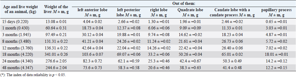
AbstractBackground: The study of the peculiarities of the anatomy of sheep and rabbits’ digestive systems is an important way to improve the efficiency of these animals’ breeding. Aim: The aim of the presented research was to study structural changes of such digestive glands as the liver and the pancreas which occur in the process of ontogenesis in sheep and rabbits. Methods: Sheep of the “Kazakh fat-tailed semi-coarse-wooled” breed (n=8) raised in the “Sayan” private peasant agriculture and rabbits of the “Grey Giant” breed (n=8), raised on the mini rabbit farm of the Agriculture Faculty of Shakarim University were used in the research. Two experimental animal groups were formed (of sheep, “Kazakh fat-tailed semi-coarse-wooled” breed, n=8; rabbits, “Grey Giant” breed, n=8). The liver and pancreas’ ontogenesis development has been studied in these animals. Results: The study presents a holistic view of the macro-microscopic structure of the liver and the pancreas of animals in crucial age periods, stages, and phases of postembryonic ontogenesis (by the example of sheep and rabbits). The authors have traced age stages of adaptive change and structural-functional change of stromal-parenchymatous structures of the liver in sheep and rabbits taking into account stages and crucial phases of development. Conclusion: Development of the liver and the pancreas are characterized by discontinuous growth in the process of postnatal ontogenesis. A crucially important period is the first months after birth, during which the weight and functionality of these organs grow rapidly. Keywords: Digestive glands, Langerhans islets, Liver, Pancreas, Microstructure. IntroductionSheep are reported to have existed in the regions of Egypt, Mesopotamia, and on the islands in the Mediterranean Sea long before Christ (Bogolyubski, 1963). Sheep breeding is a traditional trade in Kazakhstan. However, in the last years, the overall number of sheep and, subsequently, the harvest of mutton have decreased by 2.5 times, whereas the sale of wool— by 3.7 times (Kusainov and Nurov, 1992). Therefore, studying the digestive glands of sheep can help mitigate these economic downfalls. Humanity is faced with the problem of less feeding in different world regions. And a type of animal that can resolve it is a rabbit (Oryctolagus cuniculus). It has many preferences like a short reproductive period and a fast rise up. The specification in its digestive features can give us possibilities to develop this breeding branch and give the qualitative and dietical product to the world market. Rabbits are grass-eating animals but have a single-chamber stomach and can’t efficiently digest their main feed. Due to this, coprophage is a physiological norm for a rabbit. So, studying the rabbit digestive system’s features gives us an additional possibility to develop rabbit breeding in various countries at a better efficiency level (Abou-Kassem et al., 2021). Rabbit meat has always been seen as valuable due to its outstanding taste qualities as well as the content of useful and nutritive substances. For that, rabbit liver deserves special attention. It is not only a recognized dietetic foodstuff but also a delicacy. Its calorie content does not exceed 165 kcal per 100 gram of product. A 100 gram of rabbit liver contains 19 grams of protein, only 10 grams of fat, and no carbohydrates. The chemical content of rabbit liver is also unique (Mc Garry and Foster, 1980). It is explained according to Combes et al. (2013) by the specificity of the digestion of rabbits, which allows for enhancing the useful action of food. The peculiarities of the anatomy of sheep’s and rabbits’ digestive systems determine both their physiological features and their value for the agricultural sector. The study aims to identify the main peculiarities of the liver and pancreas at different stages of animal development. Materials and MethodsA study of the liver and pancreas was conducted in a special room of a veterinary clinic prosectorium and in the block of the veterinary laboratory of the Faculty of Agriculture of Shakarim University, Semey, East Kazakhstan region in the period from 2018 to 2020. Material for the research has been selected from sheep of the “Kazakh fat-tailed semi-coarse-wooled sheep” breed (n=8), raised in the “Sayan” private peasant agriculture, Tarbagataysky region, East Kazakhstan region; and rabbits of “Grey Giant” breed (n=8), raised on the mini farm of the Agriculture Faculty of Shakarim University. Before every experiment, the animal was weighed, the slaughter was conducted with an exsanguinating method, and the mass of the studied organs and their dimensions were determined: a study of the topographic position of the liver and pancreas in relation to adjoining anatomical structures, and their position in the body in relation to the elements of the skeleton were determined. Then, a visual assessment of the organ was carried out (color, consistency, presence of lobes, incisures, appendices, and pathological changes). The absolute masses of glands were determined by weighing them on electronic scales. An increase in the mass of the liver and pancreas was studied against the background of the overall growth of the body mass, where a relative mass of the organ to the body mass was identified on a percentage basis. When studying the structure, topography, and vascularization of the liver and pancreas of sheep and rabbits, a complex of morphological research methods was used: preparation, radiography of blood flow, preliminary injection with radiopaque mass, a method of interstitial injection, and production of histological preparations (Tabakayeva et al., 2021). Photographing of histological preparations was carried out with the help of a photomicrography set including a microscope MICROmed-3 LUM 2410031 and a photographic attachment with a photo camera CANON PowerShot A640 with a resolution of 3,648 × 2,736. For the study of histological structure, a 1 cm3 piece of the pancreas was taken and fixed in neutral formalin solution; rinsed in running water, and dehydrated in alcohols with increasing concentration (50%, 60%, 70%, 80%, 96%, and 100%). The researched materials were transmitted by the needed time in the ethanol and chloroform solution, chloroform solution and consolidated by pouring them with paraffin. Sections of 5–8 μm in width were prepared on a rotary microtome P/A 186. Prepared sections of 5 μm in width were colored with hematoxylin-eosin and with eosin red. Microscopy was carried out with the microscope MICROmed-3 LUM 2410032 (ТМ MICROmed, 36021, Poltava, Ukraine) and the photographic attachment with the CANON photo camera (CANON PowerShot A640 (model number: 1287B001)). For the histological studies, brightfield microscopes and biological research microscopes MBI-1 (lens 20) and Jenamed-2 (an ocular GF-10, lenses 20 and 40) were used. Structural units of the pancreas were measured with the help of an ocular micrometer MBI-15x (Yefremova et al., 2017). The information obtained as a result of the work was processed with the help of variation statistics methods (Statistica program of the Microsoft Office Word 2007 (12.0.6787.5000) SP3 MSO (12.0.6807.5000)). The reliability of the differences in figures between age groups of animals was determined using a Student’s test. Ethical approvalAnimals for the selection of the liver and pancreas for the study were slaughtered with an exsanguinating method in compliance with the European Convention on the European Convention for the Protection of Vertebrate Animals used for Experimental and other Scientific Purposes. Results and DiscussionMacroscopic analysis of extramural glands of sheep and rabbitsIn order to analyze the liver’s macroscopic parameters, we have first conducted an analysis of its mass fluctuations taking into account the structural parts of the organ (Table 1). In the study of macroscopic parameters of the “Grey Giant” rabbit liver, six anatomical parts of the liver were distinguished: left anterior, left posterior, right, quadrate, and caudate lobes with caudate and papillary processes. When comparing the values of rabbit liver mass, it was determined that the mass of the liver of a 48-month-old rabbit increased by 24.52 times compared to a 15-day-old rabbit. Subsequently, we compared the relative mass of the lobes of the rabbit liver. The comparison was conducted with consideration of the growth of the animals (in the process of ontogenesis). We measured six lobes of the liver lobes of the Grey Giant rabbits: left anterior lobe constituted 30.9% of the overall mass of the organ; left posterior—20.4%; right lobe—10%; quadrate lobe—15%; caudate with a caudate process—18.7%, and a papillary lobe—5% of the mass of the liver. The left lateral lobe is the most developed. These data comply with the data presented by other scientists. And this is in accordance with previous research by Stan (2018) and Amin and Robinson (2020). The liver of the sheep of the “Kazakh fat-tailed semi-coarse-wooled sheep” breed does not differ from the liver of the other breeds of sheep which are discovered in the study of Ubashyev (2003) and Klimenkova and Barkalova (2016). In order to obtain information on the changes in macroscopic parameters of the liver of the sheep, we analyzed the changes in the liver mass and in the structural elements of the organ (Table 2). Table 1. Changes of the liver’s biometric parameters*, “Grey Giant” breed rabbits, postnatal ontogenesis (n=8).
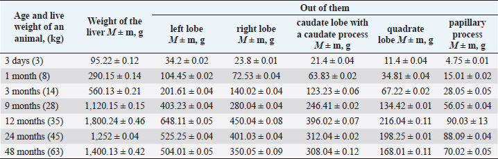
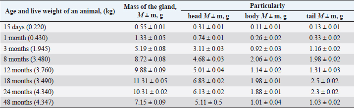
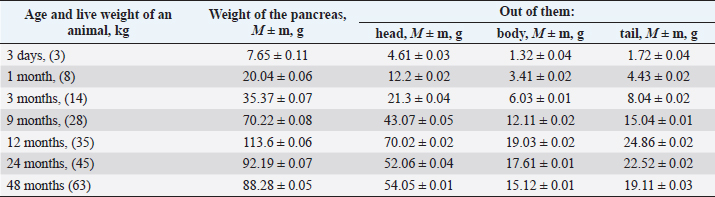
In the comparison of the values of the weight of the liver of “Kazakh fat-tailed semi-coarse-wooled sheep,” it was discovered that a 48-month-old sheep’s liver was 18.82 times bigger than that of a 3-day-old lamb. In the study of the liver, weight dynamics has been discovered the relative mass of the liver in newborn lambs is 3.16%, which significantly differs from the data obtained by other researchers: the relative mass of newborn sheep’s liver equals 2.1% (Ubashyev, 2003). According to our results, the relative mass of the liver at the age of 3 months and of an adult sheep equaled 4% and 4% of the overall weight of an animal correspondingly. The relative mass of the liver anatomical parts of the Kazakh fat-tailed semi-coarse-wooled sheep at the age of 48 months constitutes from the overall mass of the organ: left—36%; right—25%; caudate with a caudate process—22%; quadrate—12% and a papillary process—5%. Ben (2015) and Ondar (2019) noted the live weight of sheep after they are born is gradually growing, but in different periods of postnatal development, the intensity of the liver growth is not the same. Reliable enlargement of linear dimensions of all the liver lobes is recorded in the period from neonatal to the age of 6.5 months, with an exception of the length of the papillary process, later only the length of the left lobe is reliably increasing (Ben and Donkova, 2015). When studying the age dynamics of the biometric indicators of the pancreas, we observed that the weight of the pancreas increased by 20.33 times by the age of 48 months compared to the weight of the pancreas of a 15-day-old rabbit. The head of the pancreas is enlarged by 22 times. The body of the pancreas is enlarged by 17.09 times. The tail was by 19.23 times. Depending on the age of the rabbits, we observed enlargement of the head of the pancreas by 91.2%, the body of the pancreas—by 78.8%, and the tail of the pancreas—by 82.2 (Fig. 1). When determining age differences in the pancreas of sheep, it was determined that the weight of the pancreas of a 3-day-old lamb increased by 14.84 times compared to the indicator of pancreas mass growth of a 48-month lamb (Table 3). The head of the pancreas is enlarged by 11.72 times, the body of the pancreas is enlarged by 11.45 times, and the tail—by 11.11. Depending on the age of the sheep, we observed enlargement of the head part of the pancreas by 38.3%, the body of the pancreas by 64.2%, and the tail—by 63.8%. When studying the pancreas of animals of different ages, we also determined their biometric indicators at the initial stage of the research (Table 4). At that, the pancreas was tentatively divided into three anatomical parts: head, body, and tail. Shevchenko et al. (2010) identify three anatomical parts, such as the left and right lobes and the body of the pancreas. Linear dimensions of the pancreas with the age of fetuses and sheep after birth grow unevenly. Their most intense linear growth corresponds to the age of 3 and 12 months of postnatal development, besides, the indicators of the right lobe significantly surpass the left lobe and the body of the gland (Shevchenko, 2013). Table 2. Changes of biometric indicators of the sheep’s liver in the postnatal ontogenesis (n=8).
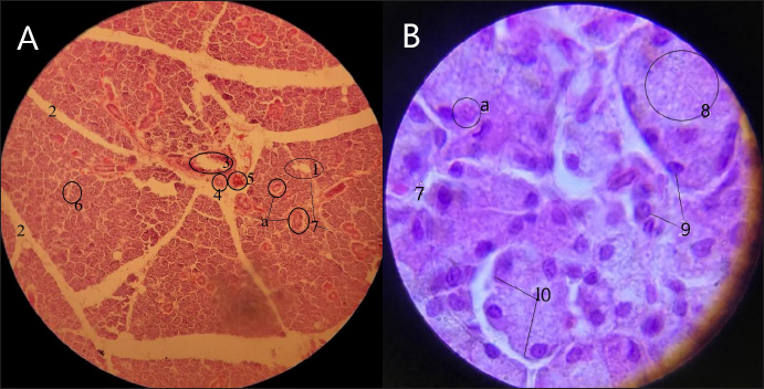
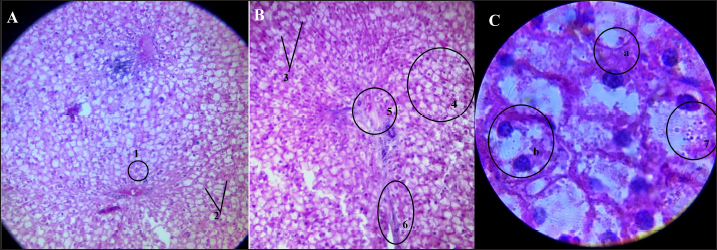
Fig. 1. Histological section of the pancreas of a 24-month-old Grey Giant rabbit. Hematoxylin-eosin coloring, (A): magnification—80; (B): magnification—320. (1): Pancreatic Langerhans islets; (2): Connective tissue interlayer; (3): Intermediate excretory duct; (a): Subsidiary excretory ducts; (4): Intermediate blood vascular artery; (5): Intermediate venous vessel; (6): Fibrous (cellular) connective tissue; (7): Stroma; (8): Apical part; (9): Acinus cells or exocrine pancreatic cells; (10): Acinus. Histological structure of the pancreas of the rabbit of the “Grey Giant” breedStudying the microstructure of the pancreas we saw pancreatic islets—Langerhans islets. This islet belongs to the endocrine system of the gland. And also we saw a connective tissue interlayer. Thanks to this tissue, the pancreas spits into the lobes. The acinus was located on the adenomeres of the pancreas. These are histostructural units of various shapes (oval, conical, round), and their borders are clearly visible. In the acinus, one can see acinus cells or exocrine pancreatic cells. The nucleus of the acinus is distinctly visible. The basal (peripheral) part takes coloring well since it contains many ribosomes secreting enzymes. The apical (intermediate) part of the organ is characterized by cells filled with zymogenic granules, which serve to excrete digestive juice enzymes. Histological structure of the liver of a rabbit of the “Grey Giant” breedThe histological structure of the liver of a 24-month-old rabbit is presented in Figure (2). In the histological study, the intralobular vein is located between trabeculae, in the parenchyma, one can see sinusoid capillaries and hepatic cords (plates) well. Inside the plate, there are distinctive hepatocytes, both uninuclear and binuclear. Hepatocytes have triangular, oval, and rounded shapes. In the center between the lobes, there are triads with a large evident excretory duct. The interlobular vein is small and complex in shape, its lumen is filled with blood corpuscles, and a venous vein is hardly noticeable. Arterial blood vessels are conspicuous, the muscular layer is easily visible in the artery vessel walls. Table 3. The results of the pancreas’s biometric studies, “Grey Giant” breed rabbits (n=8).
Table 4. Results of the pancreas biometric studies of the Kazakh fat-tailed semi-coarse-wooled” breed sheep (n=8).
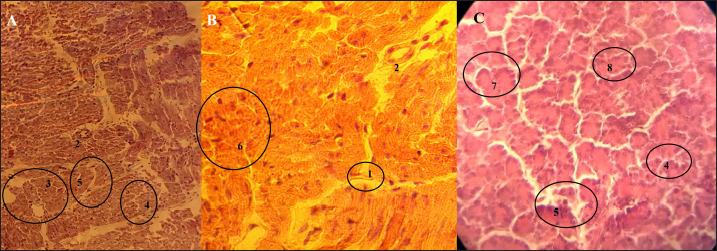
Fig. 2. Histological structure of the liver of a rabbit of the “Grey Giant” breed (aged 24 months): (A): magnification 40; (B): magnification 100; (C): magnification 400. (1): Intralobular vein; (2): Sinusoidal capillary; (3): Hepatic cord (plate); (4): Hepatocytes; (a): uninuclear; (b): binuclear; (5): Triad; (6): Trabecula; (7): Granular cytoplasm. The histological structure of the pancreas of the sheep of the “Kazakh fat-tailed semi-coarse-wooled” breed (aged 24 months) is presented in Figure (3). The connective tissue dividing the pancreas into lobes contains interlobular excretory ducts of an oval shape surrounded by nucleolus. Nearby there are clearly visible pancreatiс Langerhans islets. Langerhans islets belong to the endocrine gland. One can observe white subtle lines of fibrous (cellular) connective tissue. The pancreas consists of parenchyma, and the parenchyma consists of numerous acini. Adenomeres of acini are of different shapes. There one can easily distinguish exocrine pancreatitis from nuclei. Zymogenic granules are only slightly noticeable in the apical (intermediate) part.
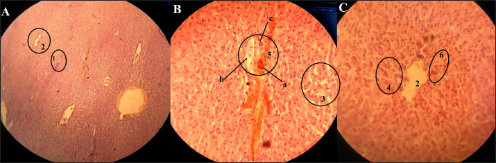
Fig. 3. Histological structure of the pancreas of a sheep of the “Kazakh fat-tailed semi-coarse-wooled” breed (aged 24 months): (A): magnification 40; (B): magnification, 100; (C): magnification, 400. (1): Connective tissue; (2): Interlobular excretory duct; (3): Pancreatic langerhans islet; (4): Fibrous (cellular) connective tissue; (5): Stroma; (6): The functional pancreas parenchyma organized in acini; (7): Terminal portion; (8): Apical part (intermediate). Histological structure of the liver of the sheep belonging to the “Kazakh fat-tailed semi-coarse-wooled” breedIn the histological structure of the sheep liver (Fig. 4), trabeculae are well visualized, and owing to them, one can easily identify the lines of the organ lobes. Inside the trabeculae, there is an interlobular central vein, and a triad including a bile duct, interlobular vein, and an artery is clearly visualized. The bile duct is irregularly shaped, bile duct walls are formed by epithelial cells, the interlobular vein is filled with blood, is of irregular shape, and has a thin wall. The artery is rounded and has a thick muscular wall. Sinusoid capillaries are visualized in Figure (4) in a form of white stripes. Near there are conspicuous hepatiс cords (plates). Inside the plates, there are well-visualized hepatocytes. Between the plates, there are elastic fibers, which are hard to distinguish. Comparison of the histological structure of the pancreas of sheep and rabbitsWhen studying age changes in the microstructure of the extramural digestive glands in the postembryonic period of sheep and rabbits, we discovered that during the period of growth from a 3-day-old lamb to a 4-year-old sheep the liver of a sheep grew by 18.82 times; the liver of a rabbit grew by up to 24.52 times at the age from 15 days to 4 years. It was determined that the size of the pancreas in sheep grew by 14.84 times, and that of a rabbit did by 20.33 times. An intermediate duct of a rabbit is irregularly shaped and the nuclei of the cells surrounding it are not visualized. The intermediate excretory duct of the pancreas of the sheep has a distinct shape, it is clearly visible as well as the nuclei of the surrounding cells. Differences between pancreatic islets in the pancreas of both studied animal species are not expressed; subsidiary excretory ducts can be seen in the pancreas of a rabbit, whereas in sheep these excretory ducts were rarely observed. Intervals of connective tissue in the pancreas of a rabbit are large, wide, and rectilinear. Thanks to this tissue the pancreas lobes are distinctly divided. Connective tissue intervals of sheep are thin and sinuous; one can notice that the pancreas lobes are located close to each other. A difference in acini is typical for both animal species, differences between stroma are not observed, and non-zymogenic granules are faintly distinct in sheep. In the process of maturation, the diameters of Langerhans islets in the pancreas of a rabbit change unevenly. Micrometric indicators of a pancreatic acinus: in some rodents major and minor diameters of the pancreatic acinus changed heterochronically (Combes et al., 2013). In the liver of rabbits, interlobular borders (trabeculae) are not distinctly visible. In the liver of sheep, on the other hand, trabeculae are clearly visible, and intermediate central veins allow identification of the liver lobes. The main peculiarity of the agricultural holdings of the republic is the numerous natural pastures. This circumstance, indubitably, promotes further development of sheep, horse, goat, camel, and rabbit breeding. Dzhusibaliyeva and Abdykerova (2018) have noted a detailed study of features of the digestive system of rabbits and sheep will allow the intensification of the agriculture of the republic with the aim of abundant production of valuable goods: wool, meat, and kumiss at affordable prices. In the course of the research, the structure of the liver and pancreas of sheep and rabbits was meticulously studied. Sheep and rabbits are animals crucial for the animal husbandry of Kazakhstan, they constitute a significant part of the consumer’s basket in Kazakhstan. Sheep are a source of mutton and wool, whereas meat of rabbit is a delicacy, and rabbit liver is a healthy dietetic foodstuff rich in microelements (Mc Garry and Foster, 1980; Aksoy et al., 2018; Amin et al., 2020).
Fig. 4. Histological structure of the liver of the sheep of the “Kazakh fat-tailed semi-coarse-wooled” breed: (A): magnification 40; (B): magnification 100; (C): magnification 400. (1): Trabecula; (2): Interlobular central vein; (3): Sinusoid capillaries; (4): Hepatic cords or plates; (5): Triad; (a): bile duct; (b): interlobular vein; (c): artery; (6): Hepatocytes. Thus, perspective plans of sheep breeding include such goals as an increase of livestock to 70% and an average slaughtering weight of sheep (considering lamb) to 40 kg. The weight of sheared wool is planned to reach 4.7 kg per sheep. Rabbit breeding, however, is perspective as a way to fill the national food basket with high-quality dietetic meat. All these goals can be achieved with a timely and proper application of agronomic, zootechnic, veterinary, and industrial engineering measures, which require a profound knowledge of the anatomy of the species under consideration. It was proven growth of extramural digestive glands occurs unevenly, heterochrony manifesting itself in the formation of their structural organization by the beginning of the productive period. By the beginning of the productive period, the organs of the digestive system function in interconnection and reinforce their correlative relationships (Tabakayeva et al., 2021). However, Ubashyev (2003) gave the data on the relative mass of newborn sheep’s liver may differ from what has been previously believed. There are also some inconsistencies in the identification of anatomical parts of the pancreas done by other scientists like Shevchenko et al. (2010) for example. ConclusionA period in postnatal ontogenesis that is the most crucial for the growth and development of extramural digestive glands is the first month of life, which is characterized by the most intensive growth of the relative weight of the organ (the liver and pancreas of a rabbit reach its development at age of 18–20 months and those of a sheep do at the age of 12–15 months). The rapidity of the organs’ growth remains stable for 2–3 at this level. After that, the organ’s growth speed becomes decreases. But the parenchyma has got its maximum development. The processes of development of glandular epithelium in extramural digestive glands occur unevenly, and they are characterized by heterochronism, which manifests itself in the formation of their structural organization. Digestive system organs function interconnectedly and enhance their correlations by the beginning of the productive period. The obtained data allow completing and deepening of the knowledge about the age morphology of extramural digestive glands and can serve as the most important interior indicators for assessment and comparative morphology, and thus, make an important contribution to morphology. Authors’ contributionM.S. Zhakiyanova has a part in forming the research concept and design, experimental data collection and analysis, and writing the main part of the article. S.M. Seilgazina and G.I. Dzhamanova have made experimental data analysis, critical review, and have made final changes in data presentation. A. Ygiyeva and K. Yu. Derbyshev had a part in data collection and statistical analysis of the experimental results and has a part in discussion of section creation. All of the authors made the approval of the final text of the manuscript. ReferencesAbou-Kassem, D.E., Mahrose, K.M., El-Samahy, R.A., Shafi, M.E., El-Saadony, M.T., Abd El-Hack, M.E., Emam, M., El-Sharnouby, M., Taha, A.E. and Ashour, E.A. 2021. Influences of dietary herbal blend and feed restriction on growth, carcass characteristics and gut microbiota of growing rabbits. Ital. J. Anim. Sci. 20(1), 896–910. Aksoy, N.H., Karaşahin, T., Dursun, Ş., Akbulutd, N.K., Haydardedeoğlue, A.E., İlgunf, R. and Büyükleblebici, O. 2018. Comparative investigation of some liver enzyme functions considering age and gender distinctions in healthy Akkaraman sheep. J. Exper. Clin. Med. 35(3), 71–75. Amin, B., Ford, K.I. and Robinson, R.A. 2020. Quantitative proteomics to study aging in rabbit liver. Mech. Ageing. Dev. 187, 111227. Amin, B. and Robinson, R.A. 2020. Dataset of quantitative proteomic analysis to understand aging processes in rabbit liver. Data. Br. 31, 105701. Ben, W. 2015. Dynamics of age-related changes in the liver of Tuvan short-fat-tailed sheep. Bulletin of the Krasnoyarsk State Agrarian Univ. 6, 204–209. Ben, V. and Donkova, N.V. 2015. Macro and micromorphology of the liver of Tuvinian short-fat tailed sheep. Vestnik KrasGAU. 2, 185–189. Bogolyubski, S.N. 1963. Sheep breeding. Nauka, Moscow, Russia, pp: 23. Combes, S., Fortun-Lamothe, L., Cauquil, L. and Gidenne, T. 2013. Engineering the rabbit digestive ecosystem to improve digestive health and efficacy. Animal 7(9), 1429–1439. Dzhusibaliyeva, A.K. and Abdykerova, G.Z. 2018. State support of occupation in agriculture of Kazakhstan. Problemy Agrorynka. 2, 215–224. Klimenkova, I.V. and Barkalova, N.V. 2016. Morphological peculiarities of the digestive system organs in sheep. Aktualnyye Problemy Intensivnogo Razvitiya Zhyvotnovodstva 19(2), 22–27. Kusainov, A.K. and Nurov, I.A. 1992. Reference-book on sheep breeding. Textbook. Almaty, Kazakhstan, pp: 32. Mc Garry, J.D. and Foster, D.W. 1980. Regulation of hepatic fatty acid oxidation and ketone body production. Annu. Rev. Biochem. 49, 395–420. Ondar, B.A. 2019. Age dynamics of internal organs in young stock of Tuvinian sheep. In Proceedings. XI Mezhdunarodnaya studencheskaya nauchnaya konferenstyya. Studencheskiy nauchnyy forum 2019 May, 23, pp: 22–23. Shevchenko, A.D. 2013. Morphofunctional characteristic of the pancreas of the Edilbay sheep. PhD Dissertation, Mordovia State University N.P. Ogareva, Saransk, Russia, p 152. Shevchenko, A.D., Seitov, M.S. and Davletberdin, D.F. 2010. Topography of the pancreas and duodensum of the sheep of the Edilbay breed. Izvestiya OGAU. 27(1), 197–199. Stan, F. 2018. Comparative study of the liver anatomy in the rat, rabbit, guinea pig and Chinchilla. Bull. Univ. Agric. Sci. Vet. Med. 75(1), 33–40. Tabakayeva, O.V., Tabakayev, A.V., Lyakh, V.A. and Schelkanov, M. 2021. Anatomy of food raw material and bioresources of animal origin. Vladivostok, Russia: Study guide, DVFU. Ubashyev, O.I. 2003. Anatomical-histologic characteristic of the liver of the Buryat coarse-hair sheep. Extended abstract of the Cand. Scien. (Biol.). Dissertation, Ulan Ude, Kazakhstan. Yefremova, Y.N., Bendersky, N.S. and Panchenko, P.C. 2017. Biometrical parameters of the liver and pancreas sizes according to the results of an ultrasonic examination. Molodoy uchenyy. 14(148), 222–225. | ||
| How to Cite this Article |
| Pubmed Style Zhakiyanova M, Seilgazina S, Ygiyeva A, Dzhamanova G, Derbyshev K. Age changes in extramural digestive glands of sheep and rabbits in the postembryonic period. Open Vet J. 2023; 13(1): 123-130. doi:10.5455/OVJ.2023.v13.i1.14 Web Style Zhakiyanova M, Seilgazina S, Ygiyeva A, Dzhamanova G, Derbyshev K. Age changes in extramural digestive glands of sheep and rabbits in the postembryonic period. https://www.openveterinaryjournal.com/?mno=51234 [Access: April 24, 2024]. doi:10.5455/OVJ.2023.v13.i1.14 AMA (American Medical Association) Style Zhakiyanova M, Seilgazina S, Ygiyeva A, Dzhamanova G, Derbyshev K. Age changes in extramural digestive glands of sheep and rabbits in the postembryonic period. Open Vet J. 2023; 13(1): 123-130. doi:10.5455/OVJ.2023.v13.i1.14 Vancouver/ICMJE Style Zhakiyanova M, Seilgazina S, Ygiyeva A, Dzhamanova G, Derbyshev K. Age changes in extramural digestive glands of sheep and rabbits in the postembryonic period. Open Vet J. (2023), [cited April 24, 2024]; 13(1): 123-130. doi:10.5455/OVJ.2023.v13.i1.14 Harvard Style Zhakiyanova, M., Seilgazina, . S., Ygiyeva, . A., Dzhamanova, . G. & Derbyshev, . K. (2023) Age changes in extramural digestive glands of sheep and rabbits in the postembryonic period. Open Vet J, 13 (1), 123-130. doi:10.5455/OVJ.2023.v13.i1.14 Turabian Style Zhakiyanova, Meiramgul, Saule Seilgazina, Akerke Ygiyeva, Gulnara Dzhamanova, and Kamil Derbyshev. 2023. Age changes in extramural digestive glands of sheep and rabbits in the postembryonic period. Open Veterinary Journal, 13 (1), 123-130. doi:10.5455/OVJ.2023.v13.i1.14 Chicago Style Zhakiyanova, Meiramgul, Saule Seilgazina, Akerke Ygiyeva, Gulnara Dzhamanova, and Kamil Derbyshev. "Age changes in extramural digestive glands of sheep and rabbits in the postembryonic period." Open Veterinary Journal 13 (2023), 123-130. doi:10.5455/OVJ.2023.v13.i1.14 MLA (The Modern Language Association) Style Zhakiyanova, Meiramgul, Saule Seilgazina, Akerke Ygiyeva, Gulnara Dzhamanova, and Kamil Derbyshev. "Age changes in extramural digestive glands of sheep and rabbits in the postembryonic period." Open Veterinary Journal 13.1 (2023), 123-130. Print. doi:10.5455/OVJ.2023.v13.i1.14 APA (American Psychological Association) Style Zhakiyanova, M., Seilgazina, . S., Ygiyeva, . A., Dzhamanova, . G. & Derbyshev, . K. (2023) Age changes in extramural digestive glands of sheep and rabbits in the postembryonic period. Open Veterinary Journal, 13 (1), 123-130. doi:10.5455/OVJ.2023.v13.i1.14 |

