


| Original Article | ||
Open Vet J. 2022; 12(5): 657-667 Open Veterinary Journal, (2022), Vol. 12(5): 657–667 Original Research Comparative study of efficacy of prepartum injection of multivitamins and selenium- vitamin E (ά-tocopherol)-combination on post-partum clinical findings, serum steroids, calf and placental weights, and milk antioxidant biomarkers changes in female dromedary camelArafat Khalphallah1, Enas Elmeligy2, Asem Mohammed Zakaria3, Rezk Said Ghallab4, Abdulrahman Abdulkarim5* and Ragab H. Mohamed61Division of Internal Medicine, Department of Animal Medicine, Faculty of Veterinary Medicine, Assiut University, Assiut, Egypt 2Veterinary Teaching Hospital, Faculty of Veterinary Medicine, Assiut University, Assiut, Egypt 3Department of Food Hygiene, Faculty of Veterinary Medicine, Aswan University, Aswan, Egypt 4Department of Theriogenology, Faculty of Veterinary Medicine, Matrouh University, Matrouh, Egypt 5Faculty of Veterinary Medicine, Omar Almukhtar University, Bayda, Libya 6Department of Theriogenology, Obstetrics, and Artificial Insemination, Faculty of Veterinary Medicine, Aswan University, Aswan, Egypt *Corresponding Author: Abdulrahman Abdulkarim. Faculty of Veterinary Medicine, Omar Almukhtar University, Bayda, Libya. Email: abdulrahman.tahir [at] omu.edu.ly Submitted: 10/06/2022 Accepted: 12/08/2022 Published: 13/09/2022 © 2022 Open Veterinary Journal
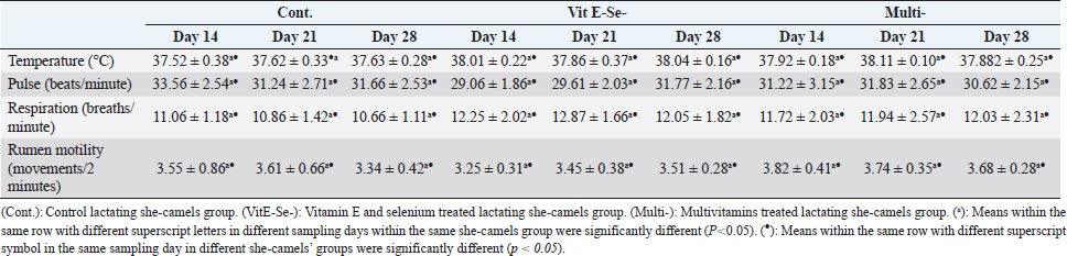
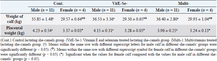
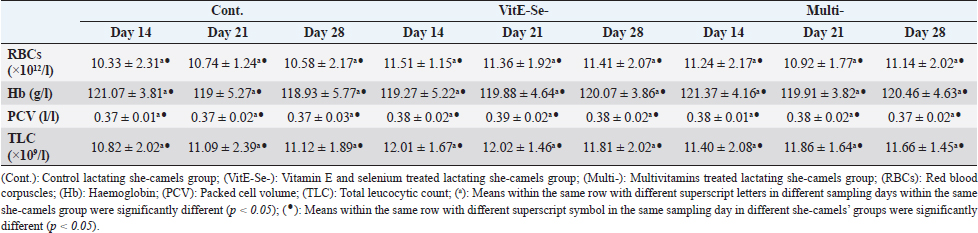
AbstractBackground: All concentrates given to camels were enriched in selenium (Se) in selenite form. The impacts of Se supplementation on lactating female health, milk, and Se/antioxidant statuses received no research interest. Aim: The current study aimed to compare the efficacy of long-term prepartum injection of Se-vitamin E combination and multivitamins on maternal post-calving clinical findings, serum steroid hormones, milk antioxidants, milk somatic cell count (SCC) status, calf body weight, placental weight (PW), and vaginal wash isolates. Methods: From three equal groups of postpartum she-camels (n=45), one group received no treatment and served as control group (Cont.; n=15). For 3 months prepartum, one group had received a combination of vitamin E (ά-tocopherol) and Se (VitE-Se-; n=15), and the third one received multivitamins (Multi-; n=15). All dams were subjected to clinical and laboratory assays including milk total antioxidant capacity (TAC), Se, vitamin E, and milk SCC on Days 14, 21, and 28 post-calving. Steroid hormones and calf and PW were estimated at birth (Day 0). Results: The study reported higher efficacy of Se-vitamin E combination comparing with that of multivitamins as a long-term prepartum injection in recently calved she-camels that was reflected through significant changes in steroids hormones (Drop), i.e., progesterone (P4) and estradiol (E2), the milk antioxidant biomarkers (Elevation), i.e., TAC, Se, vitamin E, and milk SCCs (Reduction). Both two therapeutic regimens had a more powerful effect that the control one. Conclusion: The applied therapeutic supplements had no significant effect on clinical and hematological changes as well as calves’ body weights and PWs. Body weights were significantly higher in male camel calves than those of female calves either in Cont., VitE-Se-, or Multi-. Keywords: Clinical findings, Multivitamins, Selenium-vitamin E combination, She-camel fertility, steroid hormones. IntroductionArabian camel was well suited to the desert environment due to its significant physiological and metabolic adaptation. Nutrients requirements of the camel were not well defined, particularly the trace elements. Selenium (Se) is one of the most important trace elements required for domestic animals and its metabolism was described in many farm animals (Seboussi et al., 2009) However, there were few references concerning Se in camel (Faye and Bengoumi, 1994). Only some values in blood or plasma in different areas from China (Liu et al., 1994), Morocco (Hamliri et al., 1990), and Saudi Arabia (Al-Qarawi et al., 2001) were available. Se supplementation was often recommended to treat conditions attributed to Se deficiency such as cardiomyopathy (Seboussi et al., 2009). Vitamin E (named tocopherol), a fat-soluble vitamin, had been known for a long time as a natural biological antioxidant and the first line of defense against peroxidation which severely damaged cells and tissues (Al-Qarawi et al., 2001; Seboussi et al., 2004). Normal vitamin E concentrations in serum and milk of camel and their variability had only been described during the last years (Seboussi et al., 2008; Faye et al., 2019). Vitamin E deficiency was also incriminated in sudden deaths occurring in large camelids showing myocardial degeneration and necrosis (Finlayson, 1971; Faye et al., 2019). Oxidative stress was commonly known as a cellular or individual imbalance level between oxidants and antioxidants. Oxidative damage was considered as one result of such an imbalance and involves oxidizing cellular macromolecules, cell death by necrosis or apoptosis, as well as damage to the structural tissue damage (Lykkesfeldt and Svendsen, 2007). Generally, antioxidants equalize, delay, and prevent oxidative damage (Halliwell and Gutteridge, 2007). The role of SOD and total antioxidant capacity (TAC) as antioxidant biomarkers were described in many studies (Shoieb et al., 2016; Abd El-Hamid, 2021) whereas different antioxidants support the body against the toxic effects of the reactive oxygen species. Somatic cells are cells of the immune system that were part of the natural defense mechanisms, including macrophages, lymphocytes, polymorphonuclear, and some epithelial cells (Ruegg and Pantoja, 2013). Somatic cell count (SCC) is considered as a mirror that reflects state of the udder, it can distinguish between infected and uninfected quarters, increased SCC than normal level either in bulk milk or individual quarter indicates mastitis (Pantoja et al., 2009; Carrillo-Casas et al., 2012; Sukur and Esendal, 2020). SCC was widely used in a cow for monitoring the milk quality in the dairy industry, but in camel, it was not widely used because the physiological variations and the basal levels of cells in this species were not yet established (Abdurahmann et al., 1992; Nagy et al., 2013). The interpretation of this test could be problematic. Notably, the variation along the lactation was rarely documented (Nagy et al., 2013; Saleh et al., 2013; Kaskous, 2016). Effect of vitamins and trace minerals supplementation (McDowell, 1992), especially on pregnant female camel as well as antioxidant status of the she-camels milk needed further analysis. Therefore, the current study aimed to compare between the efficacy of long-term prepartum injection of vitamin E-Selenium combination and that of multivitamins and their effects on post-partum clinical findings, serum steroids, calves and placental weight (PWs), milk antioxidants capacity, and vaginal bacterial isolates in female dromedary camel. Materials and MethodsAnimals and therapeutic treatmentThe present study included 45 apparently healthy pregnant multiparous female-camels reared in private farms in Aswan governorates, Egypt. Their body weights ranged from 550 to 700 kg. Their age ranged between 9 and 13 years. Camels were housed in an open yard. She-camels equally divided into the control group (Cont.; n=15) that did not receive any therapeutic treatment, vitamin E-Se treated group (Vit. E-Se; n=15), and multivitamins treated group (Multi-; n=15). Vit. E-Se treatment received subcutaneous injections every 10 days for 3 months’ pre-partum a combination of vitamin E (Alpha Tocopheryl Acetate 68 mg/ml) and 1.5 ml/45 kg Bwt Se (Potassium Selenate, Vitesel Emulsion for Injection, Norbrook Laboratories Ltd, Industrial Oakley Hay Estate, Corby, Oakley Way East, UK). Multi-vitamin treatment received pre-partum subcutaneous injections every ten days for 3 months of 30 ml/head multi-vitamins (Each 100 ml solution is containing Vitamin A15000 IU, D3 25 µg, E 20 mg, B1 10 mg, B2 sodium phosphate 5 mg, B6 3 mg, Nicotinamide 35 mg, Pantothenol 25 mg, B12 25 µg and Chlorocresol as a preservative 1mg for Injection; Norbrook Laboratories Ltd, Oakley Hay Industrial Estate, Corby, Oakley Way East, UK). She-camels were clinically examined and laboratory sampled at days 14, 21, and 28 post-partum. SamplesWhole blood and serum samples were collected from the jugular vein whereas all precautions for collection and preparation of samples to achieve an accurate assessment of hematological and biochemical indices were taken. Serum samples were collected and kept frozen at −20°C for subsequent hormonal analyses using commercial test kits according to the standard protocols of suppliers (Coles, 1986). For hematological analyses, the investigated camels were sampled at days 14, 21, and 28 post-partum. Vaginal swabs of mothers were collected directly after parturition for bacteriological examinations then transported immediately to the laboratory in ice box (Quinn et al., 1994). Milk samples were collected on days 14, 21, and 28 post-partum during the routine morning milking into sterile screw-capped bottles and were transferred immediately to the laboratory where they were stored overnight at +4°C before analysis or kept frozen till be examined (Hamed et al., 2010) for milk SCC, TAC, vitamin E, and Se. Clinical examinationThe clinical examinations included measurements of heart and respiratory rates and recording of rectal temperatures as well as rumen movements was done as described by Fowler (2010); Abdel-Rahman et al. (2017); Hassan et al. (2019); Mohamed et al. (2021) on Days 14, 21, and 28 post-partum. Body weight of calves and PW were obtained immediately after calving. Handling of calves and placentaAfter calving, the birth related traits (birth type, calve birth weight, and gender) were recorded within 12 hours after parturition. Each dam was left to deliver the placenta naturally, and placentas were collected immediately after delivery; care was taken to ensure that any PWs taken were of the total placenta with all fluid removed before weighing. Complete blood picture indicesComplete blood picture including red blood corpuscles (RBCs), total leucocytic count (TLC), hemoglobin (Hb), and packed cell volume (PCV) were manually estimated according to Coles (1986); Harvey (2001); Latimer et al (2011); Faye and Bengoumi (2018). Hormonal analysisSerum progesterone (P4) concentrations were measured by enzyme-linked immunosorbent assay ELISA-Sandwich Protocol (Oxford Biomedical Research commercial kits, Rochester Hills, MI). Serum cortisol and estradiol (E2) concentrations were measured using commercial radioimmunoassay kits (Parameters commercial kits, RandD Systems, MN 55413, Inc. 614 McKinley Place NE Minneapolis, Toll Free USA, Canada). Milk antioxidants bio-markers assaysTAC was determined colorimetrically using a commercial test kit (Biodiagnostic Company for Biotechnology, Egypt) according to Abdel-Hameid et al. (2018). Se was analyzed using inductively coupled plasma-mass spectrometry according to Moatkhef et al. (2020). Vitamin E concentration was determined by HPLC according to Farah et al. (1992). Milk somatic cell count (Milk SCC)Milk SCC was measured automatically by somatic cell counter according to Mohamed et al (2019). Bacteriological examinationsBacteriological examinations were done on vaginal swab. Samples were collected and transported immediately to the lab. in ice box. For bacterial isolation, vaginal swabs under complete aseptic condition were inoculated into nutrient broth (Oxoid, UK) and incubated at 37°C for 24 hours and then sub cultured into the following media; 5% sheep blood agar and MacConkey agar at 37°C for 24–48 hours. The produced colonies were identified according to Quinn et al. (1994). Statistical analysisSPSS statistical software program for Windows, version10.0.1 (SPSS Inc., Chicago, IL) was used for data analysis. The obtained data were described as mean ± SD. The data obtained from the clinical findings and laboratory analyses were analyzed by general linear model repeated measures analysis of variance and significance level of results was set at p < 0.05. The significance of differences was evaluated between the means at different sampling days within the same she-camels group or between the means at the different she-camels’ groups on the same sampling days 14, 21, or 28. The data obtained from body weight of calves or that of placenta were analyzed by independent-sample t-test, whereas the significance of differences was evaluated between the means at male calf and female calf within the same she-camel group. Ethical approvalAll experimental protocols were approved by Institutional Animal Care and Use Committee guidelines of Assiut University, Egypt that was in agreement with the Guide for Laboratory Animals Use and Care of the National Institutes of Health in USA (NIH publication No. 86-23, revised 1996). ResultsClinical findings and complete blood picture indicesTemperature, pulse, respirations, rumen motility, calves body weight and placental weight showed no significant changes between Cont., Vit. E-Se, and multi-Vit. on Days 14, 21, or 28 post-partum. Values of temperature, pulse, and respiration were within the reference ranges (Tables 1 and 2). Whole blood picture parameters including RBCs, Hb, PCV, and TLC showed no significant variations between Cont., Vit. E-Se, and multi-Vit throughout days 14, 21, or 28 post-partum (Table 3) and were within the reference ranges. calves body weight and placental weightNormal parturition was reported in all she-camels. Body weight of male calf was (p < 0.05) higher (p < 0.05) than that of female calf at time of parturition. Placental weight in case of male calf was higher (p < 0.05) than that in case of female calf (Table 2). Serum hormones levelsSerum concentrations of P4 were (p < 0.05) dropped at day of calving in Multi- comparing with P4 values in Cont. and Vit E-Se-. In contrast, serum values of E2 were (p < 0.05) elevated after calving (Day 0) in Multi- comparing with E2 values in Cont., Vit E-Se- (Table 4). Milk antioxidants bio-markers assaysMilk values of TAC, Se, and vitamin E were (p < 0.05) improved in VitE-Se- and Multi- at days 14, 21, and 28 post-partum comparing to their values in Cont. This significant (p < 0.05) raise of those milk antioxidant biomarkers values were also reported in Vit E-Se- comparing with their values in Multi- at days 14, 21, and 28. High elevations (p < 0.05) in milk values of TAC, Se, and vitamin E were observed at day 14 either in Cont., Vit E-Se- or Multi- when they compared with their values at days 21 and 28 post-calving. Antioxidant biomarkers throughout the current study were within the reference ranges (Table 5). Milk SCCMilk SCCs were significantly (p < 0.05) decreased at days 21 and 28 post-calving in Cont. when their values compared with those at day 14. In contrast, these milk SCCs were significantly (p < 0.05) improved at days 21 and 28 either in Vit E-Se- or Multi- when their values compared with those at day 14. These significant (p < 0.05) elevations in milk SCCs were also noted in Multi- at days 14, 21, and 28 post-calving when they compared with their values in VitE-Se-. On the other hand, milk SCCs were significantly raised in Cont. comparing with VitE-Se- and Multi- at day 14. This significant increase in milk SCCs was stated in both of VitE-Se- and Multi- particularly at day 28 when their values compared with those in Cont. Milk SCCs throughout the current study were within the reference ranges (Table 5). Table 1. Mean values (M ± SD) of temperature, pulse, respiration and rumen movements in Cont. (n=15), VitE-Se- (n=15), and Multi- (n=15) she-camels.
Bacteriological examinations of vaginal swabsMost common bacterial isolates from vaginal swab of she-camels after calving included 7 coli and Staphylococcus spp followed by Proteus spp and Pseudomonas spp either in Cont., VitE-Se-, or Multi- (Table 6). DiscussionClinical findings, calves body weight, and placental weightThe prepartum injections of Se vitamin E combination or multivitamins had no prominent effect on clinical findings, calves body weights, and placental weight after calving in investigated she-camels. Temperature, pulse, respirations, rumen motility, calves body weight, and placental weight showed no significant changes between Cont., VitE-Se- and Multi- either at days 14, 21, or 28 post-partum. No significant changes were observed between days 14, 21, and 28 either in Cont., VitE-Se-, or Multi-. Values of temperature, pulse, and respiration were within normal range Fowler (2010); Hassan et al. (2019); Bhatt et al. (1960); Nielsen (1964); Hamad et al. (2017); Kamr et al. (2020). Normal parturition was reported in all she-camels. Body weight of male calf was significantly higher than that of female calf at time of parturition. Placental weight in case of male calf was also significantly higher than that in case of female calf. On the other side, previous reports mentioned that camel could eat 20% less than other domestic animals according to its production performance and live weight to maintain its metabolic functions Seboussi et al. (2009). The ability of the camel to recycle minerals, water, and nitrogen was described by Seboussi et al. (2009). Therefore, physiology of the camel should be taken in consideration during Se supplementation. Moreover, the results reported by Seboussi et al. (2009) confirmed that the camel is highly sensitive to Se supplementation and increase of Se level in milk and serum is very important. However, the increase in Se level could reflect a high sensitivity to toxicity, the tolerance level of camel to Se toxicity was shown in the current study. Faye and Seboussi (2009) concluded that the metabolism of Se in camel and other herbivores was quite comparable, with similar signs in case of toxicosis or deficiency, comparable mode of excretion and comparable level in organs and serum. According to dietary supplement of Se and mean body weight of the animal, selenosis occurred with 0.05 mg/kg LW Se supply only. Severe intoxication occurred with 16 mg Se supplementation, i.e., 0.10 mg/kg LW. These values were five times lower than those for sheep and cattle. Based on these results, it seemed essential to limit Se supplementation in camel at 0.01–0.02 mg/kg LW, i.e., approximately 4–8 mg per day for adult animals or 0.5–1 ppm in the diet. Table 2. Mean values (M±SD) of calving weight of calf and placental weight at calving time either for male or female calf in Cont. (n=15), VitE-Se- (n=15) and Multi- (n=15) she-camels.
Complete blood picture indicesWhole blood picture parameters in the present work including RBCs, Hb, PCV, and TLC showed no significant variations either between Cont., VitE-Se-, and Multi- either at days 14, 21, or 28 post-partum or within the same she-camels group (Cont., VitE-Se-, or Multi-) between days 14, 21, and 28. Values of RBCs, Hb, PCV, and TLC were within the reference ranges reported by Fowler (2010). Prepartum long-term treatment of pregnant she-camels with either vitamin E-Se combination or multivitamins reported no clear effect on blood pictures parameters either between different she-camels’ groups or within each she-camel group. On other hand, Se concentration in serum was positively correlated to WBC and eosinophil (E), and negatively correlated with RBCs and lymphocyte. Except with WBC, there was no link with vitamin E (Seboussi et al.,2009). The negative correlation with lymphocytes was observed on adult camels only and could be explained by the interferences between Se level in organism and cellular events responsible for an immune response. Elevated Se had been shown to promote peroxidative damage in in vitro and in vivo systems. Lymphocyte cell membranes were especially susceptible to free radical damage (Björnstedt et al., 1996). Serum concentrations of ovarian hormonesThe previous literature mentioned that serum estrogen and cortisol concentrations were elevated clearly before parturition and have coincided with a decrease in serum P4 values that might support their roles in triggering parturition in she-camels. Furthermore, it could be concluded that together with other parameters, cortisol, estradiol-17B and P4 could be used as good indicators to predict the time of parturition in she-camel (Abdel-Rahman et al., 2017). Referring to the present study, steroids hormones including P4 and E2 showed significant changes in their serum values at calving time (Day 0) in Multi- comparing to their values at Cont. and VitE-Se-. Serum concentrations of P4 were significantly dropped meanwhile serum values of E2 were highly improved at day of calving (Day 0) in Multi- comparing with their values in Cont. and VitE-Se-. Their values were higher than the reference ranges had been reported by Mohamed et al. (2021) and Ayoub et al. (2003). Ayoub et al. (2003) reported that the concentrations of sex steroid hormones varied according to different physiological conditions with higher concentrations in the pregnant she-camels in compare to the nonpregnant ones. Moreover, the source of high P4 reported in nonpregnant lactating she-camels in the present study would be either ovarian or adrenal. Moreover, luteal activity was found even in nonmated she-camels in some cases due to spontaneous ovulation possibly enhanced (Elias, 1990; Ayoub et al., 2003;). Serum concentrations of P4 in she-camels were constantly low (Homeida et al., 1988), and its concentration started to rise after mating. Following mating, at least one corpus luteum was formed that secreted a significant amount of P4. Camels showed serum P4 levels above 2.0 and 3.0 ng/ ml at 20 and 30 day of mating should be considered as pregnant one (Quzy et al., 2013). Agarwal et al. (1987); Abdel-Rahman et al. (2017) added that P4 levels elevated significantly in pregnant she-camels after conception and during the first and the second trimester of pregnancy then dropped significantly during the 3rd trimester. Moreover, P4 levels in she-camels declined significantly during periparturient period. Milk antioxidants bio-markers assaysSeveral studies explored camel’s milk as a potential protein substrate for generating bioactive protein hydrolysates with antioxidant activities (Al-Saleh et al., 2014; Shori et al., 2014). One reference only was available on Se content of camel milk (Al-Awadi and Srikumar, 2001), and the reported value (13.9 ± 2.4 ng/ml) was quite lower than in Seboussi et al. (2009) study, but the authors did not mention the lactation stage. In dairy cow, the milk Se concentration varied from 19.4 to 53.7 ng/ml with Se dietary Se between 0.15 and 0.40 ppm (Juniper et al., 2006). It should underline the richness of camel milk in Se. On average, Se intake by camel calves on the first three months of lactation, was 8 μg/kg LW in the control group (Seboussi et al.,2009). Table 3. Mean values (M±SD) of whole blood picture indices in Cont. (n=15), VitE-Se- (n=15), and Multi- (n=15) she-camels.
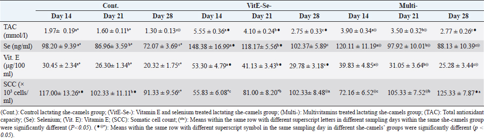
During the current study, the antioxidant capacity biomarkers were greatly affected with prepartum treatment either with Se-vitamin E combination or with multivitamins. A significant improvement of antioxidant biomarkers levels of she-camels milk, i.e., TAC, Se, and vitamin E, due to long-term treatment with either Se-vitamin E combination or multivitamins in investigated she-camels during last pregnancy, was stated in VitE-Se- and Multi- at days 14, 21, and 28 post-partum comparing to their values at Cont. This elevations of milk antioxidant biomarkers values were reported also at VitE-Se- comparing with Multi-. Moreover, milk values of TAC, Se, and vitamin E were significantly increased at day 14 either in Cont., VitE-Se-, or Multi- when they compared with their values at days 21 and 28 post-calving. Milk values of these antioxidant biomarkers were within the reference range reported by Stahl et al. (2006); Seboussi et al. (2009); Sboui et al. (2016); Alavi et al. (2017); and Abdel-Hameid et al. (2018). Some authors reported the absence of vitamin E in camel milk (Ronald de Almeida, 2011), information confirmed again in a recent book published in 2017 (Alavi et al., 2017). In fact, vitamin E concentration in camel milk was nearly similar to that present in cow milk: 56 and 60 μg/100 ml, respectively (Farah et al., 1992; Faye et al., 2019), but Haddadin et al. (2008) reported that the vitamin E concentration in camel milk was 1.78 ± 0.58 μg/100 ml only. Stahl et al. (2006) reported higher values in colostrum (136.9 ± 98.4) compared to milk (32.7 ± 12.8 μg/100 ml). However, this concentration was significantly lower than that present in cow milk, which was used as a control (171 ± 114 μg/100 ml). Moreover, as for other fat-soluble vitamins, α-tocopherol content varied according to seasonal fat concentration in milk, i.e., the reverse to the lactation curve (Faye et al., 2019). Se concentration increased in similar proportion in milk in lactating she-camels (86.4 ± 39.1 ng/ml in the control group vs. 167.1 ± 97.3 ng/ml in treated group), Vitamin E did not change significantly and was, on average, 1.14 ± 0.89 and 1.17 ± 0.72 ng/ml in the treated and control groups, respectively Seboussi et al. (2009). The reported data of Se in camel milk were rare. Al-Awadi and Srikumar (2001) reported a much lower Se value (13.9 ± 2.4 ng/ml) than Seboussi et al. (2009), but without the reference to the stage of lactation. In a meta-analysis performed on cattle’s data (Ceballos et al., 2009), Se was supplemented at a dose of less than 3 mg/day under selenite form and the increased Se level in milk was on average of 12.6 ng/ml only after supplementation. In comparison, the apparent good efficiency of Se transfer in camel milk had to be confirmed. Table 4. Mean values (M ± SD) of serum P4 and E2 in Cont. (n=15), VitE-Se- (n=15), and Multi- (n=15) she-camels at day of calving (Day 0).
Milk SCCOne of the important reasons of raising Camelus dromedarius was its unique milk composition. However, milk production losses could be lowered by lowering SCC levels. At this point, applying hygienic and proper milking processes and routine subclinical mastitis examinations in the herds could firstly be advised to camel breeders (Atasever and Koç, 2016). Regarding to Cont., milk SCCs were significantly dropped at days 21 and 28 comparing to their values at day 14. In contrast, either in VitE-Se- or Multi-, this significant reduction in milk SCCs was reported at day 14 when they compared with their values at days 21 and 28. Moreover, milk SCCs values were significantly increased at day 14 then significantly dropped at day 28 in Cont. when they compared with their values in VitE-Se- or Multi-. Throughout the present work, Multi- had higher values of milk SCCs than those in VitE-Se- throughout the current study at days 14, 21, and 28. Milk SCCs throughout the current study were within the reference ranges mentioned by Hamed et al (2010) and Saleh et al (2013). Regarding to SCC variation, SCC in milk was widely used as an indicator of the degree of inflammation of the udder and to predict udder infection as well as subclinical mastitis since long time in cattle (Poutrel and Rainard 1982). The references in camel were more recent (Merin et al., 2004; Kaskous, 2019) and the variation factors, except in case of intramammary infections were not widely studied. In spite of the highest level had been recommended as 400 × 1,000 cells/ml for cow milk by EU directives, this level was reported to be 250 × 1,000 cells/ml for camels (Abbood, 2016). Up until now, many studies had been conducted to determine SCC of raw milk in camels (Wernery et al., 2008; Saleh and Faye, 2011; Nagy et al., 2013). However, wide variations in SCC values were informed among the investigations. Also, there was still a lack of information reported on the association of milk production and losses with SCC (Atasever and Koç, 2016). Table 5. Mean values (M ± SD) of TAC, Se, Vit. E and SCC in camel’s milk in Cont. (n=15), VitE-Se- (n=15), and Multi- (n=15) she-camels.
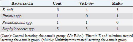
Table 6. Bacterial isolates from vaginal swabs after parturition in Cont., VitE-Se-, and Multi-.
Bacteriological examinations of vaginal swabs of mothersIt had been discovered that camel’s milk had antidiabetic, bactericidal activities, and hostile to hepatitis (Agrawal et al., 2009). To various degrees, it resisted the contamination with microorganisms due to its characteristic inhibitory frameworks such as the lactoperoxidase/thiocyanate/ hydrogen peroxide framework, lactoferrins, lysozyme, immunoglobulins, and free greasy acids (EL-Fakharany et al., 2012). Regarding to the present work, most common bacterial isolates from vaginal swab of she-camels after calving included E. coli and Staphylococcus spp followed by Proteus spp and Pseudomonas spp either in Cont., VitE-Se-, or Multi-. ConclusionThe study reported higher efficacy of Se-vitamin E combination comparing with that of multivitamins as a long-term prepartum injection in recently calved she-camels that was reflected through significant changes in steroids hormones, i.e., estradiol (E2) and progesterone (P4) the milk antioxidant biomarkers and SCCs. Both two therapeutic regimens had more powerful effect than that the control one. The applied therapeutic supplements not significantly affect the clinical and hematological changes as well as calves’ body weights and placental weights. No significant changes in the clinical findings and blood pictures parameters as well as placental weight and calves body weight were demonstrated between Cont., VitE-Se-, and Multi-. Body weights were significantly higher in male camel calves than those of female calves either in Cont., VitE-Se-, or Multi-. AcknowledgementsNot Applicable. Authors’ contributionsRHM, EE, and AK prepared conception and design of study. AK, RHM, MSS, and EE conducted the field study and camel examination. AMZ, MSS, and EE collected laboratory samples and conducted biochemical analyses. AK, AA, MSS, and EE manipulated and statistically analyzed the data. AMZ, RHM, and EE performed analysis, data curation and interpretation of data. All authors drafted the manuscript. EE, RHM, AA, and AK carried out final writing, critical review and revision. All authors have read and approved the final manuscript. Availability of data and materialsThe datasets used and/or analyzed during the current study are available from the corresponding author on reasonable request. Conflict of interestThe authors declare that they have no competing interests. ReferencesAbbood, A.S. 2016. Compare between somatic cell count (SCC) in she camel and cow milk and genetic study. Indian J. Res. 5(7), 145–146. Abdel-Hameid, A.A., Saad, N.M., Wahba, N.M. and Sayed, R.G. 2018. Nutritional Value and Antioxidant Activity of Camel’s Milk. J. Adv. Vet. Res. 8(4), 90–94. Abd El-Hamid, I.S. 2021. Blood constituents, antioxidant activities and hormonal profile in she-camels (Camelus dromedarius) during different physiological statuses in the Northwestern Coast of Egypt. I.J.V.S. 10(4), 247–258. Abdurahmann, O.A.S., Cooray, R. and Bornstein, S. 1992. The ultrastructure of cells fragments in mammary secretions of Camelus bactrianus. J. Vet. Med. A. 39, 648–655. Abdel-Rahman, H.M.A., Ibrahim, M.A. and Elmetwaly, H.A. 2017. Hormonal profile, antioxidant status and some biochemical parameters during pregnancy and periparturient period in Dromedary she camel. Egypt. J. Vet. Sci. 48(2), 81–94. Agarwal, S.P., Khanna, N.D., Agarwal, V.K. and Dwaraknath, P.K. 1987. Circulating levels of estrogen and progesterone in female camels (Camelus dromedarius) during pregnancy. Theriogenology 28, 849–859. Agrawal, R.P., Dogra, R., Mohta, N., Tiwari, R., Singhal, S. and Sultania, S. 2009. Beneficial effect of camel’s milk in diabetic nephropathy. Acta Biomed. 80, 131–134. Alavi, F., Salami, M., Emam-Djomeh, Z. and Mohammadian, M. 2017. Neutraceutical properties of camel milk. In Nutrients in dairy and their implications for health and disease. Eds., Watson, R., Collier, R., and Preedy, V. New York, NY: Academic Press, pp: 451–468. Al-Awadi F.M. and Srikumar, T.S. 2001. Trace elements and their distribution in protein fractions of camel milk in comparison to other commonly consumed milks. J. Dairy Res. 68(3), 463–469. Al-Qarawi, A., Abbas, A., Haroun, B., Mahmoud, E. and Al-Hawas, M. 2001. Clinicopathological investigation of Selenium responsive myopathy in young adult camels. J. Camel Pract. Res. 8, 23–27. Al-Saleh, A.A., Metwalli, A.A., Ismail, E.A. and Alhaj, O.A. 2014. Antioxidative activity of camel’s milk casein hydrolysates. J. Camel Pract. Res. 21(2), 229–237. Atasever, S. and Koç, A. 2016. Milk yield losses caused by high somatic cell count in dromedary camels (Camelus dromedarius). In Proceeding of first international Selçuk-Ephesus symposium on culture of camel-dealing and camel wrestling, Selçuk, İzmir-Turkey. Ayoub, M.A., El-Khouly, A.A. and Mohamed, T.M. 2003. Some hematological and biochemical parameters and steroid hormone levels in the one-humped camel during different physiological conditions. Emir. J. Agric. Sci. 15(1), 44–55. Bhatt, F.L., Kholi, R.N. and Rathore, U.S. 1960. The normal body temperature, respiratory frequency and heart rate of the camel. Indian Vet. J. 37, 456–462. Björnstedt, M., Odlander, B., Kuprin, S., Claesson, H. and Holmgren, A. 1996. Selenite incubated with NADPH and mammalian thioredoxin reductase yields selenide, which inhibits lipoxygenase and changes the electron spin resonance spectrum of the active site iron. Biochemistry 35(26), 8511–8517. Carrillo-Casas, E.M. and Miranda-Morales, R.E. 2012. Bovine mastitis pathogens: prevalence and effects on somatic cell count. In Milk production–an up-to-date over view of animal nutrition, management and health. Ed., Chaiyabutr, N. . London, UK: In Tech; doi: 10.5772/51032. Ceballos, A., Sanchez, J., Stryhn, H., Montgomery, J.B., Barkema, M.H. and Wichtel, J.J. 2009. Metaanalysis of the effect of oral selenium supplementation on milk selenium concentration in cattle. J. Dairy Sci. 92, 324–342. Coles, E.H. 1986. Veterinary clinical pathology, 4th ed. Philadelphia, PA: W.B. Saunders Company, pp: 132–139. EL-Fakharany, E.M., Abedelbaky, N., Haroun, B.M., Sánchez, L., Redwan, N.A. and Redwan, E.M. 2012. Anti-infectivity of camel polyclonal antibodies against hepatitis C virus in Huh7.5 hepatoma. Virol. J. 201, 1–9. Elias, E. 1990. Early weaning in the one humped camel. “Is it possible to improve reproductive performance in camel”. Proceeding UCDEC, Paris. Farah, Z., Rettenmaier, R. and Atkins, D. 1992. Vitamin Content of Camel Milk. Int. J. Vit. Nutr. Res. 62, 30–33. Faye, B. and Seboussi, R. 2009. Selenium in camel—a review. Nutrients 1(1), 30–49. Faye, B. and Bengoumi M. 1994. Trace-element status in camels. A review. Biol. trace Element Res. 41, 1–11. Faye, B. and Bengoumi, M. 2018. Camel clinical biochemistry and hematology. Cham, Switzerland: Springer International Publishing. https://doi.org/10.1007/978-3-319-95562-9 Faye, B., Konuspayeva, G. and Bengoumi, M. 2019. Vitamins of camel milk: a comprehensive review. J. Camelid Sci. 12, 17–32. Finlayson, R. 1971. Calcific cardiomyopathy in young camels (Camelus spp.). J. Comp. Pathol. 81(1), 71–76. Fowler, M.E., 2010. Medicine and surgery of camelids, 3rd ed. Ames, IO: Blackwell Publishing Ltd, pp: 89–109, 408. Haddadin, M.S., Gammoh, S. and Robinson, R. 2008. Seasonal variations in the chemical composition of camel milk in Jordan. J. Dairy Res. 75, 8–12. Halliwell, B. and Gutteridge, J.M.C. 2007. Gutteridge. Free radicals in biology and medicine, 4th ed. New York: Oxford University Press. Hamad, B., Aggad, H., Hadef, L. and Adaika, A. 2017. Effect of cold and hot seasons on thermoregulation and hemogram blood parameters of dromedary camel (Camelus dromedarius) in Algeria. Livestock Res. Rural Dev. 29(7), 1–8. Hamed, H., Gargouri, A., Hachana, Y. and El Feki, A. 2010. Comparison between somatic cell and leukocyte variations throughout lactation in camel (Camelus dromedarius) and cow's milk. Small Rumin. Res. 94, 53–57. Hamliri, A., Khallaayoune, K., Johnson, D.W. and Kessabi, M. 1990 The relationship between the concentration of selenium in the blood and the activity of glutathione peroxidase in the erythrocytes of the dromedary camel (Camelus dromedarius). Vet. Res. Comm. 14, 27–30. Harvey, J.H. 2001. Atlas of veterinary hematology. Philadelphia, PA: Elsevier, WB Saunders Company, pp: 3–74. Hassan, H.Y., Gadallah, S., Kamr, A. and Abdelazeim, A. 2019. Serum iron, calcium, phosphorus and magnesium concentrations and their effects on hemato-immune dynamics in diseased camels (Camelus dromedarius). EC Vet. Sci. 4(10), 1–11. Homeida, A.M., Khalil, M.G.R. and Taha, A.A. 1988. Plasma concentrations of progesterone, oestrogens, testosterone and LH activity during the estrous cycle in camel (Camelus dromedarius). J. Reprod. Fert. 83, 593–598. Juniper, D.T., Phipps, R.H., Jones, A.K. and Bertin, G. 2006. Selenium supplementation of lactating dairy cows: effect on selenium concentration in blood, milk, urine, and feces. J. Dairy Sci. 89(9), 3544–3551. Kamr, A., Gadallah, S., Arbaga, A. and Hassan, H.Y. 2020. Oxidant and antioxidant biomarkers and the risk factor of age on their concentrations in pneumonic Arabian camels (Camelus dromedarius). J. Camelid Sci. 13, 40–48. Kaskous, S. 2016. Importance of camel milk for human health. Emir. J. Food Agric. 28(3), 158–163. Kaskous, S. 2019. Camel milk composition, udder health and effect of different storage times and temperatures on raw milk quality using camel milking machine "StimuLactor". Agric. Food Sci. Res. 6(2), 172–181. Latimer, K.S., Mahaffey, E.A. and Prasse, K.W. 2011. Duncan and Prasse's Veterinary Laboratory medicine: clinical pathology, 5th ed. Ames, IA: Blackwell Publishing Ltd, Wiley-Blackwell, pp: 3–82. Liu, Z.P., Ma, Z. and Zhang, Y.J. 1994. Studies on the relationship between sway disease of bactrian camels and copper status in Gansu Province. Vet. Res. Comm. 18, 251–260. Lykkesfeldt, J. and Svendsen, O. 2007. Oxidants and antioxidants in disease: Oxidative stress in farm animals. Vet. J. 173, 502–511. McDowell, L.R. 1992. Minerals in animal and human nutrition. San Diego, CA: Academic Press. Merin, U., Sela, S., Rosen, B., Pinto, R. and Leitner, G. 2004. Standards for camel milk. In Proc. Intern. Workshop, Desertification combat and food safety: the added value of camel producers”. Ashkabad (Turkmenistan), 19-22 April 2004. “Vol. 362 NATO Sciences Series, Life and Behavioural Sciences”. Eds., Faye, B. and Esenov, P. Amsterdam, The Netherlands: IOS press Publ., pp: 152–158. Moatkhef, F, Ismail, H., Agamy, N. and Aborhyem, S. 2020. Quantitative determination of selenium in the most common food items sold in Egypt. J. Egypt Public Health Assoc. 95, 15. Mohamed, R.H., Khalphallah, A., Nakada K., Elmeligy, E., Hassan, D., Ebissy, E.A., Ghandour, R.A., Mousa, S.A. and Hassaneen, A.S.A. 2021. Clinical and correlated responses among steroid hormones and oxidant/antioxidant biomarkers in pregnant, non-pregnant and lactating CIDR-pre-synchronized dromedaries (Camelus dromedarius). Vet. Sci. 8(11), 247. Mohamed, R.H., Zakaria, A.M., Keshta, H.G. and Ghallab, R.S. 2019. Milk composition, ovarian hormones and serum biochemical profile of apparently healthy female dromedary camels during early lactation. J. Biosci. Res. 16(S1–2), 15–21. Nagy, P., Faye, B., Marko, O., Thomas, S., Wernery, U. and Juhasz, J. 2013. Microbiological quality and somatic cell count in bulk milk of dromedary camels (Camelus dromedarius): descriptive statistics, correlations, and factors of variation. J. Dairy Sci. 96(9), 5625–5640. Nielsen, K.S. 1964. Desert animals. adaptation and environment. Oxford, UK: Oxford University Press, pp: 277. Pantoja, J.C., Reinemann, D.J. and Ruegg, P.L. 2009. Associations among milk quality indicators in raw bulk milk. J. Dairy Sci. 92(10), 4978–4987. Poutrel, B. and Rainard, P. 1982. Predicting the probability of quarter infection (by major pathogens) from somatic cell concentration. Am. J. Vet. Res. 43(7), 1296–1299. Quinn, P.J., Carter, M.E., Markey, B. and Carter, G.R. 1994. Clinical veterinary microbiology. London, UK: Mosby Europe Limited. Quzy, I., Suhel, A. and Purohit, G.N. 2013. Hormonal management of ovarian activity in breeding camels two months ahead of the natural breeding season. Camel Int. J. Vet. Sci. 1(1), 37–49. Ronald de Almeida, C. 2011. Camel milk: characteristics and perspectives for use in clinical practice. Rev. Chil. Nutr. 38(2), 211–218. Ruegg, P.L. and Pantoja, J.C.F. 2013. Understanding and using somatic cell counts to improve milk quality. I.J.A.F.R. 52(2), 101–117. Saleh, S.K. and Faye, B. 2011. Detection of subclinical mastitis in dromedary camels (Camelus dromedaries) using somatic cell counts, California mastitis test and udder pathogen. Emir. J. Food Agric. 23(1), 48–58. Saleh, S.K., Al-Ramadhan, G. and Faye, B. 2013. Monitoring of monthly SCC in she-camel in relation to milking practice, udder status and microbiological contamination of milk. Emir. J. Food Agric. 25(5), 403–408. Sboui, A., Djegham, M., Belhadj, O. and Khorchani, T. 2016. Camel milk: nutritional qualities and effect on blood sugar variations. Options Mediterranean, A. 115, 487–492. Seboussi, R., Faye, B. and Alhadrami, G. 2004. Variation Factors of Some Trace Elements (Selenium, Copper and Zinc) and Enzymes Indicators of Muscular Fatigue in the Serum of Camels (Camelus dromedarius) in the United Arab Emirates. Rev. Elev. Méd. Vét. Pays Trop. 57, 87–94. Seboussi, R., Faye, B., Alhadrami, G., Askar, M., Ibrahim, W., Hassan, K. and Mahjoub, B. 2008. Effect of different selenium supplementation levels on selenium status in camel. Biol. Trace Elem. Res. 123, 124–138. Seboussi, R., Alhadrami, G., Askar, M. and Faye, B. 2009. Selenium status and supplementation in dromedary camels. J. Camelid Sci. 2, 8–14. Shoieb, S.M., Ibrahim, H.M.M., Sayed-Ahmed, M. and El-khodery, S.A. 2016. Antioxidant trace elements and oxidative stress levels associated with pasteurellosis in camel-calves (Camelus dromedarius). J. Vet. Sci. Technol. 7, 393. Shori, A.B. and Baba, A.S. 2014. Comparative antioxidant activity, proteolysis and in vitro a-amylase and a-glucosidase inhibition of Allium sativum-yogurts made from cow and camel’s milk. J. Saudi Chem. Soc. 18, 456–463. Stahl, T., Sallmann, H.P., Duehlmeier, R. and Wernery, U. 2006. Selected vitamins and fatty acid patterns in dromedary milk and colostrum. J. Camel Pract. Res. 13(1), 53–57. Sukur, H. and Esendal, O. 2020. Presence and antimicrobial resistance of coagulase-negative staphylococci isolated from animals in a Veterinary Teaching Hospital in Cyprus. Vet. Med. 65, 191–198. Wernery, U., Fischbach, St., Kletzka, S., Johnson, B. and Jose, Sh. 2008. Evaluation of some camel milk parameters used in mammary health. J. Camel Pract. Res. 15(1), 49–53. | ||

| How to Cite this Article |
| Pubmed Style AK, EE, Zakaria AM, Ghallab RS, Abdulkarim A, Mohamed RH. Comparative study of efficacy of peripartum injection of multivitamins and selenium (ά-tocopherol)-vitamin E combination on post-partum clinical findings, serum steroids, calf and placental weights and milk antioxidant biomarkers changes in female dromedary camel. Open Vet J. 2022; 12(5): 657-667. doi:10.5455/OVJ.2022.v12.i5.10 Web Style AK, EE, Zakaria AM, Ghallab RS, Abdulkarim A, Mohamed RH. Comparative study of efficacy of peripartum injection of multivitamins and selenium (ά-tocopherol)-vitamin E combination on post-partum clinical findings, serum steroids, calf and placental weights and milk antioxidant biomarkers changes in female dromedary camel. https://www.openveterinaryjournal.com/?mno=43229 [Access: April 18, 2024]. doi:10.5455/OVJ.2022.v12.i5.10 AMA (American Medical Association) Style AK, EE, Zakaria AM, Ghallab RS, Abdulkarim A, Mohamed RH. Comparative study of efficacy of peripartum injection of multivitamins and selenium (ά-tocopherol)-vitamin E combination on post-partum clinical findings, serum steroids, calf and placental weights and milk antioxidant biomarkers changes in female dromedary camel. Open Vet J. 2022; 12(5): 657-667. doi:10.5455/OVJ.2022.v12.i5.10 Vancouver/ICMJE Style AK, EE, Zakaria AM, Ghallab RS, Abdulkarim A, Mohamed RH. Comparative study of efficacy of peripartum injection of multivitamins and selenium (ά-tocopherol)-vitamin E combination on post-partum clinical findings, serum steroids, calf and placental weights and milk antioxidant biomarkers changes in female dromedary camel. Open Vet J. (2022), [cited April 18, 2024]; 12(5): 657-667. doi:10.5455/OVJ.2022.v12.i5.10 Harvard Style , A. K., , . E. E., Zakaria, . A. M., Ghallab, . R. S., Abdulkarim, . A. & Mohamed, . R. H. (2022) Comparative study of efficacy of peripartum injection of multivitamins and selenium (ά-tocopherol)-vitamin E combination on post-partum clinical findings, serum steroids, calf and placental weights and milk antioxidant biomarkers changes in female dromedary camel. Open Vet J, 12 (5), 657-667. doi:10.5455/OVJ.2022.v12.i5.10 Turabian Style , Arafat Khalphallah, Enas Elmeligy, Asem Mohammed Zakaria, Rezk Said Ghallab, Abdulrahman Abdulkarim, and Ragab H. Mohamed. 2022. Comparative study of efficacy of peripartum injection of multivitamins and selenium (ά-tocopherol)-vitamin E combination on post-partum clinical findings, serum steroids, calf and placental weights and milk antioxidant biomarkers changes in female dromedary camel. Open Veterinary Journal, 12 (5), 657-667. doi:10.5455/OVJ.2022.v12.i5.10 Chicago Style , Arafat Khalphallah, Enas Elmeligy, Asem Mohammed Zakaria, Rezk Said Ghallab, Abdulrahman Abdulkarim, and Ragab H. Mohamed. "Comparative study of efficacy of peripartum injection of multivitamins and selenium (ά-tocopherol)-vitamin E combination on post-partum clinical findings, serum steroids, calf and placental weights and milk antioxidant biomarkers changes in female dromedary camel." Open Veterinary Journal 12 (2022), 657-667. doi:10.5455/OVJ.2022.v12.i5.10 MLA (The Modern Language Association) Style , Arafat Khalphallah, Enas Elmeligy, Asem Mohammed Zakaria, Rezk Said Ghallab, Abdulrahman Abdulkarim, and Ragab H. Mohamed. "Comparative study of efficacy of peripartum injection of multivitamins and selenium (ά-tocopherol)-vitamin E combination on post-partum clinical findings, serum steroids, calf and placental weights and milk antioxidant biomarkers changes in female dromedary camel." Open Veterinary Journal 12.5 (2022), 657-667. Print. doi:10.5455/OVJ.2022.v12.i5.10 APA (American Psychological Association) Style , A. K., , . E. E., Zakaria, . A. M., Ghallab, . R. S., Abdulkarim, . A. & Mohamed, . R. H. (2022) Comparative study of efficacy of peripartum injection of multivitamins and selenium (ά-tocopherol)-vitamin E combination on post-partum clinical findings, serum steroids, calf and placental weights and milk antioxidant biomarkers changes in female dromedary camel. Open Veterinary Journal, 12 (5), 657-667. doi:10.5455/OVJ.2022.v12.i5.10 |

