


| Original Article | ||
Open Vet J. 2022; 12(1): 69-74 Open Veterinary Journal, (2022), Vol. 12(1): 69–74 Original Research First phalanx exostosis in traditional equestrian horses in Western LibyaMohamed H. Abushhiwa1*, Taher N. Elmeshreghi1, Abdulrhman M. Alrtib2, Emad M. Bennour3 and Aiman H. Oheida21Department of Surgery and Theriogenology, Faculty of Veterinary Medicine, University of Tripoli, Tripoli, Libya 2Department of Anatomy, Histology and Embryology, Faculty of Veterinary Medicine, University of Tripoli, Tripoli, Libya 3Department of Internal Medicine, Faculty of Veterinary Medicine, University of Tripoli, Tripoli, Libya *Corresponding Author: Mohamed H. Abushhiwa. Department of Surgery and Theriogenology, Faculty of Veterinary Medicine, University of Tripoli, Tripoli, Libya. Email: m.abushhiwa [at] uot.edu.ly Submitted: 03/12/2021 Accepted: 06/01/2022 Published: 25/01/2022 © 2022 Open Veterinary Journal
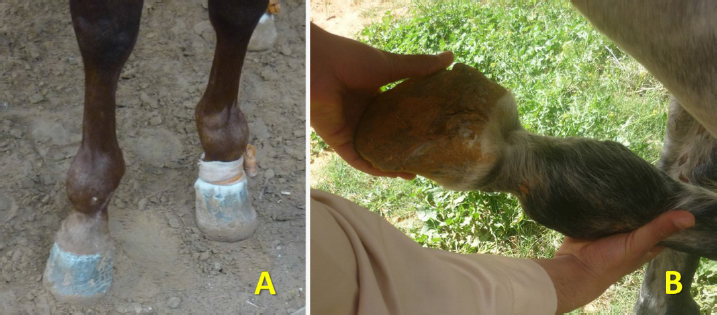
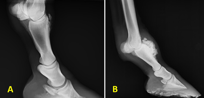
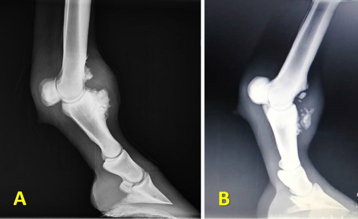
AbstractBackground: Orthopedic diseases involving the forelimb fetlock joint of horses other than those used in professional racing have not been widely reported in the literature. One of these problems is the exostosis on the proximal forelimb phalanx (P1), which has not been reported previously in Western Libya. Aim: The study aimed to investigate the prevalence of P1 exostosis in traditional equestrian horses (known locally as Sarj horses) of different breeds in Libya that participated in a special social event and described its clinical findings radiographic appearance. Methods: The current study involved 60 horses admitted to private veterinary clinics with varied fetlock orthopedic problems. The studied horses were aged between 6 and 11 years old and galloped on hard grounds. Clinical and radiographic examinations were carried out on all horses. Lateromedial radiographs for one or both forelimb fetlock joints were acquired for each horse. Results: Clinical examination revealed that 21 horses (35%) had hard non-painful swelling on the dorsal aspect of fetlock with joint stiffness during flexion. In 19 of them, the swelling was bilateral. No forelimb lameness was noticed. Radiographically, hard swelling was diagnosed as bone exostosis, with various sizes, on the proximo-dorsal aspect of P1. There was also a new bone formation on the disto-dorsal part of third metacarpal bone in two cases. Conclusion: This study has documented the first report on bone exostosis on the proximo-dorsal aspect of the forelimb proximal phalanx as a common problem in Sarj horses that galloped on hard grounds in Western Libya. Although this bone lesion did not cause lameness in all reported cases, further investigations are warranted to identify its histopathological nature, potential etiology, and proper treatment. Keywords: Exostosis, Fetlock, Forelimb proximal phalanx, Horse, Radiography. IntroductionExostosis is defined as a benign bony proliferation on the surface of a bone (Bertoni et al., 2012). It might also be benign bony neoplasms such as osteochondromas and osteomas (Pool, 1990; Chew and Weissleder, 1991; Bertoni et al., 2012). Exostosis is considered uncommon in horses (Butler et al., 2017). In long bones of horses, the lesion is reported in radius (Nixon et al., 2004), third metacarpal/metatarsal bone (Bertoni et al., 2012), second and fourth metacarpal/metatarsal bones (Honnas, 1992; Zubrod et al., 2004), proximal forelimb phalanx (Seghrouchni et al., 2019a; Shaw and Rosanowski, 2019), forelimb middle phalanx (Easter et al., 1998), and tibia (Collins, 1998; Barton et al., 2020). The new bone formation has been attributed to a number of causative agents and associated factors. They included, for instance, the periosteal response to direct trauma, tearing of soft tissues during joint hyperextension, and cyclic strains (Zubrod et al., 2004; Bertoni et al., 2012). However, the association between potential causes and each type of bone exostosis has received less attention and is not certainly identified, especially in horses. Bone exostosis usually does not cause clinical lameness and could not be discovered unless specific diagnostic tools are used for some reason. For example, Barton et al. (2020) identified bone exostosis incidentally at the caudo-proximal aspect of the tibia during a routine pre-purchase radiographic examination of yearling horses (not at racing age) that showed no lameness. However, if the new overgrowth bone disrupted the surrounding soft tissue structures such as tendons and ligaments (Honnas, 1992; De Souza et al., 2021), then a variable degree of lameness would be seen and ranged from mild to moderate (Honnas, 1992; Bramlage, 2009; De Souza et al., 2021). In exostosis of the proximal phalanx (P1), in particular, the affected horse may show transient to mild lameness with a significant reduction in the fetlock joint flexion (Shaw and Rosanowski, 2019). Although the performance of these horses was not considerably affected, the lesion overgrowth and/or chronic and severe fetlock pain could end their career (Shaw and Rosanowski, 2019). The developed lesion was described as a firm and non-painful swelling on one or both fetlock joints (Seghrouchni et al., 2019b; Shaw and Rosanowski, 2019). The radiographic examination revealed that the bony reaction was initially progressed on the dorsolateral aspect of the proximal extremity of P1 and then extended to involve its dorsomedial side and the distal dorsal part of the third metacarpal bone (Mc3) (Shaw and Rosanowski, 2019). Grossly, the lesion mass, which was larger laterally, occupied almost the entire proximo-dorsal aspect of P1 with a varied size (diameter/depth) between 0.5 and 5.1 cm (Seghrouchni et al., 2019a). Its shape differed among the unilateral cases but was similar when the lesion developed bilaterally in a horse (Seghrouchni et al., 2019b). The surrounding soft tissues, including the joint capsule, were clearly affected by the exostosis (Seghrouchni et al., 2019a), which seemed to remain extra-articular (Bramlage, 2009; Shaw and Rosanowski, 2019). There are few available reports about horses’ P1 exostosis in the literature. In recent studies, P1 exostosis exhibited a high incidence among horses practicing different exercise types in different countries. For instance, in Southeast Asia, where P1 exostosis is known as “tropical joint syndrome (TJS),” the lesion was identified in almost 16.6 per 1,000 thoroughbred racing horses at Singapore Turf Club (Shaw and Rosanowski, 2019). This study has pointed out the potential effect of some factors, such as hot and humid climate, in inducing P1 exostosis and whether tropical and warmer climates would have similar influences. In a different region, Morocco, where a traditional equestrian (Tbourida) is popular, the lesion was found in 25 out of 127 Tbourida horses diagnosed with osteoarticular abnormalities (Seghrouchni et al., 2019b). The authors attributed the lesion occurrence to the improper preparation of horses for the Tbourida event, during which they would not be able to accommodate the huge biomechanical stress resulting in fetlock joint hyperextension, and to the hard surface, which exposes the distal limb to repetitive trauma, which might end up with a periosteal reaction (Seghrouchni et al., 2019a). Based on the above studies, it seems reasonable to state that P1 exostosis is more than just an uncommon lesion, and its prevalence should be reconsidered among racing and traditional equestrian horses rather than being overlooked. In Western Libya, there is a local type of equestrian sport. The participating horses known as Sarj horses could be from different breeds, and their number could also be varied in each start line. Galloping details in this sport are quite similar to those of Tbourida in Morocco, except not having a sudden stop at the end of the fast galloping. Although this local sport is very popular and thousands of horses are involved all around the region, there is no published data about the prevalence of P1 exostosis or even its radiographic description. Therefore, the current study was firstly aimed to estimate the prevalence of P1 exostosis in the horses that practice in the local equestrian sport in Western Libyan. The secondary objective was to describe the clinical and radiographic features of the lesion on P1. Materials and MethodsSixty traditional equestrian horses aged between 6 and 11 years were admitted to private veterinary clinics in the western part of Libya with orthopedic problems in the fetlock region. Twenty-one of them presented with a hard swelling involving the dorsal aspect of the forelimb fetlock with no history of lameness. A thorough clinical examination was carried out on all the horses with a dorsal fetlock swelling. This examination included a general health exam and a lameness examination (Reed et al., 2017). The later exam included local palpation around the fetlock area and flexion test to the fetlock, followed by walking and trotting on soft and hard ground. A radiological examination was carried out to investigate bony structures of articular involvement. For most cases, radiographic images were taken of the affected limb with the horse standing squarely on four limbs. Based on the owner’s request, both forelimbs were radiographed in some cases. For all the horses, lateromedial views for the fetlock were acquired. However, dorsomedial views were obtained for a small number of horses because of the low value of this viewing, such as lesions and financial limitations. Ethical approvalAs the work was conducted on clinical cases following usual diagnostic procedures and techniques, no ethical approval was requested. ResultsThe results showed that P1 exostosis was diagnosed in 21 out of 60 cases of the identified orthopedic problems in Sarj horses, with a prevalence of 35%. The clinical examination revealed that 19 horses showed bilateral and only 2 horses had unilateral swelling of different sizes (Fig. 1A). Flexion of the affected joints was obviously painful, and the flexion angle was variably affected or even had some kind of stiffness (Fig. 1B). Most horses showed mild or even no lameness during walking and trotting. The radiological examination revealed a new bone formation involving the dorso-proximal aspect of P1 (Fig. 2A and B). The size of the newly formed bone was variable among cases. No other abnormal radiographic findings were noted in the horses. In two cases, the exostosis was extended proximally to include the dorsal aspect of the distal extremity of Mc3 (Fig. 3A). In one of the two cases, there were multiple fragments in both P1 and Mc3 exostosis (Fig. 3B).
Fig. 1. Sarj horses presented with a fetlock joint swelling. (A) Photo shows dorsal swelling involving the fetlock region bilaterally. (B) Photo shows the fetlock joint stiffness during the flexion test.
Fig. 2. Distal limb radiographs for Sarj horses presented with hard swellings involving the dorsal aspect of the fetlock region. (A) Lateromedial view of forelimb fetlock shows small new bone formation on the dorso-proximal part of P1. (B) Lateromedial radiograph for another horse with a larger dorsal P1 exostosis. P1: first phalanx. DiscussionP1 exostosis has not yet been reported in Libya, although the firm non-painful swelling at the fetlock region seems a fairly common lesion. The current study has found that P1 exostosis was common in Sarj horses, with a prevalence of 35% from the identified orthopedic problems in the fetlock area. The current result is comparable, in many aspects, to the few available studies in the literature. The lesion was also commonly recognized in Morocco, with an approximate prevalence of 20% of the osteoarticular findings (Seghrouchni et al., 2019b). However, Shaw and Rosanowski (2019) reported, in their survey study about the prevalence of TJS in Southeast Asia and Australia, that this lesion was estimated between 1% and 10% in training racehorses. Despite the difference in the prevalence in the aforementioned studies, it could be assumed that a new bone formation on P1 of racing and traditional equestrian horses remains possible as long as several underlying factors exist.
Fig. 3. Radiographic findings in two different Sarj horses with forelimbs P1 and Mc3 bone exostosis. (A) Lateromedial radiograph shows a new bone formation involving the dorso-proximal P1 and disto-dorsal Mc3. (B) Lateromedial radiograph a new bone formation involving the dorso-proximal P1 and the disto-dorsal Mc3. There are multiple bone fragments close to the newly formed bone lesions, which seem to be from the lesion. P1: first phalanx bone; Mc3: third metacarpal bone. Although the histopathological examination of the lesion has not been involved in the current study, it might be suggested that the P1 exostosis is likely to be an osteochondroma. This is because of the very high similarity in the clinical and radiographic features and lesion location in Libyan Sarj horse and Moroccan Tbourida horses (Seghrouchni et al., 2019a, 2019b). However, histopathological examination of the lesion, in addition to computerized tomography and magnetic resonance imaging scans, are indeed required to confirm the diagnosis (Seghrouchni et al., 2019a; De Souza et al., 2021). Sarj horses are traditional equestrian sport horses. Unlike professional racing horses, they do not rigorously fulfil the criteria for having an eligible body and/or limb conformation, and the training regimen implies professional racing. Their physical preparation and training regimen and period simply coincide with an event. Thus, Sarj horses are mostly seen as overweight and physically not certainly fit enough for the stressful sport. With respect to their riders, who are usually their owners, no strict requirements and rules are followed, unlike in professional races. Consequently, it can easily note some riders with overweight. Overweight horses (O’Sullivan, 2007) and increased riders’ weight (Gunnarsson et al., 2017) have been reported to have a negative effect on the horse locomotion due to increasing load. Such load, which would also be increased due to increasing the horse speed during gallop (Davies et al., 1993), might force the fetlock joint to be overloaded and even hyperextended. Hyperextension has also been attributed to the type of sport in which the body status applies more mechanical stress leading to the fetlock hyperextension. In Tbourida, for instance, the fetlock joint was imposed to be hyperextended to tolerate the specific body posture that followed the sudden final stop after galloping (Seghrouchni et al., 2019a). Accordingly, the ability of the joint and the surrounding structures to safely accommodate the resultant hyperextension in these insufficiently prepared Sarj horses seems to be questionable. Another fundamental issue in this local equestrian sport is the type of track ground. The track ground was suggested to significantly influence the distal limb kinematics. In soft tracks, it was found that the hoof can rotate forward, minimizing the pressure on the navicular area, and at the same time, decreasing the fetlock extension, thus reducing the joint load (Scheffer and Back, 2001). This means that the hard surface has probably a negative impact on fetlock joints. Firstly, it would prevent the hoof from the forward rotation and thus overload and possibly hyperextend the fetlock joint during the galloping. Secondly, the hard surface could induce a repetitive trauma on the distal limb and particularly, in this study, on the dorsal aspect of P1. The repetitive stress and strain were suggested to cause damage (Kumar, 2001; Lin et al., 2004) and enthesopathy of the tendons, leading to an osseous proliferation in bones (Bramlage et al.,1980; Widmer and Blevins, 1994). This seems to be one of the potential pathogenic events of developing P1 exostosis in Sarj horses in this study. It might be started from damaging, by the repetitive trauma, the insertion of the lateral digital extensor tendon and one of the common digital extensor tendon insertions on P1 and ended with a new bone proliferation. If this was the case, then this might be reasonable to consider the hard surface as one of the main underlying factors in developing P1 exostosis. Seghrouchni et al. (2019b) have also described the negative effect of the joint hyperextension and the hard surface in generating repetitive trauma. This could also be supported by the absence of P1 exostosis cases among Sarj horses galloping on a soft (sandy) track. Another potential reason for developing P1 exostosis in Sarj horses in this study is related to the proximal growth plate of the bone. It has been suggested that when the repetitive trauma is applied on P1, this might cause separation of the proximal growth plate, which might then be developed to the lesion (Seghrouchni et al., 2019b). Developing the overgrowth bone could normally stop when the growth plate closed (De Souza et al., 2021). In addition, the possible factors responsible for P1 exostosis in that Sarj horses might be the improper trimming and shoeing usually seen in Sarj horses because of the relatively high cost of such services in Libya. Improper trimming and shoeing may result in imbalanced feet and subsequently uneven foot loading (Horan et al., 2021). Other than the mechanism of overloading and hyperextension, the P1 exostosis seems to appear at a certain age. The new bone formation in this study was identified in Sarj horses aged between 6 and 11 years old. Almost a similar age range was recorded in Tbourida horses (6–10 years) (Seghrouchni et al., 2019b). According to Butcher and Ashley-Ross (2002), there is a significant relationship between the horse age and its fetlock joint dorsal flexion resulting from the impact of the mid-stance stage during moderate galloping. They reported that at 5 years of age, thoroughbred racehorses’ fetlock joints experienced the greatest acute angle of the joint dorsal flexion at the mid-stance than younger ages. Although the previously mentioned study did not involve horses older than 5 years age, its result might be reasonable to expect how acute the fetlock joint extension will be in insufficiently/improper prepared and overweigh horses, considering such an acute fetlock joint extension in the highly trained racehorse. This study showed that more than 90% of P1 exostosis cases were bilateral. Perhaps this would not be unusual because Sarj horses are galloping along a temporarily prepared straight course. P1 was reported to be exposed to balance forces during walking and trotting (Merritt et al., 2010). Hence, racing without turn might also generate balanced or equal loading on the right and the left P1. Consequently, when the fetlock joint was overloaded, the resultant repetitive trauma on both right and left P1s would be the same and likely ended up with a bilateral new bone formation. Such assumption might be supported by the nearly similar result found in Tbourida horses that galloped in the same way, where 80% of the cases had a bilateral lesion (Seghrouchni et al., 2019b). Exostosis has generally been associated with varied clinical lameness, ranging from no to moderate (Honnas, 1992; Bramlage, 2009; Vanderperren and Saunders, 2009; Shaw and Rosanowski, 2019; Barton et al., 2020; De Souza et al., 2021). In this study, most of the Sarj horses diagnosed with P1 exostosis showed mild or even no lameness. This low degree of lameness might reflect the limited effect of disrupting the surrounding tissues by the lesion, which was detected radiographically on the dorso-proximal aspect of P1. The affected tissues included the lateral and common digital extensor tendons, the extensor branch of the suspensory ligament, and the fetlock joint capsule (Seghrouchni et al., 2019a). At this stage of the lesion, the performance of horses would not be affected significantly (Shaw and Rosanowski, 2019), and they might still have time to more sports practice. However, it should be taken into account that a progression of the lesion, over time, from a minor to a severe bone reaction to include more than the insertion of the tendon and culminate in an incurable case was reported (Shaw and Rosanowski, 2019). Therefore, it may be difficult to guarantee the participation of such affected Sarj horses in the future, considering the chronic and severe fetlock pain in particular. ConclusionExostosis on the proximo-dorsal aspect of P1 is a common lesion among Sarj horses in certain places in Libya. This lesion appears to be a multifactorial problem that may arise due to the irregular training program, the heavyweight of both horses and riders, bad shoeing, and galloping on hard ground. Future studies involving a more significant number of cases and covering wider geographic locations are highly recommended to determine the histopathological characteristics of this lesion, main causes, and the proper treatment. Conflict of interestThe authors declare that there is no conflict of interest. AcknowledgmentThe authors would like to thank the owners of the horses involved in this study for their cooperation. ReferencesBarton, C.K., Sandow, C.B. and Rodgerson, D.H. 2020. Radiographic description of a bone exostosis lesion on the caudal aspect of the proximal tibia in three thoroughbred yearlings (2014-2019). J. Equine Vet. Sci. 95, 1–3. Bertoni, L., Forresu, D., Coudry, V., Audigie, F. and Denoix, J.M. 2012. Exostoses on the palmar or plantar aspect of the diaphysis of the third metacarpal or metatarsal bone in horses: 16 cases (2001–2010). JAVM 240, 740–747. Bramlage, L.R. 2009. Part 1: Operative orthopedics of the fetlock joint of the horse: traumatic and developmental diseases of the equine fetlock joint. Proc. Am. Ass. Equine Practnrs. 55, 96–143. Bramlage, L.R. Gabel, A.A. and Hackett, R.P. 1980. Avulsion fractures of the origin of the suspensory ligament in the horse. J. Am. Vet. Med. Assoc. 176, 1004–1010. Butcher, M.T. and Ashley-Ross, M.A. 2002. Fetlock joint kinematics differ with age in thoroughbred racehorses. J. Biomech. 35, 563–571. Butler, J.A., Colles, C.M., Dyson, S.J., Kold, S.E. and Poulos, P.W. 2017. Clinical radiology of the horse. John Wiley & Sons, Ltd., Oxford, UK. Chew, F. and Weissleder, R. 1991. Radiation-induced osteochondroma. Am. J. Roentgenol. 157(4), 792. Collins, J.A. 1998. Ossifying fibroma/osteoma in the proximal tibia of a mature gelding. Vet. Rec. 43, 367–368. Davies, H.M.S., McCarthy, R.N. and Jeffcott, L.B. 1993. Surface strain on the dorsal metacarpus of Thoroughbreds at different speeds and gaits. Acta Anatomica. 146, 148–153. De Souza, T., Jones, R., Foote, A. and Suthers, J. 2021. Computed tomographic (CT) arthrogram contributes to the diagnosis of an osteochondroma of the distal calcaneus in a horse. Equine Vet. Educ. 33, 186–191. Easter, J.L., Watkins, J.P., Berrige, B. and Homco, L.D. 1998. A digital osteochondroma as the course of lameness in a foal. Vet. Comp. Ortho. Traumatol. 11, 49–51. Gunnarsson, V., Stefánsdóttir, G.J., Jansson, A. and Roepstorff, L. 2017. The effect of rider weight and additional weight in Icelandic horses in tölt: Part II. Stride parameters responses. Animals 11, 1567–1572. Honnas, C.M. 1992. Surgical treatment of selected musculoskeletal disorders of the forelimb. In Equine surgery. Ed., Auer, J.A. Philadelphia, PA: WB Saunders Co, pp: 985–1051. Horan, K., Coburn, J., Kourdache, K., Day, P., Harborne, D., Brinkley, L., Carnall, H., Hammond, L., Peterson, M., Millard, S. and Pfau, T. 2021. Influence of speed, ground surface and shoeing condition on hoof breakover duration in galloping thoroughbred racehorses. Animals 11, 2588. Kumar, S. 2001. Theories of musculoskeletal injury causation. Ergonomics 44, 17–47. Lin, T.W., Cardenas, L. and Soslowsky, L.J. 2004. Biomechanics of tendon injury and repair. J. Biomech. 37, 865–877. Merritt, J.S, Pandy, M.G., Brown, N.A., Burvill, C.R., Kawcak C.E., McIlwraith, C.W. and Davies, H.M. 2010. Mechanical loading of the distal end of the third metacarpal bone in horses during walking and trotting. Am. J. Vet. Res. 71, 508–514. Nixon, A.J., Schachter, B.L. and Pool, R.R. 2004.Exostoses of the caudal perimeter of the radial physis as a cause of carpal synovial sheath tenosynovitis and lameness in horses: 10 cases (1999–2003). J. Am. Vet. Med. Assoc. 224, 264–270. O’Sullivan, C.B. 2007. Injuries of the flexor tendons: focus on the superficial digital flexor tendon. Clin. Tech. Equine Pract. 6, 189–197. Pool, R.R. 1990. Tumors of bone and cartilage. In Tumors in domestic animals, 3rd ed. Ed., Moulton, J.E Berkeley, CA: University of California Press, pp: 181–195. Reed, S.M., Bayly, W.M. and Sellon, D.C. 2017. Equine internal medicine. Elsevier Health Sciences, St. Louis, Missouri, USA. Scheffer, C.J.W. and Back, W. 2001. Orthopaedics effects of navicular’shoeing on Equine distal forelimb kinematics on different track surface. Vet. Quart. 23, 191–195. Seghrouchni, M., Bollo, E., Piro, M., Alyakine, H., Bouayad, H., Chakir, J., Azrib, R. and El Allali, K. 2019a. Osteochondroma of the first phalanx in Tbourida horses. Front. Vet. Sci. 5, 328. Seghrouchni, M., Elkasraoui, H., Piro, M., Alyakine, H., Bouayad, H., Chakir, J., Tligui, N., Elallali, K. and Azrib, R. 2019b. Osteoarticular radiographic findings of the distal forelimbs in Tbourida Horses. Heliyon 5, e02514. Shaw, D.J. and Rosanowski, S.M. 2019. Tropical joint syndrome: exostosis on the dorsal aspect of the proximal phalanx in racing Thoroughbreds in Asia. Equine Vet. Educ. 32, 60–65. Vanderperren, K. and Saunders, J.H. 2009. Diagnostic imaging of the equine fetlock region using radiography and ultrasonography. Part 2: The bony disorders. Vet. J. 181, 123–136. Widmer, R.W. and Blevins, E.W. 1994. Radiographic evaluation of degenerative joint disease in horses. Compend. Contin. Educ. Pract. Vet. 16, 907–920. Zubrod, C.J., Schneider, R.K. and Tucker, R.L. 2004. Use of magnetic resonance imaging to identify suspensory desmitis and adhesions between exostoses of the second metacarpal bone and the suspensory ligament in four horses. J. Am. Vet. Med. Assoc. 224, 1815–1820. | ||
| How to Cite this Article |
| Pubmed Style Abushhiwa M, TE, Alrtib AM, Bennour EM, Oheida AH, . First Phalanx Exostosis in Traditional Equestrian Horses in Western Libya. Open Vet J. 2022; 12(1): 69-74. doi:10.5455/OVJ.2022.v12.i1.8 Web Style Abushhiwa M, TE, Alrtib AM, Bennour EM, Oheida AH, . First Phalanx Exostosis in Traditional Equestrian Horses in Western Libya. https://www.openveterinaryjournal.com/?mno=33996 [Access: April 18, 2024]. doi:10.5455/OVJ.2022.v12.i1.8 AMA (American Medical Association) Style Abushhiwa M, TE, Alrtib AM, Bennour EM, Oheida AH, . First Phalanx Exostosis in Traditional Equestrian Horses in Western Libya. Open Vet J. 2022; 12(1): 69-74. doi:10.5455/OVJ.2022.v12.i1.8 Vancouver/ICMJE Style Abushhiwa M, TE, Alrtib AM, Bennour EM, Oheida AH, . First Phalanx Exostosis in Traditional Equestrian Horses in Western Libya. Open Vet J. (2022), [cited April 18, 2024]; 12(1): 69-74. doi:10.5455/OVJ.2022.v12.i1.8 Harvard Style Abushhiwa, M., , T. E., Alrtib, A. M., Bennour, E. M., Oheida, A. H. & (2022) First Phalanx Exostosis in Traditional Equestrian Horses in Western Libya. Open Vet J, 12 (1), 69-74. doi:10.5455/OVJ.2022.v12.i1.8 Turabian Style Abushhiwa, Mohamed, Taher Elmeshreghi, Abdulrhman M. Alrtib, Emad M. Bennour, Aiman H. Oheida, and . 2022. First Phalanx Exostosis in Traditional Equestrian Horses in Western Libya. Open Veterinary Journal, 12 (1), 69-74. doi:10.5455/OVJ.2022.v12.i1.8 Chicago Style Abushhiwa, Mohamed, Taher Elmeshreghi, Abdulrhman M. Alrtib, Emad M. Bennour, Aiman H. Oheida, and . "First Phalanx Exostosis in Traditional Equestrian Horses in Western Libya." Open Veterinary Journal 12 (2022), 69-74. doi:10.5455/OVJ.2022.v12.i1.8 MLA (The Modern Language Association) Style Abushhiwa, Mohamed, Taher Elmeshreghi, Abdulrhman M. Alrtib, Emad M. Bennour, Aiman H. Oheida, and . "First Phalanx Exostosis in Traditional Equestrian Horses in Western Libya." Open Veterinary Journal 12.1 (2022), 69-74. Print. doi:10.5455/OVJ.2022.v12.i1.8 APA (American Psychological Association) Style Abushhiwa, M., , T. E., Alrtib, A. M., Bennour, E. M., Oheida, A. H. & (2022) First Phalanx Exostosis in Traditional Equestrian Horses in Western Libya. Open Veterinary Journal, 12 (1), 69-74. doi:10.5455/OVJ.2022.v12.i1.8 |

