


| Original Article | ||
Open Vet J. 2022; 12(5): 649-656 Open Veterinary Journal, (2022), Vol. 12(5): 649–656 Original Research Comparative morphological study of the male and female metacarpal and metatarsal bones of the dromedary camel with special reference to the blood vasculatureGamal Mounir Allouch*, and Fahad Abdullah AlshanbariDepartment of Veterinary Medicine, College of Agriculture and Veterinary Medicine, Qassim University, Buraydah, Saudi Arabia *Corresponding Author: Gamal Mounir Allouch. Department of Veterinary Medicine, College of Agriculture and Veterinary Medicine, Qassim University, Buraydah, Saudi Arabia. Email: g.alloush [at] qu.edu.sa Submitted: 02/02/2022 Accepted: 10/08/2022 Published: 12/09/2022 © 2022 Open Veterinary Journal
AbstractBackground: It is vital to understand the gross anatomy and dimensions of the metacarpal and metatarsal bones in camels in order for veterinarians to identify fraud cases between males and females by carefully distinguishing between them Aim: It is to make comparisons of the morphological characteristics and measurements of the metacarpus and metatarsus bones of male and female camels. Methods: Forty metacarpus and metatarsus of adult camels of both sexes were collected from a typical Burydah slaughterhouse in KSA. The bones were treated according to the established methods of boiling, drying, and bleaching to study morphology. The measurement of the bones in this study has been taken by using digital vernier calipers. Results: The metacarpus and metatarsus consisted of two large and two small bones. The large metacarpal and metatarsal bones consisted of fused III and IV. Except for the distal side in which the two bones diverge more from each other. The metacarpal bone is similar to the metatarsus, except that it is smaller in measurement majority. The small Mc-Mt II and Mc-Mt V were smaller and present on the palmo-lateral or planto-lateral aspect of the large bones, respectively. The length of the metacarpus and metatarsus is almost equal nearly in camels unlike the rest of the animals as well as the metacarpus bone was unlike the metatarsus in form and measurements generally. Conclusion: The large metacarpus and metatarsus bones are distinguished by the fusion of the third and fourth bones along the length of the bone. Except for the distal side in which the two bones diverge more from each other like the rest of the animals. The morphologically characterized majority of the metacarpal bone was similar to the metatarsus, except that it was proximal extremity, cross-section, and measurement. Keywords: Camel, Metacarpus, Metatarsus, Anatomy, Morphometrics. IntroductionThe morphology and morphometry of the metacarpal and metatarsal bones of male and female camels have received little scientific attention. In the dromedary camel, the metacarpus and metatarsus III and IV were merged except in the distal region, where they diverge (Smuts and Bezuidenhout, 1987). According to Dyce et al. (2010) and Budras and Habel (2011), the metacarpal and metatarsal bones in bovines are made up of fusion of Mc. III and IV and the lateral small metacarpal bones. The third metacarpal (III) bone is the only fully grown bone in the horse, while the second and fourth metacarpal (II–IV) bones are referred to as small or splint bones (Getty, 1975; Dyce et al., 2010; Budras et al., 2013). The proximal extremity of the metacarpus in buffalo appears slightly concave (Waad, 2007) and nearly flattened in the horse (Dyce et al., 2010; Budras et al., 2013). The metacarpal bones were quadrilateral in outline in ox, the palmar surface was flat and broad as described by Raghavan (1964). The distal extremity of the metacarpus in the camels differed from that of the horse and ruminant in that the condyles were angulated according to Smuts and Bezuidenhout (1987). In the horse, the distal extremity is made up of two condyles separated by a sagittal ridge and articulated with one digit (Budras et al., 2013). In calves and cows, the lateral condyle was larger than the medial condyle (Nacambo et al., 2007). The purpose of this study is to describe the morphology of these bones, as well as to examine the metacarpus and metatarsus morphometric data. Materials and MethodsForty of metacarpus and metatarsus of 10 adult females and males’ dromedary camels and similar ages (2–3 years) were used for this study. The specimens were collected from a typical Burydah slaughterhouse post-slaughtering in the Al Qassim region, KSA. Each limb was cut off at the level of the carpus and tarsus bones identified as the right or left fore hind limb. The skin and subcutaneous structures (tendons, arteries, veins, and nerves) were removed, then, the bones of the limbs were boiled in water with sodium hydroxide 30% (NaOH). Next, the bones were dried at room temperature for 2 days. After that, they were whitened by dipping them in hydrogen peroxide solutions and sundried for 2–3 days (Gofur, and Khan, 2010; Allouch, 2014; Choudhary et al., 2019; Bharti et al., 2020). measurements were taken from the cannon bone epiphysis to the tip of the pedal bone. The biometrical measurements of metacarpus and metatarsus were recorded by using digital vernier calipers using non-stretchable thread (Muggli et al., 2011). Ethical approvalThe study protocol was approved by the Committee of Animal Welfare and Ethics, Faculty of Agriculture and Veterinary Medicine, Qassim University, Saudi Arabia. ResultsCamel fore and hind limbs are distinguished by long bones able to support the heavy weight of the camel body and prevent sinking in the desert sands. The third and fourth metacarpal and metatarsal bones are fused except in the distal part where they diverge to form separate articulations with the corresponding digits. The results indicated that, in camels, there are two fully developed Mc and Mt (III) and (IV) and small Mc and Mt (II–V) bones. Metacarpal bonesThe metacarpal bone (Figs. 1A; 2B; 3C; and 4D) is similar to the metatarsal bone, especially the length. The metacarpus is flattened and compressed craniocaudally. It has dorsal longitudinal groove between III and IV Mc bones (Figs. 1,4 and 3,4). It is made up of four bones: two large ones, Mc (III and IV) (Figs. 1A,6,7; 2B,6,7; 3C,6,7; and 4D,6,7), and two small bones, Mc (II and V) which were totally welded without any details. The Mc III and IV are quadrilateral in shape and are united except at the distal end, where they ramify to generate distinct articulation aspects with the corresponding digits. The proximal articular surface of the Mc III (Figs. 1A,3; 2B,2; 3C,3; 4D,3; and 5A) is on the same level or a little higher than that of Mc IV (Fig. 1,1). The two bones are separated by a small notch in the front and with a large notch behind. The third metacarpal bone (Mc III) (Fig. 1,1) has a large tuberosity dorso-proximally (Figs. 1A,2; 2B,3; 3C,2; and 4D,2) and a proximal interosseous foramen. The cranio-lateral facet of the third metacarpus (III Mc) articulates with the third carpal bone, while the palmar-lateral facet articulates with the second carpal bone, while the proximal articular surface of Mc IV articulates with the fourth carpal bone (Fig. 5A). The line of fusion between Mc III and IV is determined by a thin line on the dorsal surface. The proximal two-thirds of the palmar surface is concave, with a proximal interosseous foramen. A rough zone near the proximal articular surface is present where ligaments might be attached. In the proximal half, the medial border is flattened and rectangular; in the distal half, it becomes slender and has a rounded border. The lateral border looks similar to the medial border; however, it is narrower and thinner.
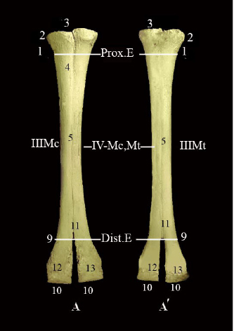
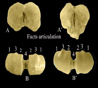
Fig. 1. (A) Dorsal view of the metacarpus, (Aʹ) Metatarsus of male.
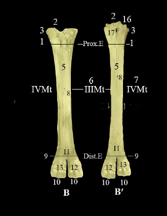
Fig. 2. (B) Palmer view of metacarpus, (Bʹ) Planter view of metatarsus of male.
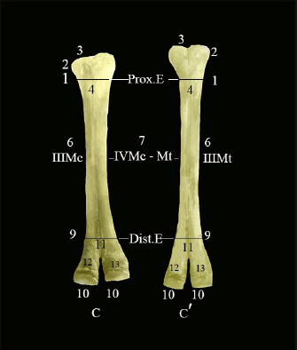
Fig. 3. (C) Dorsal view of the metacarpus, (Cʹ) Metatarsus of female.
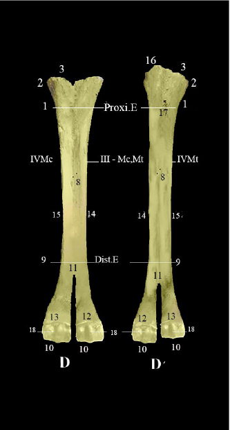
Fig. 4. (D) Palmer view of metacarpus, (Dʹ) planter view of metatarsus of female. Showing (1) proximal extremity of large metacarpus (III and IV Mc), (2) articular surfaces, (3) tuberosity of the third metacarpus, (4) dorsal longitudinal groove, (5) shaft of metacarpal bone (III Mc, IV Mc), (6) third metacarpus–metatarsus, (7) fourth metacarpus and fourth metatarsus, (8) nutrient foramens, (9) distal extremity of large metacarpus (III and IV Mc), (10) articular surfaces, (11) intertrochlear split, (12) medial condyle (13), lateral condyle (14) second metacarpus (II Mc)-metatarsus (II Mt), (15) fifth metacarpus (IV Mc)-metatarsus (IV Mt), (16) prominence process, (17) proximal canal, (18) inter-condyloid ridge. The distal extremity of the metacarpus of Mc III and IV (Figs. 1A; 2B; 3C; and 4D,9) are larger than the metatarsus bones and are situated on some level. The sagittal notch is intertrochlear split (Figs. 1A,11; 2B,11; 3C,11; and 4D,11) divided the distal extremity into two trochlea, each trochlea divides into two condyles (Figs. 1A; 2B; 3C; 4D, 12,13; and 5,3), each condyle is smooth dorsally and divides by the intercondylar ridge; palmary (Fig. 4D,18) into two area abaxial (Fig. 5,1) and axial areas (Fig. 5,2). The small metacarpal Mc II and V are represented by two ridges on either side of the palmar surface of the large metacarpal bones. The metatarsal bonesThe metatarsal bone (Figs. 1Aʹ; 2Bʹ; 3Cʹ; and 4Dʹ): is similar to the metacarpal bone. It had a quadrilateral form with four sides. There are four metatarsal bones: two long metatarsal bones, Mt (III and IV) (Figs. 1Aʹ,6,7; 2Bʹ,6,7; 3Cʹ,6,7; and 4Dʹ,6,7) fused except in the distal part where they were splay to form independent articulation surfaces for III and IV digits. The two bones were totally welded metatarsal bones, Mt II and V without details. The proximal articular surface of the metatarsal bones (Figs. 1Aʹ,3; 2Bʹ,2; 3Cʹ,3; 4Dʹ,3; and 5Aʹ) is characterized by a pointed process planetary (Figs. 3Cʹ,16 and 4Dʹ,16). It has two main bean-shaped facets for the tarsal articulation. The lateral one is the largest of the two surfaces, for articulation with the fourth tarsal bone (T4), while other facet is small oval surface, situated medially to articulate with the second tarsal bone (T2). The proximal articular surface of Mt III is slightly higher than that of Mt IV. The medio-planter facet on the proximal articular surface of Mt III articulates with the first tarsal bone planetary and the fused second and third tarsal and fourth bones (Figs. 1Aʹ,3; 2Bʹ,2; 3Cʹ,3; 4Dʹ,3; and 5Aʹ), while the proximal articular surface of Mt (I–IV) it articulates with (T4). The shaft of the metatarsus is slenderer and squarer in diameter. The Mt III has a prominent and large tuberosity proximo-laterally bones (Figs. 1Aʹ,2; 2Bʹ,3; 3Cʹ,2; and 4Dʹ2), as well as an extended raised area, while Mt IV has a minor tuberosity proximo-medially, as well as the rough oval area along the lateral face. The dorsal surface has a shallow vascular groove that is wider and deeper (Fig. 3Cʹ,4) than the dorsal groove of the metacarpus) which determines the line fusion between Mt III and IV bones. The planter surface is concave and flanked on each side by a rough border except in its distal part. It has the proximal foramen (Figs. 2Bʹ,17 and 4Dʹ,17) in the proximal extremity that passes obliquely to the proximal articular surface. Along the proximal two-thirds of the shaft, the lateral border is slightly concave. It thins out and becomes a rounded border. The medial border of the metatarsal bone at the proximal half is oblique, flattened, and rectangular, it becomes slenderer and has a rounded border distally.
Fig. 5. (A) Proximal extremity of metacarpus showing facet for articulates with second, third and fourth carpal bones. (Aʹ) Proximal extremity of metatarsus, showing facet for articulates first bone and second, third fused and fourth tarsus bones. (B) Distal extremity of metacarpus, (Bʹ) metatarsus showing (1) abaxial articular area, (2) axial articular area, (3) inter-condyloid ridge, and (4) inter-condyloid for articulates with first phalanx.
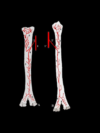
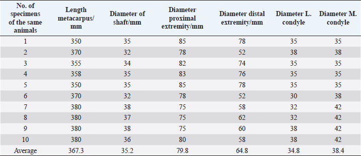
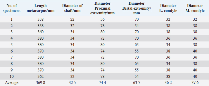
Fig. 6. Presented blood supply of metacarpus (A), metatarsus (B) showing, (1) common metacarpal artery, (1ʹ) common metatarsal artery, (2) nutrient artery of metacarpus, (2ʹ) nutrient artery of metatarsus, (3) proximal nutrient branch, (4) distal nutrient branch, (5) metaphyseal branch, and (6) epiphyseal branch. The distal extremity of metatarsal bones of Mc III and IV (Figs. 1Aʹ; 2Bʹ; 3Cʹ; and 4Dʹ,9) is smaller than the metacarpal bones and are situated on some level. The sagittal notch is intertrochlear split (Figs. 1Aʹ; 2Bʹ; 3Cʹ; and 4Dʹ,11) divided the distal extremity into two trochlea, each trochlea divides into two condyles (Figs.1Aʹ; 2Bʹ; 3Cʹ; and 4Dʹ, 12,13; 5,3), each condyle is smooth dorsally and divides by the intercondylar ridge; palmary (Fig. 4Dʹ,18) into two area abaxial (Fig. 5,1) and axial areas (Fig. 5,2). The small metacarpal Mc II and V are represented by two ridges on either side of the planter surface of the large metatarsal bones. The biometrical observations on the different parameters of the metacarpus and metatarsus showed few simple differences between the male and female camel. The average length of the metacarpal and metatarsal in males was 367 mm, while in females was 370 mm. The maximum diameter of the proximal extremity, shaft, and distal extremity in the male metacarpal bone was 80, 35, and 65 mm, respectively, while it was 75, 32, and 64 mm. The maximum diameter of the proximal extremity, shaft, and distal extremity in the male metatarsal was 72, 35, and 53 mm, while in females the measurements were about 64, 32, and 55 mm. The maximum width of the lateral condyle and medial condyle in the male metacarpal bone was 30 and 35mm and in females was about 36 and 38 mm. The maximum width of the lateral condyle and medial condyle in males’ metatarsal bone was 32 and 34 mm and was 32 and 35 mm in females (Tables 1, 2, 3 and 4). Table 1. Morphometric metacarpus of camel males.
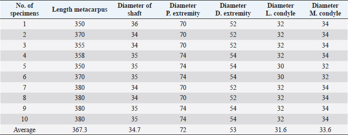
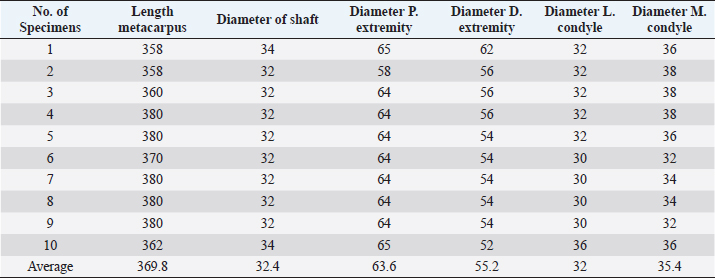
Table 2. Morphometric metatarsus of camel males.
The one-humped camels’ metacarpus-metatarsus were supplied by two nutrients arteries from common palmar-planter digital artery, which is a direct continuation of the median artery. The nutrient artery enters through two nutrient foramens proximal and distal nutrient arteries at the middle part of the bones where they lead into a nutrient canal. This canal extends into the bone body where it gives inside the bone metaphyseal artery to the shaft of the bone, and the epiphyseal artery to supply the spongy bones of the proximal and distal parts of the bones. DiscussionThe anatomical and physiological features of the camel adapt to extreme climatic desert environments (Al Juboori et al., 2010). It is necessary to understand the anatomy of the metacarpus and metatarsus in camels to recognize occurrences of lameness. This could be caused to anatomical flaws (Al-Ani, 2004; Gahlot, 2007). In the present study, the metacarpal and metatarsal bone was four in number, two large III and V which were quadrilateral in form, compressed from side to side, and two small fused metacarpal and metatarsal II and V which were in accordance to Smuts and Bezuidenhout (1987) and Al-Redah and Hussin (2016) in camel, Raghavan (1964) and Budras and Habel (2011) in ox, Yadav et al. (2015) in Indian spotted deer and Choudhary (2015) in blackbuck deer. In contrast to horse, Getty (1975) and Budras et al. (2013) who revealed that the metacarpal and metatarsal bones were three in number, one large III and two smalls were II and IV. The present study revealed that the metacarpus in camels consisted of the fusion of the Mc. III and IV, Fusion of two bones which extended along the whole length except at the distal end where they diverge forming V shape, this was in agreement with the report of Smuts and Bezuidenhout (1987) on camel. Table 3. Morphometric metacarpus of camel female.
Table 4. Morphometric metatarsus of camel female.
Similar studies were recorded by Budras and Habel (2011) and Dyce et al. (2010) in bovine who stated the metacarpus and metatarsus consists of a large metacarpal bone (fusion of Mc. III and IV). They also added the lateral small metacarpal bone. In contrast to horse, Getty (1975) and Buders et al. (2013) reported that the third metacarpal bone is the only completely developed, with the other two being the 2nd and 4th metacarpal bones, which are as small metacarpal bones. This fusion in the metacarpal bones creates a strong bone that acts that works to counteract the forces involved in directly standing that agree with Siddiqui et al. (2008). Siddiqui et al. (2008) in Black Bengal goat, Raghavan (1964) in ox, Getty (1975) in sheep and Yadav et al. (2015) in Indian spotted deer stated that the small bone had a quadrilateral disc-like dorsal face attached with large bones. In this study, the small metacarpal or metatarsal II and V were fusion completely with large bones on the lateral and medial borders. The vascular line and groove in the metacarpal and metatarsal bones of camels were wider and deeper, this agrees with Smuts and Bezuidenhout (1987) and Al-Redah and Hussin (2016) in camel. According to the proximal extremity of the (Mc and Mt III) in camels, we found contradict those of Waad (2007) in buffalo, who stated that the proximal end was slightly concave for articulation with a distal row of carpal bones. However, in horses, the proximal extremity of (Mc III) bears an undulating articular surface adapted to the distal row of the carpus (Getty 1975; Dyce et al., 2010). The present study showed that the Mc III has a significant tuberosity dorso-proximally, whilst the Mt III has a prominent and large tuberosity proximo-laterally. On the other hand, the Mt IV has a large tuberosity proximo-medially, as well as the roughly oval area along the lateral face of the Mt IV, according to the current study. These findings were in line with those of earlier research in ruminant it has on the medial borer two tuberosities, dorsal and palmar (Raghavan, 1964; Metais and Vislobokova, 2007), Choudhary (2015) in blackbuck. While in horses, the metacarpal tuberosity lies on the medial side of the dorsal surface of the Mc. III (Getty 1975; Dyce et al., 2010). The current study presented that the length of the metacarpus and metatarsus is almost equal nearly, unlike the rest of the animals but differed in that the shaft was slenderer, the medial border is flattened and rectangular. The lateral border looks similar to the medial border; however, it is narrower and thinner. The plantar surface was concave. while the planter surface was flat and broad as described by Smuts and Bezuidenhout (1987) and Al-Redah and Hussin (2016) in camel and by Raghavan (1964) and Budras and Habel (2011) in ox. The present study showed that the dorsal surface had a faint line on the Mc and it had a shallow groove on Mt which indicates the fusion line between Mc (III and IV) and Mt (III and IV) respectively, this differs in horses which are represented by smooth and convex from side to side and nearly straight in its length (Budras et al., 2013), while in bovine the dorsal surface was characterized by a dorsal longitudinal groove (Getty 1975; Budras and Habel, 2011). In this study, the distal extremity of Mc and Mt in camel was divided by a dorso-palmar or plantar cleft into two condyles. In comparison with the ruminant this cleft was more separated from the articular surface of Mc. III and IV and each articular surface prepared to articulate with proximal phalanx (Raghavan, 1964; Budras and Habel, 2011) in ox, (Choudhary, 2015) in blackbuck. However, the divided condyles were not straight but angulated as reported by Smuts and Bezuidenhout (1987) in camel. In contrast, in horse, the distal end was different from camel and ruminant. It was undivided and composed of two condyles separated by a sagittal ridge to articular with one digit (Getty, 1975; Dyce et al., 2010; Burders et al., 2013). The lateral condyle was larger than the medial condyle in calves and cows (Nacambo et al., 2007) while the lateral condyle was equal in size with the medial condyle in camel according to our study. In contrast to Ismail et al. (2008), who stated that the metacarpal bone was larger than the metatarsal bone in width and diameter in camels, our study found that the metacarpal and metatarsal bones were approximately similar in length and diameter. Budras et al. (2013) in horse, Budras and Habel (2011) in ox, they found that the metatarsus was longer than the metacarpus. In this study, the Biometrical observation of similar parameters of the metacarpus and metatarsus as shown in the tables indicated that the lateral and medial condyles of females’ metacarpus were longer than that of males. On the other hand, females have larger metatarsal dimensions than males in the length of the metatarsal bone, the diameter of the distal extremity, and the condyles. the proximal extremity and shaft diameters of males are longer than those of female. These results disagree with Al-Redah and Hussin (2016) in camel who stated the metacarpal bone is similar to the metatarsus, except that it is larger and its body is more compressed and Choudhary (2015) reported it in blackbuck. According to the blood supply of the metacarpus and metatarsus bones, this study found that the bones take the blood supply from two nutrient arteries not previously described. ConclusionThe results showed that the large metacarpus and metatarsus are distinguished by the fusion of the third and fourth bones along the whole length except for the distal end in which the two bones diverge more from each other compared to the rest of the animals for the purpose of distributing the weight of the camel over a larger area. The metacarpal bone is similar to the metatarsal bone, but it is smaller in most measurements. Both bones receive their blood supply, from two nutrient arteries which are distributed throughout the bone. Authors contributionsG.A.: planned and conceived the search. G.A.: interpreted the results and designed the figures. Wrote the manuscript. FA performed part of the practical section and revised the final manuscript. AcknowledgmentsThe researchers would like to thank the Deanship of Scientific Research, Qassim University for financially aiding in the publication of this project. The authors acknowledge the support of the Department of Veterinary Medicine, College of Agriculture and Veterinary Medicine, Qassim University, Buraydah, Saudi Arabia. Conflict of interestsThe authors declare that there is no conflict of interest. ReferencesAl-Ani, F.K. 2004. Camels: management and diseases, 1st ed. Milton, Canada: Dar Ammar Book Publisher, pp: 30–45. Al Juboori, A., Naseer, A. and JiJi, K. 2010. Management and healthcare of racing camels. Santiago, Chile: XXVI world Buiatrics Congress, pp: 14–18. Allouch, G.M. 2014. Scientific technique for dkeletons preservation and preparation of anatomical models to promote veterinary anatomy. J. Vet. Anat. 7(2), 133–139. Al-Redah, S.A. and Hussin, A.M. Anatomical study of bone of camel foot. In Proceeding of the International Scientific Conference, College of Veterinary Medicine University of Basrah, Basrah, Iraq, 2016. Bharti, S.K., Singh, I. and Choudhary, O.P. 2020. Gross morphological and morphometrical studies on the metacarpals of blue bull (Boselaphus tragocamelus). Indian. J. Vet. Anat. 32, 16–18. Budras, K.D., Sack, W.O., Horowitz, A. and Berg, R. 2013. Anatomy of the horse. Hanover, Germany: Schlütersche Verlagsgesellschaft mbH and Co. KG, pp: 3–25. Budras, K.D. and Habel, R.E. 2011. Bovine anatomy, an illustrated text, 1st ed. Hannover, Germany: Schlütersche, Verlag Gesellschaft. Choudhary, O.P., Kalita, P.C., Konwar, B., Doley, P.J., Kalita, G. and Kalita, A. 2019. Morphological and applied anatomical studies on the head region of local Mizo pig (Zovawk) of Mizoram. Int. J. Morphol. 37, 196–204. Choudhary, O.P. 2015. Osteo-morphological studies of skull and appendicular skeleton of Indian blackbuck (Antilope cervicapra). Ph.D. Thesis, GBPUAT, Pantnagar, India. Dyce, K.M., Sack, W.O. and Wensing, C.J. 2010. Textbook of veterinary anatomy. Philadelphia, PA: W.B. Saunders Company, Inc., pp: 370–739. Gahlot, T.K. Proceedings of the International Camel Conference “Recent trends in Camelids research and Future strategies for saving Camels”, Rajasthan, India, 2007, p 158–165. Getty, R. 1975. Sisson and grossman’s the anatomy of the domestic animal, 5th ed. Philadelphia, PA: W. B. Saunders Company, pp: 748–762. Gofur, M.R. and Khan, M.S.I. 2010. Development of a quick, economic and efficient method for preparation of skeleton of small animals and birds. Int. J. Bio. Res. 2(7), 13–17. Ismail, Z.B., Alzghoul, M.B., Daradka, M., Al-Shiyab, A.H. and Tashman, O.G. 2008. Morphometric measurements of digital bones in juvenile male camels (Camelus dromedarius). J. Camel. Pract. Res. 15, 117–120. Metais, G. and Vislobokova, I.A. 2007. Basal ruminants. In The evolution of artiodactyls. Eds., Prothero, D.R. and Foss, S.E. Baltimore, MA: The Johns Hopkins University Press, pp: 189–212. Muggli, E., Sauter-Louis, C., Braun, U. and Nuss, K. 2011. Length asymmetry of the bovine digits. Vet. J. 188(3), 295–300. Nacambo, S., Hassig, M., Lischer, C. and Nuss, K. 2007. Difference in the length of the medial and lateral metacarpal and metatarsal condyles in calves and cows a post-mortem study. Anat. Histol. Embryol. 36, 408–412. Raghavan, D. 1964. Anatomy of ox. New Delhi, India: Indian Council of Agricultural Research. Siddiqui, M.S.I., Khan, M.Z.I., Moon, S., Islam, M.N. and Jahan, M.R. 2008. Macro-anatomy of the bones of the fore limb of black Bengal goat (CAPRA HIRCUS). Bangl. J. Vet. Med. 6(1), 59–66. Smuts, M. and Bezuidenhout, A.J. 1987. Anatomy of the dromedary. Oxford, UK: Clarendon Press. Waad, S.K. 2007. Anatomical and histological study of the foot of endogenous buffaloes bubalus bubalis. M. Sc. Thesis, University of Al-Basra, Basrah, Iraq. Yadav, S.C., Joshi, S., Mathur, R. and Choudhary, O.P. 2015. Morphometry of tarsal and metatarsal of Indian spotted deer (Axis axis). Indian. Vet. J. 92, 43–46. | ||
| How to Cite this Article |
| Pubmed Style Allouch GM, El-Shambari FAEA. Comparative morphological study of the male and female metacarpal and metatarsal bones of the dromedary camel with special reference to the blood vasculature. Open Vet J. 2022; 12(5): 649-656. doi:10.5455/OVJ.2022.v12.i5.9 Web Style Allouch GM, El-Shambari FAEA. Comparative morphological study of the male and female metacarpal and metatarsal bones of the dromedary camel with special reference to the blood vasculature. https://www.openveterinaryjournal.com/?mno=24373 [Access: April 20, 2024]. doi:10.5455/OVJ.2022.v12.i5.9 AMA (American Medical Association) Style Allouch GM, El-Shambari FAEA. Comparative morphological study of the male and female metacarpal and metatarsal bones of the dromedary camel with special reference to the blood vasculature. Open Vet J. 2022; 12(5): 649-656. doi:10.5455/OVJ.2022.v12.i5.9 Vancouver/ICMJE Style Allouch GM, El-Shambari FAEA. Comparative morphological study of the male and female metacarpal and metatarsal bones of the dromedary camel with special reference to the blood vasculature. Open Vet J. (2022), [cited April 20, 2024]; 12(5): 649-656. doi:10.5455/OVJ.2022.v12.i5.9 Harvard Style Allouch, G. M. & El-Shambari, . F. A. E. A. (2022) Comparative morphological study of the male and female metacarpal and metatarsal bones of the dromedary camel with special reference to the blood vasculature. Open Vet J, 12 (5), 649-656. doi:10.5455/OVJ.2022.v12.i5.9 Turabian Style Allouch, Gamal Mounir, and Fahad Abdulla El-Shambari Abdulla El-Shambari. 2022. Comparative morphological study of the male and female metacarpal and metatarsal bones of the dromedary camel with special reference to the blood vasculature. Open Veterinary Journal, 12 (5), 649-656. doi:10.5455/OVJ.2022.v12.i5.9 Chicago Style Allouch, Gamal Mounir, and Fahad Abdulla El-Shambari Abdulla El-Shambari. "Comparative morphological study of the male and female metacarpal and metatarsal bones of the dromedary camel with special reference to the blood vasculature." Open Veterinary Journal 12 (2022), 649-656. doi:10.5455/OVJ.2022.v12.i5.9 MLA (The Modern Language Association) Style Allouch, Gamal Mounir, and Fahad Abdulla El-Shambari Abdulla El-Shambari. "Comparative morphological study of the male and female metacarpal and metatarsal bones of the dromedary camel with special reference to the blood vasculature." Open Veterinary Journal 12.5 (2022), 649-656. Print. doi:10.5455/OVJ.2022.v12.i5.9 APA (American Psychological Association) Style Allouch, G. M. & El-Shambari, . F. A. E. A. (2022) Comparative morphological study of the male and female metacarpal and metatarsal bones of the dromedary camel with special reference to the blood vasculature. Open Veterinary Journal, 12 (5), 649-656. doi:10.5455/OVJ.2022.v12.i5.9 |

