


| Case Report | ||
Open Vet J. 2022; 12(4): 434-438 Open Veterinary Journal, (2022), Vol. 12(4): 434–438 Case Report Surgical repair of indirect inguinal hernia in bonnet macaque (Macaca radiata)Inderjeet Yadav1,2* and Ravi Kumar11National Brain Research Centre (NBRC), Manesar, Gurugram, Haryana,122052, India 2Amity Institute of Biotechnology (AIB), Amity University, Gurugram Campus, Haryana, 122412, India Submitted: 24/01/2022 Accepted: 16/06/2022 Published: 09/07/2022 *Corresponding Author: Inderjeet Yadav. National Brain Research Centre (NBRC), Manesar, Gurugram, Haryana, 122052, India. Email: inderjeet.nbrc [at] gmail.com © 2022 Open Veterinary Journal
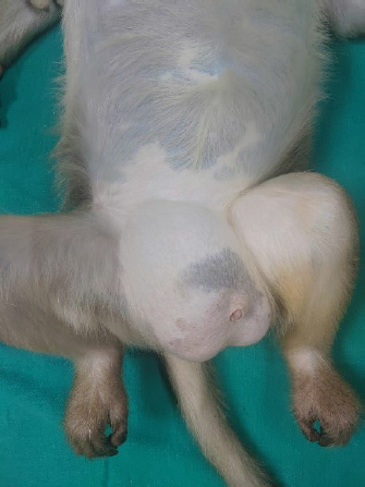
AbstractBackground: Hernia is a protrusion of an organ from the wall of the cavity bearing it. Omentum, adipose tissue, and intestinal loops are the most frequently involved organs. The present case report is a rare case of indirect inguinal hernia having omentum engaged in scrotum through hernial ring in a bonnet macaque (Macaca radiata) and its surgical management. Case Description: A 19-year-old male bonnet macaque was presented with distension of the right inguinal and scrotal region. Physical examination revealed an inguinal hernia. Surgical intervention was deemed necessary considering the state of the animal. The herniation was surgically corrected by the removal of the protruded fatty mass. The monkey was kept in strict confinement till complete healing. The animal recovered uneventfully in 2 weeks without any post-operative complications. This case report details a successful surgical repair of an indirect inguinal hernia in a bonnet macaque. Conclusion: Based on the present case study, it is concluded that surgery can be an ideal and effective option for the treatment of inguinal hernias in primates. Keywords: Bonnet macaque (Macaca radiata), Inguinal hernia, Indirect inguinal hernia, Herniorrhaphy, Omentectomy. Introduction“Hernia” is a Latin word that translates into “rupture”, referring to the protrusion of an organ from the wall of the bearing cavity. The most frequently involved organs are the omentum, adipose tissue, and intestinal loops (Bali et al., 2011; Abee et al., 2012). Inguinal hernia is a type of hernia wherein such contents protrude through a weak area in the lower abdominal wall. Inguinal hernias can be classified into a direct and indirect hernia. Direct inguinal hernia is caused by a weakness or defect in the floor of Hesselback’s triangle which is a zone of muscular weakness in the abdominal posterior wall. Indirect inguinal hernias develop laterally to the Hesselback’s triangle, from the deep inguinal ring down the inguinal canal. These are common in primates and asymptomatic hernias generally do not need repair (Abee et al., 2012). Inguinal hernias have been reported in several monkey species (Graham-Jones, 1962; Starzynski, 1965; Rawlings et al., 1971; Chaffee and Shehan, 1973; Carpenter and Riddle, 1980; Kumar and Raj, 2012; Berg et al., 2017; Sadoughi et al., 2018; Pizarro et al., 2019; Ambar et al., 2020). Indirect inguinal hernia has been reported in nonhuman primates mainly in macaques as well as in baboons (Rawlings et al., 1971; Chaffee and Shehan, 1973; Sadoughi et al., 2018). Males are more susceptible to indirect inguinal hernias due to incomplete removal of the processus vaginalis into the scrotum at the time of testicular descent (Van Veen et al., 2007; HerniaSurge Group, 2018). We have reported a new case of indirect inguinal hernia having omentum engaged in scrotum though hernial ring in a bonnet macaque (Macaca radiata) and its surgical management. Case DetailsCase proceedings in chronological orderOn November 3, 2002, the bonnet macaque was born which was initially acquired from the Committee for the Purpose of Control and Supervision of Experiments on Animals (Government of India) registered facility and was maintained at Animal Facility of National Brain Research Centre from 2004 onwards. The animal was kept in an individual stainless-steel cage with all enrichments available. On December 19, 2019, during morning rounds by the attending veterinarian, it was observed that animal was sitting in an unusual posture and on approaching the animal it was found to be having an unusual bulge in the inguinal region. A thorough clinical examination revealed healthy afebrile and unstressed animal. Surgical intervention was not required as the animal was behaving normally with no sign of distress and pain, and was passing feces and urine normally at that time. On February 23, 2020, during a routine morning round animal was found to be lethargic and dull with very less movement. Animal also refused feed and water. There was sudden increased bulging in the right testicular area of the animal. Physical examinationPhysical examination under anesthesia revealed a hard, non-reducible swelling on the right side of the inguinal area measuring 10 cm in diameter with a ruler, which extended from the lower groin to the scrotum (Fig. 1). Abdominal wall was observed to be mildly congested. The content or nature of the swelling could not be accurately determined by the palpation. Surgical planningGiven the size of the hernia, surgical intervention was scheduled the next day morning. All surgical instruments were sterilized before the start of the surgery. AnesthesiaThe animal was fasted overnight before inducing anesthesia. Atropine sulfate as preanesthetic was injected at 0.05 mg/kg body weight (0.6 mg/ml) to decrease saliva production and respiratory tract secretion during surgery. Anesthesia was induced with a mixture of ketamine at 10 mg/kg body weight (50 mg/ml) and xylazine at 0.5 mg/kg body weight (20 mg/ml). Anesthetic drugs were injected after squeezing the animal in a squeeze cage. The depth of anesthesia was monitored by monitoring heart rate, muscle tension, pinch reflex, and eye blink reflex. The animal was intubated with a cuffed endotracheal tube having a 5 mm internal diameter and 2% lidocaine was sprayed on the larynx before endotracheal tube intubation. The animal was maintained under general anesthesia with 0.5% to 3% isoflurane in 100% oxygen.
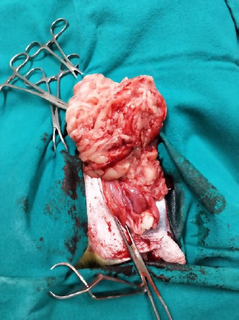
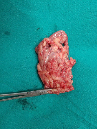
Fig. 1. Inguinal hernia in bonnet macaque. Surgical procedureThe animal was placed in dorsal recumbency and an incision was made vertically on the skin, starting from the lower groin and ending to the mid scrotum medially to the swollen area and exposing the hernial sac by blunt dissection. Hernial sac was also incised which revealed the omentum partially engaged in the scrotum through the hernial ring suggestive of an indirect inguinal hernia. On visual examination no adhesions were detected and the omentum was also normal in color. Vascular supply appeared normal and there were no signs of necrosis. Attempts were made to manually reduce the hernia first, but as our attempts to manually reducing the hernia failed, the inguinal ring had to be enlarged by incising the musculature. Another attempt was made for a manual reduction but it was also unsuccessful due to the large size of the hernial content (Fig. 2). To reduce the content of hernia, partial omentectomy was done with the help of electrocautery to avoid chances of bleeding (Fig. 3). After omentectomy, it was possible to reduce the contents into the abdominal cavity. The right testicle was removed after ligating the right spermatic cord at the level of the inguinal canal to prevent post-surgical complications. The hernial sac was sutured followed by suturing of subcutaneous tissue with a continuous pattern using absorbable suture material (Vicryl–Ethicon, 3-0). During the entire surgical procedure, the vital signs of the monkey (heart rate, oxygen saturation, core body temperature) were monitored. It was also ensured that the animal was in the surgical plane of anesthesia during an entire surgical procedure. The suturing was done in a way to ensure no dead space was left. To prevent subsequent suture failure, the skin was sutured using a simple interrupted suture with silk. Post-operative careThe animal was continuously monitored till recovery from anesthesia. Animal was carefully observed for any signs of pain, and food and fluid intake for the whole postsurgical period. The animal urinated after 5 hours of recovery from anesthesia and started taking feed and water normally. During the post-surgical period, animal was given treats like banana and apple on a daily basis to induce eating. Animal was passing dry faces so it was given liquid cremafin leading to normal passing of faces subsequently. In the post-operative care, the animal was given injection enrofloxacin at 5 mg/kg body weight IM, SID and injection ketoprofen at 5mg/kg IM, BID for 5 days. Butorphanol (0.05 mg/kg IM, TID) was also injected for 3 days after surgery. The animal was closely monitored during the postoperative period for signs of pain, lethargy, decreased appetite, and manipulation of the surgical site. Animal was anesthetized to inspect the recovery. The incision was closely monitored for evidence of swelling, discharge, infection, or dehiscence. All the sutures were intact and no pus formation was there. All the surgical wounds healed without any complications and after 14 days sutures were removed. Animal was monitored regularly for any abnormality. The animal was pronounced healed after a further 4 weeks of observation.
Fig. 2. Hernial content.
Fig. 3. Removed herniated omentum. DiscussionInguinal hernia repair is the most common surgical procedures in humans with more than 20 million procedures done annually (Kingsnorth and LeBlanc, 2003; HerniaSurge Group, 2018). Inguinal hernias are more common in males as compared to females (Van Veen et al., 2007; Chang et al., 2016; HerniaSurge Group, 2018). Apart from these various other factors such as age and race, obesity are also considered to predispose to inguinal hernia (Ruhl and Everhart, 2007; HerniaSurge Group, 2018). In the present case study, animal had hernia from a long period of time but was asymptomatic previously, thus surgical intervention was not considered necessary, which was in agreement with the study of Abee et al. (2012). But when the animal stopped feeding and became lethargic, surgical intervention was thought to be necessary. Males are more prone to indirect inguinal hernia because of the incomplete obliteration of the processus vaginalis at the time of testicular descent into the scrotum (Van Veen et al., 2007). As the Monkey acquired the inguinal hernia after birth, so contradicted any possibility of congenital anomaly. It was suspected that the reason for the disorder could have been any mechanism which resulted in sudden or repeated increases in abdominal or pelvic pressure. In the present case, the animal was having an indirect unilateral inguinal scrotal herniation in a male bonnet macaque. The inguinal hernia can be diagnosed by observing presence of bulge in groin region during visual inspection and linking with the history of the animal, however its very challenging for the veterinarians to diagnose the strangulation and evaluate its complexity. To identify the type of hernia, the contents of hernial sac and the integrity of the anatomical structure, plain or contrast radio-imaging, ultrasonography, Computed Tomography (Towfigh and Shafik, 2017) and laparoscopy (Kumar and Raj, 2012; Pizarro et al., 2019) can be used. In this case, hernia was extended from the abdominal area to the scrotal region, but it was not possible to confirm the diagnosis only by palpation. Omentum was found to be partially engaged in scrotum through the hernial ring in the present case report. This was in agreement to the cases reported in canine inguinal hernias wherein the most common organ present in inguinal hernia is omentum as reported by Strande (1989). In present case, the animal was asymptomatic initially which may be due to the presence of non-incarcerated omentum accounting for asymptomatic and benign nature of inguinal hernia. However, there are chances that omentum may become incarcerated, resulting in appearance of clinical signs that include pain and depression (Waters et al., 1993). Inguinal hernias are treated surgically either using suture-repair or mesh-repair techniques. Present case was successfully attempted with suture-repair technique. In veterinary practice, management of the hernial content by suturing the hernial ring is common regardless of body size (Al-Sobayil and Ahmed, 2007; Whitfield-Cargile et al., 2011), whereas herniation in large animals usually requires meshes (Kassam et al., 2014; Haupt et al., 2015). Depending upon bowel strangulation, severity of inguinal hernia varies from benign to extreme emergency (Ruhl and Everhart, 2007). Similar observations were recorded in present case study. In contrast with the present case study, abdomino-pelvic surgeries have been reported to have postoperative complications such as infection, evisceration, fistula, rupture of sutures, and peritonitis (Waters et al., 1993). In order to minimize mechanical constraints applied to the groin region and to avoid wound dehiscence, postoperative movements need to be reduced. However, minimal mobility is also important to prevent adhesions at the site of surgery. No data is available on prevalence of inguinal hernia in macaques, which is an alarming concern for laboratory animal facilities and zoological institutions housing non-human primates. The present case recovered uneventfully without any post-operative complication and was brought on normal appetite after 1 week. Sutures were removed after 2 weeks and the surgical incision got healed spontaneously, on dressing with topical betadine application. The present case study concluded that case of inguinal hernia should be given conventional medicines in initial stages to cope with clinical sign and symptoms. But in case of no improvement, go for surgical intervention after proper diagnosis. As observed in present study, surgery can be an ideal and effective solution for the treatment of inguinal hernias in primates. Postoperative care can be a challenging task in macaques because of possible site manipulation. Regular checkup should be done to ensure proper healing. In this case study for the first time, the emergency case of inguinal hernia was successfully managed by surgical intervention. Further epidemiological studies are required on the inguinal hernia prevalence in non-human primates. AcknowledgmentsThe authors would like to thank National Brain Research Centre (NBRC, India) for providing all the necessary support. The authors would like to thank Mr Mukesh for assisting in post-operative care related to this case study. Conflict of interestThe authors declare that there is no conflict of interest. Authors’ contributionIY investigated and treated the case and wrote the manuscript. RK contributed to the manuscript writing, literature review and revised the manuscript. Both authors read and approved the final manuscript. ReferencesAbee, C., Mansfield, K., Tardif, S. and Morris, T. 2012. Nonhuman primates in biomedical research: biology and management. Cambridge, MA: Academic Press. Al-Sobayil, F.A. and Ahmed, A.F. 2007. Surgical treatment for different forms of hernias in sheep and goats. J. Vet. Sci. 8(2), 185. Ambar, N., Levi, O., Martin, H., Lee, L., Fahie, M. and Eshar, D. 2020. Surgical management of an inguinal Hernia in an infant captive eastern hoolock gibbon (Hoolock leuconedys). Isr. J. Vet. Med. 75(2), 22–27. Bali, C., Tsironis, A., Zikos, N., Mouselimi, M. and Katsamakis, N. 2011. An unusual case of a strangulated right inguinal hernia containing the sigmoid colon. Int. J. Surg. Case Rep. 2(4), 53–55. Berg, M.R., Allister, R.P. Mac and Martin, L.D. 2017. Nonreducible inguinal hernia containing the uterus and bilateral adnexa in a rhesus macaque (Macaca mulatta). Comp. Med. 67(6), 537–540. Carpenter, R.H. and Riddle, K.E. 1980. Direct inguinal hernia in the cynomolgus monkey (Macaca fascicularis). J. Med. Primatol. 9(3), 194–199. Chaffee, V. and Shehan, T. 1973. Indirect inguinal hernia in two baboons. J. Am. Vet. Med. Assoc. 163(6), 638. Chang, S.J., Chen, J.Y.C., Hsu, C.K., Chuang, F.C. and Yang, S.S.D. 2016. The incidence of inguinal hernia and associated risk factors of incarceration in pediatric inguinal hernia: a nation-wide longitudinal population-based study. Hernia 20(4), 559–563. Graham-Jones, O. 1962. Surgical repair of an umbilical hernia in a gorilla (Gorilla gorilla). Int. Zoo Yearb. 3(1), 109–110. Haupt, J., García-López, J.M. and Chope, K. 2015. Use of a novel silk mesh for ventral midline hernioplasty in a mare. BMC Vet. Res. 11(1), 58. HerniaSurge Group. 2018. International guidelines for groin hernia management. Hernia 22(1), 1–165. Kassam, M., Elkammer, M., Korittum, A. and AbdelWahed, A. 2014. Using of polypropylene mesh for hernioplasty in calves. Alexandria J. Vet. Sci. 40(1), 112. Kingsnorth, A. and LeBlanc, K. 2003. Hernias: inguinal and incisional. Lancet 362, 1561–1571. Kumar, V. and Raj, A. 2012. Surgical management of unilateral inguino-scrotal hernia in a male rhesus macaque. Res. Rev. J. Vet. Sci. Technol. 1(1), 1–4. Pizarro, A.I., Amarasekaran, B., Brown, D. and Pizzi, R. 2019. Laparoscopic repair of an umbilical hernia in a Western chimpanzee (Pan troglodytes verus) rescued in Sierra Leone. J. Med. Primatol. 48(3), 189–191. Rawlings, C.A., Kirk, J.H., Harwell, J.F. and Capps, W.F. 1971. Indirect inguinal hernia in two rhesus monkeys. J. Am. Vet. Med. Assoc. 159(5), 621–622. Ruhl, C.E. and Everhart, J.E. 2007. Risk factors for inguinal hernia among adults in the US population. Am. J. Epidemiol. 165(10), 1154–1161. Sadoughi, B., Dirheimer, M., Regnard, P. and Wanert, F. 2018. Surgical management of a strangulated inguinal hernia in a Cynomolgus Monkey (Macaca fascicularis): a case reporTraitement chirurgical d’une hernie inguinale étranglée chez un macaque cynomolgus (Macaca fascicularis) : cas clinique et discussion de la dém. Rev. Primatol. 25(9). DOI: https://doi.org/10.4000/primatologie.3372 Starzynski, W. 1965. Surger for abdominal hernia in a pig-tailed macaque Maraca nemestrina. Int. Zoo Yearb. 5(1), 184–185. Strande, A. 1989. Inguinal hernia in dogs. J. Small Anim. Pract. 30(9), 520–521. Towfigh, S. and Shafik, Y. 2017. Diagnostic considerations in inguinal hernia repair. In: Textbook of hernia, Springer International Publishing, pp: 35–39. Van Veen, R.N., Wessem, K.J.P. Van, Halm, J.A., Simons, M.P., Plaisier, P.W., Jeekel, J. and Lange, J.F. 2007. Patent processus vaginalis in the adult as a risk factor for the occurrence of indirect inguinal hernia. Surg. Endosc. Other Interv. Tech. 21(2), 202–205. Waters, D.J., Roy, R.G. and Stone, E.A. 1993. A retrospective study of inguinal hernia in 35 Dogs. Vet. Surg. 22(1), 44–49. Whitfield-Cargile, C.M., Rakestraw, P.C., Hardy, J., Cohen, N.D. and Davis, B.E. 2011. Comparison of primary closure of incisional hernias in horses with and without the use of prosthetic mesh support. Equine Vet. J. 43(Suppl. 39), 69–75. | ||
| How to Cite this Article |
| Pubmed Style Yadav I, Kumar R, . Surgical Repair of Indirect Inguinal Hernia in Bonnet Macaque (Macaca radiata). Open Vet J. 2022; 12(4): 434-438. doi:10.5455/OVJ.2022.v12.i4.3 Web Style Yadav I, Kumar R, . Surgical Repair of Indirect Inguinal Hernia in Bonnet Macaque (Macaca radiata). https://www.openveterinaryjournal.com/?mno=14810 [Access: April 20, 2024]. doi:10.5455/OVJ.2022.v12.i4.3 AMA (American Medical Association) Style Yadav I, Kumar R, . Surgical Repair of Indirect Inguinal Hernia in Bonnet Macaque (Macaca radiata). Open Vet J. 2022; 12(4): 434-438. doi:10.5455/OVJ.2022.v12.i4.3 Vancouver/ICMJE Style Yadav I, Kumar R, . Surgical Repair of Indirect Inguinal Hernia in Bonnet Macaque (Macaca radiata). Open Vet J. (2022), [cited April 20, 2024]; 12(4): 434-438. doi:10.5455/OVJ.2022.v12.i4.3 Harvard Style Yadav, I., Kumar, R. & (2022) Surgical Repair of Indirect Inguinal Hernia in Bonnet Macaque (Macaca radiata). Open Vet J, 12 (4), 434-438. doi:10.5455/OVJ.2022.v12.i4.3 Turabian Style Yadav, Inderjeet, Ravi Kumar, and . 2022. Surgical Repair of Indirect Inguinal Hernia in Bonnet Macaque (Macaca radiata). Open Veterinary Journal, 12 (4), 434-438. doi:10.5455/OVJ.2022.v12.i4.3 Chicago Style Yadav, Inderjeet, Ravi Kumar, and . "Surgical Repair of Indirect Inguinal Hernia in Bonnet Macaque (Macaca radiata)." Open Veterinary Journal 12 (2022), 434-438. doi:10.5455/OVJ.2022.v12.i4.3 MLA (The Modern Language Association) Style Yadav, Inderjeet, Ravi Kumar, and . "Surgical Repair of Indirect Inguinal Hernia in Bonnet Macaque (Macaca radiata)." Open Veterinary Journal 12.4 (2022), 434-438. Print. doi:10.5455/OVJ.2022.v12.i4.3 APA (American Psychological Association) Style Yadav, I., Kumar, R. & (2022) Surgical Repair of Indirect Inguinal Hernia in Bonnet Macaque (Macaca radiata). Open Veterinary Journal, 12 (4), 434-438. doi:10.5455/OVJ.2022.v12.i4.3 |

