


| Original Article | ||
Open Vet J. 2022; 12(2): 212-220 Open Veterinary Journal, (2022), Vol. 12(2): 212–220 Original Research Farm and animal-level risk factors associated with rectal prolapse in bovine and buffalo calves in Egypt with special reference to the optimal treatment strategyAlaa Samy1, Eman Abo Elfadl2, Naglaa Gomaa3, Mohamed A. Hamed4, Abdelnaser Ahmed Abdallah5 and Mohamed Abdo Rizk6*1Department of Surgery, Anesthesiology, and Radiology, Faculty of Veterinary, Medicine, Mansoura University, Mansoura, Egypt 2Department of Animal Husbandry and Development of Animal Wealth, Faculty of Veterinary Medicine, Mansoura, Mansoura University, Mansoura, Egypt 3Department of Internal Medicine, Infectious and Fish Diseases, Faculty of Veterinary Medicine, Kafr Elshiekh University, Kafr Elshiekh, Egypt 4Department of Surgery, Anesthesiology, and Radiology, Faculty of Veterinary, Medicine, Aswan University, Aswan, Egypt 5Veterinary Teaching Hospital, Faculty of Veterinary Medicine, Department of Internal Medicine and Infectious Diseases, Mansoura University, Mansoura 35516, Egypt 6Department of Internal Medicine and Infectious Diseases, Faculty of Veterinary Medicine, Mansoura University, Mansoura, Egypt *Corresponding Author: Mohamed Abdo Rizk. Department of Internal Medicine and Infectious Diseases, Faculty of Veterinary Medicine, Mansoura University, Mansoura 35516, Egypt. Email: dr_moh_abdo2008 [at] mans.edu.eg Submitted: 08/01/2022 Accepted: 17/03/2022 Published: 02/04/2022 © 2022 Open Veterinary Journal
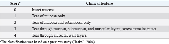
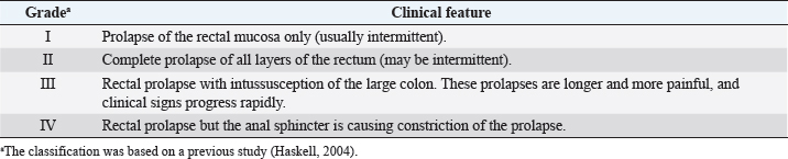
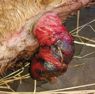
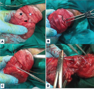
AbstractBackground: Rectal prolapse (RP) is a serious illness of the rectum and small intestine causing serious health problems in domestic animals. However, there is paucity in the estimation of the risk factors associated with this problem in calves. Aim: In the present study, we investigated the prevalence and risk factors associated with the rectal prolapse in both bovine and buffalo calves in Egypt, highlighting the most appropriate treatment strategy. Methods: Forty-two calves (23 bovine and 19 buffalo) suffering from varying degrees of rectal prolapse were used. From the owners’ anamnesis, the farm- and animal-level risk factors associated with each animal were collected. Fisher’s exact tests were used to determine the distribution of frequencies in the different rectal prolapse grades. Descriptive statistics were calculated in the form of mean ± standard deviation (SD) using one-way analysis of variance. Crosstabs were used to determine Spearman’s correlation between variables. According to the disease severity, the appropriate treatment strategy was accomplished either by medicinal or surgical interferences. Results: The final logistic regression form demonstrated that the statistical test, Hosmer and Lemeshow’s goodness of fit, indicates a significant result (χ2=8.91). Body score was the potential risk factor for the occurrence of RP in calves. Medicinal management along with dietary modification was sufficient to treat 70% of grade I in a successful manner, while 33.3% (grade I and grade II) were effectively treated surgically with reduction and application of purse-string sutures. Conclusion: The current study advocates the valid role of resection of rectal mucosa combined with manual reduction and retention in treating calves suffering from grade II rectal prolapse. The final multivariate logistic regression model indicates that the calf’s body score is a potential risk factor for the occurrence of RP. Keywords: Rectal prolapse, Calves, Mucosal resection, Rectal amputation. IntroductionRectal prolapse (RP) is an important diseased condition of the rectum and small colon in the domestic animals (Anderson and Miesner, 2008). Although RP can occur in all species of domestic animals, its common occurrence was recoded in cattle and small ruminants (Anderson and Miesner, 2008). Prolapse of the rectal mucosa occurs following straining, which may be associated with tenesmus (as occurs with coccidiosis and colitis), dysuria (as a complication of cystitis, urolithiasis, dystocia, and neoplasia), chronic coughing (as a complication of bovine respiratory disease), or may be associated with other factors including neoplasia, diet (e.g., clover and high estrogenic compound feedstuff, such as soybean meal), and various toxins (Steiner, 2016). RP may be partial (incomplete) where rectal mucosa is everted externally beyond the level of the anus or complete, in which a full thickness layer of the rectum is protruded (Kashyap et al., 2019). It is seen more frequently in younger calves in association with severe diarrhea, tenesmus, and Eimeria infection (Gopalakrishnan et al., 2017). Indeed, the treatment and prognosis of RP depend on the grade of the prolapse severity and the physiological state of the exposed tissues (Anderson and Miesner, 2008). Various techniques for the management of RP have been described. The initial treatment is usually directed toward conservative management that includes fluid replacement therapy for dehydration and electrolyte disturbances, symptomatic control of diarrhea, and administration of appropriate medication in the case of parasitic or bacterial enterocolitis (Čech et al., 2010). The surgical intervention represents the main treatment in recurring or long-standing RP (Rubin, 2013). To the best of the authors’ knowledge, risk factors associated with the RP in either bovine or buffalo calves have not been studied. Therefore, the present study aimed to investigate the prevalence and risk factors associated with rectal prolapse in both bovine and buffalo calves in Egypt, highlighting the most appropriate treatment strategy. Materials and methodsAnimalsA total of 42 claves (23 bovine and 19 buffalo) with a mean age of (mean ± SD) 21.09 ± 11.24 weeks old and mean body weight (BW) of 275.76 ± 16.48 kg were admitted to Mansoura Veterinary Teaching Hospital, Mansoura University, Mansoura, Egypt, between October 2016 and January 2020 with a complaint of various size protruded masses through their anal openings. History and physical examinationFrom the owners’ anamnesis, the farm- and animal-level risk factors associated with each animal were collected. Farm risk factors included vaccination, deworming programs, previous diseased conditions, interferences for management, and the number of management trials. Additionally, animal risk factors including the species, age, gender, and body score of affected calves according to to Momont and Pruitt (1988) and Ezenwa et al. (2009), concomitant clinical signs, the age of the prolapse, length of the prolapsed part, presence of rectal tears, edema, and amenability of the prolapsed part for reduction were recorded. Physical examination included heart and respiratory rates, and meanwhile, local examination of the anal region was made carefully for grading the prolapse according to Haskell (2004) (Table 1). Management of rectal prolapseCalves were exposed to one of the following techniques, depending upon the severity of symptoms (Table 2). Treatment 1 (medical) comprised calves with a recent degree of rectal prolapse of grade I (n=7) which were treated by intravenous administration of a dose of hyoscine butylbromide (Buscopan® Ampoules 20 mg/ml, Boehringer Ingelheim, Germany) at the dose rate of 0.4 mg/kg intravenously to control contractions of GIT smooth musculature, and thus tenesmus in calves, followed by reduction of prolapsed rectal mucosa by applying digital pressure gently and then, it was replaced into its proper anatomical location. While old cases of grade I (n=3) and grade II (n=11) were treated via reduction and retention using the purse-string suturing (RR) technique under caudal epidural analgesia (treatment 2). Seven calves with grade II were surgically treated by local mucosal resection, followed by RR (treatment 3). Meanwhile, in calves with grade III (n=6) and IV (n=4), rectal amputation was performed (treatment 4). Surgical proceduresOn lateral recumbency, calves were tranquilized using intramuscular injection of xylazine HCl at a dose rate of 0.05 mg kg-1 (Xyla-Ject, 20 mg/ml; Adwia). After strict aseptic preparation, caudal epidural analgesia was conducted. After analgesia, the tail was wrapped and isolated from the operative field and the prolapsed part was rinsed with warm physiological saline and povidone-iodine (Betadine antiseptic). The visible dirt materials were removed carefully using a piece of gauze and then the prolapse was immersed in enough amounts sugar for 20–30 minutes. After that, the sugar was rinsed away from the prolapse. Then, the prolapse was treated by one of the following surgical techniques: (i) Reduction and retention purse-string suturing (RR) technique: This technique was performed with reducible rectal prolapse grade I (n=3) and II (n=11) where the mucosa was viable and no laceration was found on close inspection. By the palm of both hands, gentle pressure was applied to reposition the prolapse back into its normal anatomical position after lubrication with Faktu ointment (each 1 g containing Policresulen 50 mg, cinchocaine HCl 10 mg, Takeda, Nycomed, Philippines). Finally, a purse-string suture was placed through the skin and the deep fascia around the anus where two fingers can easily be passed through the anus. The knot was tied in a bow. (ii) Local mucosal resection, followed by reduction and retention purse-string suturing (LM-RR) technique: This technique was performed with reducible rectal prolapse grade II (n=7) with localized damaged nonviable mucosa. Localized elliptical incisions were performed enclosing the damaged areas of the mucosa (tears, necrosis, and ulcerations) and then the mucosa was carefully dissected free from the muscular layer of the rectum. The mucosa edges were sutured back together using 2-0 triclosan-coated polyglactin 910 (Vicryl plus Ethicon, Johnson and Johnson Company, Somerville, NJ). Then, the prolapse was reduced and a purse-string retention suture was applied. (iii) Rectal amputation (RA) technique: This technique was performed with nonreducible rectal prolapse III (n=6) and IV (n=4, Fig. 1) and in cases with grade II (n=4) with extensive rectal tears where a suitably sized firm rubber tube was passed into the lumen of the prolapse. Then, a circumscribed incision (Fig. 2) was made 3–5 cm away from the mucocutaneous anorectum, to separate the mucosa, submucosa, and muscularis (both the inner circular and the outer longitudinal smooth muscle layers) of the prolapsed part. Serosa was easily distinguished by its blood supply. Minute bleeding was controlled by an electro-cauterization. Using 2-0 chromic catgut sutures, all serosal blood supplies were double ligated and transected in between. After that, the first quadrant of the serosa was resected. Using 2-0 triclosan-coated polyglactin 910 (Vicryl plus Ethicon, Johnson and Johnson, Somerville, NJ), the outer layers (mucosa to the serosa) and inner layers (serosa to the mucosa) of the resected part were sutured together (Fig. 3), using simple interrupted sutures 5 mm apart. The other quadrants of the prolapse are then resected similarly. After amputation and suturing of all quadrants have been completed, the stump retracted manually. Table 1. Classification of rectal tears.
Table 2. Classification of rectal prolapse by structure and anatomic involvement (Haskell, 2004).
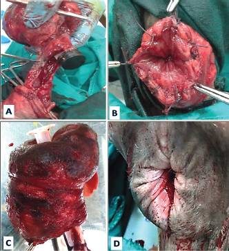
Fig. 1. Cattle calf aged 4 weeks old suffering from a rectal prolapse of grade IV.
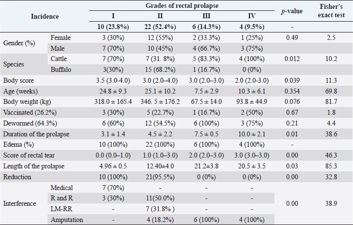
Fig. 2. (A) A circumscribed incision was made to separate the mucosa, submucosa, and muscularis (white star) of the prolapsed part from the serosa (black star). N.B: serosa was easily distinguished by its blood supply (black arrow). (B) Minute bleeding was controlled by electrocauterization. (C) Using chromic catgut sutures, all serosal blood supply were double ligated and transected in between. (D) Using coated polyglactin 910, the rectal wall was sutured together.
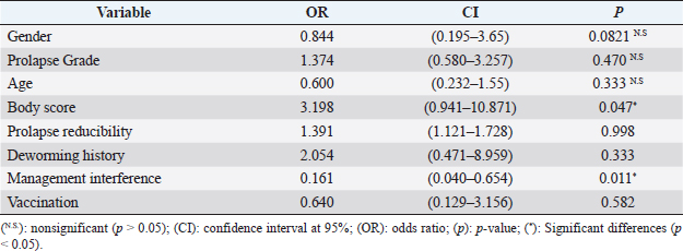
Fig. 3. (A) The prolapsed rectum was resected. (B) The rectal wall was sutured using simple interrupted suture pattern. (C) Resected prolapsed rectum. (D) The stump was retracted manually. Postoperative care and follow-upStrict dietary rest was advised for a day, followed by feeding on green easily digested food from day 7 onward and then gradually changed to normal food. Calves were rehydrated using parenteral fluids (Ringer’s lactate 25 ml/kg BW, intravenously) for the first 3 days of therapy. To counteract Eimeria infection, calves received a combination of sulfadimidine sodium in a powdered form (Aveco, Egypt) at the dose rate of 1.0 g/10 kg BW orally or sulfadimidine 33.33% (VETWIC, Egypt) at the dose rate of 15 ml/100 kg BW intravenously and metronidazole tablets (El Nile Company, Egypt) at the dose rate of 500 mg/40 kg bodyweight for 5 days; and 20% amprolium powder (ADWIA, Egypt) at the dose rate of 5 g/100 kg BW orally for 5 days. Statistical analysisData were tabulated, coded, and then analyzed using the computer program Statistical Package for the Social Science (SPSS) version 23.0. Fisher’s exact test was used to determine the distribution of frequencies (incidence, gender, species, vaccinated animals, dewormed animals, the amenability of the prolapse to reduction, and the interference technique) in the different rectal prolapse grades. Descriptive statistics (age, BW, duration of the prolapse, and the length of the prolapse) were calculated in the form of mean ± standard deviation (SD) using one-way analysis of variance, which is mean analysis of variance used to determine whether there are any statistically significant differences between the means of three or more independent (unrelated) groups. Crosstabs which are a matrix table typically used to analyze the relationship between variables in two or more categories were used to determine Spearman’s correlation between variables. Positive r-values indicate a positive correlation, and the values of both variables tend to increase together, while negative r-values indicate a negative correlation, where the values of one variable tend to increase when the values of the other variable decrease. The magnitude of the correlation coefficient indicates the strength of the association between the assessed variables and is usually interpreted as follows: greater than 0.5 is large, 0.5–0.3 is moderate, 0.3–0.1 is small, and anything smaller than 0.1 is insubstantial. The p-value of the correlation helps to determine whether the correlation is significant or not and can be used to measure the intensity of the relationship. The logistic regression analysis was accomplished to test the association between the healing status of the rectal prolapse and other risk factors. Also, univariate logistic regression statistics were performed. In such a process, the category of healing status (pass or failed) was the dependent dichotomous variable, but the other risk factors were the independent variables. The result indicated that the model’s estimates fit the data at an acceptable level in a multivariable test. For each variable, the parameters included in the results were p-value, odds ratio (OR), confidence interval (CI: 95%), regression coefficient (β), and standard error. In every statistical analysis, the results were considered to be significant at p < 0.05. Ethical approvalThe declaration of the animal owner was taken before entering the study. This inquiry was permitted by the Animal Care Committee of the Mansoura University, in compliance with the Egyptian ethical codes for studies on experimental animals (Approval number 09-021). ResultsPrevalence, univariate statistics, and multivariate statisticsOn animal and farm levels, the results of the univariate analysis for the suggested risk factors of RP are summarized in Tables 3 and 4. On clinical examination, calves revealed anorexia, rough hair coat, weakness, dehydration, frequent straining, and difficulty in defecation. The masses that protruded from the anal opening were diagnosed as RP of different grades (I–IV). RP of grade II exhibited a high incidence (52.4%, n=22), meanwhile grade IV exhibited a low incidence (9.5%, n=4). The mean ± SD length of the prolapses (12.67 ± 6.5 cm) was significantly (p=0.001) different between the different grades. 73.8% of the calves in grades I and II had a reducible RP (n=31: grade I=10 and grade II=21). All RP showed edema, and the median score of rectal tears was 1.0 (0.0–3.0). There were significant differences between the prolapse reducibility frequencies (exact=46.3, p=0.00) and the rectal tears score distribution (exact=32.8, p=0.00) in the different grades. Table 3. Incidence of rectal prolapse in calves.
Table 4. Classification of calves as rectal prolapse positive or negative with respect to different risk factors.
Regarding the anamnesis, 57.1% of the admitted calves were male (n=24) and 42.9% were female (n=18). The median (range) of their body score was 3.0 (2.0–4.0). Unvaccinated calves were 73.8% (n=31) of the total calves; 64.3% (n=27) received regular deworming programs; meanwhile, 33.3% (n=14) of these animals were neither vaccinated nor dewormed. There were significant differences (p < 0.05) between the frequencies of the disease between cattle and buffalo calves (exact=10.2, p=0.012) and the body score (exact=11.3, p=0.039) in the different grades. In general, the incidence of RP in cattle was higher than buffaloes, but in grade II of RP, the frequencies in buffaloes were higher than cattle. Body conditions score was the highest (3.5) in grade I, while it was the lowest one (2.0) in grade IV. Meanwhile, there were nonsignificant differences between the distributions of gender, age, BW, vaccination, and deworming states (exact=2.5, p=0.49; exact=69.8, p=0.354; exact=81.7, p=0.076; exact=1.8, p=0.67; and exact=4.4, p=0.21, respectively) in the different grades. Before admission, calves suffered from straining and tenesmus before prolapsing of the rectum and they were diagnosed and treated by the veterinarians. Their diagnosis was heavy worm infestation with constipation (23.8%), coccidiosis with bloody diarrhea (14.3%), salmonellosis (11.9%), nutritional causes (11.9%), and urolithiasis (4.8%). Meanwhile, the cause was not unknown in 33.3% of these animals. There was a significant difference between the frequencies of the diseases in the different prolapse grades (exact=25.7, p=0.002). The mean ± SD of the prolapse age (5.12 ± 2.75 days) was significantly (p=0.000) different between the different prolapse grades. Owners stated that 59.5% (n=25) of the prolapses were not managed by veterinarians before. There were several trials in 40.5% (n=17) from them to reduce these protruded masses which failed to be treated with RR after one trial (n=7), two trials (n=8), and three trials (n=2). Of all the admitted calves, there was a significant difference between the frequencies of the management in the different prolapse grades (exact=38.9, p=0.00). 16.7% (n=7, 70% RP grade I) were successfully cured with the medicinal management along with dietary modification, while 33.3% (grade I=3 and grade II=11) were effectively treated surgically with the RR technique. LM-RR technique was performed in 16.7% of the calves (grade II=7), while RA technique was mandatory in 14 calves (33.3%, grade II=4, III=6 and IV=4) with a successful outcome. There was a significant association (p < 0.05) between the occurrence of RP and both the animal body score and interferences for management (Table 4). On the other hand, other estimated animal- and farm-level risk factors did not affect significantly (p > 0.05) the occurrence of RP. The final logistic regression form (Table 5) demonstrated that the statistical test, Hosmer and Lemeshow’s goodness of fit, indicates a significant result (χ2=8.91). Body score was the potential risk factor for the occurrence of RP in calves. Correlation between animal- and farm-level risk factorsThere were nonsignificant correlations between gender, species, vaccination, and anti-worming state of the calves with the grade of the RP (r=0.004, p=0.978; r=−0.146, p=0.355; r=0.020, p=0.902; and r=0.201, p=0.201, respectively). There were negative significant (p=0.000–0.001) correlations between the grade of the RP with the animal age, BW, as well as body score (r=−0.510; r=−0.506; and r=−0.482, respectively). Meanwhile, there were very strong positive significant (p=0.000) correlations between the grade of RP and the prolapse age, rectal tears, and the length of the prolapse (r=0.669; r=0.817; and r=0.854, respectively). A positive correlation was present between the species with the age of the animal as well as the prolapse reducibility (r=0.259, p=0.047 and r=0.433, p=0.004, respectively). Additionally, age had a significant positive correlation with the prolapse reducibility (r=0.622, p=0.000), and a significant negative correlation with the rectal tears score (r=−0.528, p=0.000). There were significant (p=0.000) negative correlations between the prolapse reducibility and the prolapse age (r=−0.694), rectal tears score (r=−0.666), and the length of the prolapse (r=- 0.671). The prolapse age had a negative correlation with the body score (r=−0.431, p=0.004) and positive correlation with both rectal tears score and the length of the prolapse (r=0.756, p=0.000 and r=0.737, p=0.000, respectively). Furthermore, rectal tears of the prolapse had a negative correlation on the body score (r=- 0.579, p=0.000). There was a positive correlation between the length of the prolapse and the rectal tears score (r=0.889, p=0.000) and negative correlation with body score (r=- 0.431, p=0.004). Finally, the surgical interference was positively correlated with the grade (r=0.838, p=0.000) and was negatively correlated with both the age (r=−0.511, p=0.001) and reduction (r=0.718, p=0.000). DiscussionThe current study was carried out on 42 indoor reared calves suffering from RP during the last 4 years. Indeed, before admission, all the studied calves were suffering from straining due to either constipation or diarrhea for a long time. Rao et al. (2006) mentioned that the abnormal contraction of rectal muscles during straining on defecation is the major cause of RP as it results in trauma and compression of the rectal wall on the anal canal. Our results revealed that age, gender, vaccination, and deworming of calves did not affect the incidence of RP. The incidence of RP in cattle was higher than buffalo calves and the incidence of PR grade II (54.2%) was higher than other grades; the percentage of buffalo calves (68.2%) was higher in this grade than other grades. Our results showed that the grade of RP was in a high positive significant correlation with the duration of prolapse and it was significantly negatively correlated with the animal age. This could be explained by the long duration of rectal prolapse in our concerned calves (mean=5.12 days) which was too long to aggravate the condition, especially in young calves due to both continuous straining and the local ischemia exhibited by the pressure of the anal sphincter which had narrow anal orifices (Gopalakrishnan et al., 2017). This aggravation was represented by an increase in the length of the prolapse and the scores of rectal tears which were correlated significantly with the grade of the prolapse and negatively correlated with the ability to the reduction of prolapse. This could explain the positive significant correlation between age and the ability of prolapse reduction. Table 5. Final logistic regression form for risk factors associated with rectal prolapse in calves on the animal level
Generally, the treatment of RP depends upon recognition of the type and the length of the prolapse, the involved tissues, the degree of tissue damage that has taken place, and the amenability of the prolapse for reduction. Sometimes temporary protrusion of a portion of the rectal mucosa occurs during defecation or coughing, herein dietary modification along with medical therapy was effective in calves with mild symptoms and with the absence of significant mucosal prolapse as recommended by Schwandner and Schrinner (2014). Therefore, in this study, using antispasmodic drugs to control GIT contractions and tenesmus in calves, together with rehydration, the affected animals using parenteral fluids therapy, anthelmintics, and laxatives for continuous 5 days, gave a good response. Policresulen, a topical hemostatic and antiseptic dressing, was used in this study as recommended by Orescanin and Rodic (2018) to promote selective coagulation of necrotic tissues by leaving the healthy tissues intact and accompanied by re-epithelialization of the mucosal wound tissues (Kim et al., 2015). This assertion is in agreement with the findings as to the symptoms significantly decreasing during the treatment period. However, that conservative approach was less useful when significant mucosal prolapse associated with edema and swelling was observed. Therefore, in calves whose symptoms are resistant to those conservative measures, surgery remains the only option, and reduction of the prolapse is considered the first prognostic key in the treatment of such cases. The present study found significantly negative correlations between the ability of reduction with both the duration and the length of the prolapse and the score of rectal tears which aggravated the condition with edema, severe swelling, necrosis, and adhesions, and subsequently, amputation is indicated. In this study, sugar (high osmotic material) was used to minimize the edema and the swelling of the prolapse. Previous study by Muhammd (2010) recommended potassium permanganate but its tincture is not accepted by many surgeons, requiring long contact time and irritating to skin and mucous membranes (Saganuwan et al., 2008). Rectal tears and mucosal ulcers were observed in all claves with RP of grades II, III, and IV with different degrees. The underlying etiology and pathogenesis of the ulcers are not fully understood, but multiple factors may be involved as the local ischemia. Such ischemia is exhibited by the pressure of the anal sphincter which subsequently impairs the blood flow to the affected area, leading to venous congestion and edema in the mucosal lining of the rectum with resultant ulceration. Rao et al. (2006) explained that the cause of ischemia may also be related to fibroblasts replacing blood vessels. Ulcers may also occur due to the direct trauma from owner-digitation maneuver to reduce RP or to evacuate impacted feces. Zhu et al. (2014) confirmed that uncoordinated defecation with excessive straining over time plays a key role in the rectal ulcer formation. Body condition score is a more reliable indicator of the nutritional status of a cow than is BW as it describes the relative fatness of the animal through the use of a scale and is an effective management tool to evaluate the nutritional status of the herd. According to both Momont and Pruitt (1988) and Ezenwa et al. (2009), the low body condition score of these calves indicated physical weakness, poor performance, and moderate to slight muscle atrophy. In this study, the lowest body condition scores were evidenced in grade IV. This confirmed that body score is the potential risk factor for the occurrence of RP in calves where with the abnormal contraction of the weak rectal muscles during straining on defecation rectal prolapse is an expected result. Moreover, the negative correlation between the body score and rectal tears confirmed this result where poor body performance and impairment of rectal blood supply are risk factors in rectal ulcer formation. In the present study, 40.5% of the admitted calves failed to be treated with manual reduction and application of a retention purse-string suture. Straining and tenesmus are the major obstacles to achieving that goal of healing. Zhu et al. (2014) mentioned that rectal ulceration assists in increasing rectal hypersensitivity that subsequently leads to a persistent desire to defecate and/or feeling of incomplete evacuation and excessive straining. Therefore, resection of these ulcers was mandatory in all calves with reducible RP. But resection is not recommended if extensive ulcerations of the prolapsing segment have occurred as the inflammatory changes; fibrosis and adhesion of the ulcer bed make its separation from the underlying muscle layer difficult, if not impossible, and can lead to perforation (Li et al., 2015). In this study, MR-RR was applied just in seven calves with reducible RP of grade II, while amputation was mandatory in four calves with reducible grade II rectal prolapse. Haskell (2004) mentioned that long prolapses are probably intussusceptions of the rectum and/or the small colon, and the potential for mesocolic tearing increases with the length of the intussuscepted bowel. They suggest that prolapses of more than 20 cm often demand surgical resection. In this study, amputation was performed in calves with rectal prolapses of grades II, III, and IV, depending on the degree of rectal tears not the length of the prolapse. During rectal amputation, the incision was 2 cm away from the retroperitoneal portion of the rectum because it lacks the outer serosal layer and can have implications for surgical healing (Zhou et al., 2018). Triclosan-coated polyglactin 910 (Vicryl Plus) was used in this study because of its antimicrobial activity (Samra et al., 2018). The commonly used technique for rectal amputation recommended resection of the prolapse in quadrants where bleeding is annoying (Sadan, 2019). However, in the present study, a circumscribed incision was performed involving the mucosa, submucosa, and all the muscularis layers permitting visualization of all serosal blood supplies, which were easily ligated and subsequent surgical amputation with high surgeon satisfaction and a successful outcome was reached. ConclusionThe present investigation draws attention to the prevalence and risk factors associated with the occurrence of RP in bovine and buffalo calves in Egypt. The results indicate a significant association (p < 0.05) between the occurrence of RP and both the animal body score and interferences for management. The final multivariate logistic regression model indicates that the calf’s body score is a potential risk factor. Thus, these factors may be the actual explanations for variation in RP prevalence rates in Egypt. Further investigations are required to be carried out to include large sample size, a wide range of risk factors, and wide geographic areas, to establish the appropriate control methods. Authors’ ContributionsConceptualization: A.S. and M.A.R. Data curation: A.S., E.A. and M.A.H. Formal analysis: E.A. Funding acquisition: A.S., A.A.A. and M.A.R. Investigation: M.A.R., Methodology: A.S., M.A.H. and M.A.R. Project administration: M.A.R. Resources: A.S., N.G. and M.A.R. Software: E.A, N.G. and A.A. Supervision: M.A.R. Validation: A.S. and M.AR. Visualization: A.S. Writing – original draft: A.S., M.A.H. and M.A.R. All authors read and agreed to the published version of the manuscript. FundingThis study was supported financially by the Ministry of Education, Egypt. Data availability statementOn reasonable request, the corresponding author will provide the data sets created and/or analyzed during the current work. AcknowledgmentThe authors would like to thank the owners for their cooperation. Conflict of interestThe authors declare that there is no conflict of interest. ReferencesAnderson, D.E. and Miesner, M.D. 2008. Rectal prolapse. Vet. Clin. North Am. Food Anim. Pract. 24, 403–408. Čech, S., Jan, Z., Malá, E. and Doležel, R. 2010. Innovation of surgical correction of rectal prolapse in sows. Acta Vet. Brno 79, 121–125. Ezenwa, V.O., Jolles, A.E. and O’Brien, M.P. 2009. A reliable body condition scoring technique for estimating condition in African buffalo. Afr. J. Ecol. 47(4), 476-481. Gopalakrishnan, A., Dimri, U., Joshi, V., Kundave, V., Ajith, Y. and Yatoo, M. 2017. A clinically rare occurrence of rectal mucosal prolapse associated with tenesmus in a calf caused by Eimeria sp. J. Parasit. Dis. 41, 723–725. Haskell, S. 2004. Surgery of the sheep and goat digestive system. In Farm animal surgery., eds. Fubini S.L. and N.G. Ducharme, N.G., St Louis (MO), Saunders, pp: 521–526. Kashyap, D.K., Dewangan, G. and Giri, D.K. 2019. Rectal prolapse in crossbred buffalo (Bubalus bubalis) calf. Buffalo Bull. 38, 173–177. Kim, E.-C., Park, J.B., Hong, J.-Y. and Kang, K.L. 2015. Extensive gingival necrosis and sequestration of the alveolar bone caused by methimazole-induced neutropenia and three-year follow-up. J. Period. Impl. Sci. 45, 76–80. Li, J.Z., Kittmer, T., Forbes, S. and Ruo, L. 2015. Case report: Sigmoid strangulation from evisceration through a perforated rectal prolapse ulcer–An unusual complication of rectal prolapse. Int. J. Surg. Case Rep. 10, 238–240. Muhammd, S., Akhtar, M., Farooq, M., Kashif, M., Shahzad, A. and Sajid, M. 2010. Clinical management of rectal prolapse associated with pyometra and use of estrogenic compounds in non-descript buffalo. Proceed. Pakistan Cong. Zool. 30, 238–240. Momont, P.A. and Pruitt, R.J. 1988. Effects of Cow Body Condition and Calving Date on Calf Performance. South Dakota State University, 13, 1-3. Orescanin, V. and Rodic, J. 2018. Bioapifit anti-hemorrhoidal ointment efficiency - a comparison with standard approach. Glob. J. Res. Anal. 7, 49–53. Rao, S.S., Ozturk, R., De Ocampo, S. and Stessman, M. 2006. Pathophysiology and role of biofeedback therapy in solitary rectal ulcer syndrome. Am. J. Gastroenterol. 101(3), 613–618. Rubin, S. 2013. Rectal prolapse: Diseases of the rectum and anus. Merck veterinary manual. Kenilworth, NJ: Merck Sharp and Dohme Corp., Merck and Co.. Sadan, M. 2019. Rectal prolapse in camels (Camelus dromedarius): clinical findings and treatment outcomes. J. Camel Pract. Res. 26, 99–103. Saganuwan, S., Ahur, V. and Yohanna, C. 2008. Acute toxicity studies of potassium permanganate in Swiss albino mice. Niger. J. Physiol. Sci. 23(1–2), 31–35. Samra, S.S., Jagad, V., Mahajan, M., Randhawa, M. and Trehan, C. 2018. Impact of using triclosan-impregnated sutures on incidence of surgical site infection: a real world Indian study. Int. Surg. J. 5, 647–652. Schwandner, O. and Schrinner, B. 2014. Rectal mucosal prolapse in males: surgery is effective for fecal incontinence but not for obstructed defecation. Tech. Coloproctol. 18, 907–914. Steiner, A. 2016. Surgery of the colon. In: Farm Animal Surgery. Springer. Zhou, D.-F., Zhai, W.-K., Sun, Y., Han, S., Huang, L.-M., Xin, X.-G., Li, Z. and Yu, X.-F. 2018. Differences in dielectric properties between mucosal and serosal surface of malignant colorectal tissues, adjacent tissues at 1 cm and 3 cm and normal colorectal tissues. J. Southern Med. Univ. 38, 434–442. Zhu, Q.-C., Shen, R.-R., Qin, H.-L. and Wang, Y. 2014. Solitary rectal ulcer syndrome: clinical features, pathophysiology, diagnosis and treatment strategies. World J. Gastroenterol. 20, 738. | ||
| How to Cite this Article |
| Pubmed Style Samy A, EAE, Gomaa N, Hamed M, Abdallah A, MAR, . Farm and animal levels risk factors associated with rectal prolapse in bovine and buffalo calves in Egypt with special reference to the optimal treatment strategy. Open Vet J. 2022; 12(2): 212-220. doi:10.5455/OVJ.2022.v12.i2.9 Web Style Samy A, EAE, Gomaa N, Hamed M, Abdallah A, MAR, . Farm and animal levels risk factors associated with rectal prolapse in bovine and buffalo calves in Egypt with special reference to the optimal treatment strategy. https://www.openveterinaryjournal.com/?mno=140346 [Access: April 25, 2024]. doi:10.5455/OVJ.2022.v12.i2.9 AMA (American Medical Association) Style Samy A, EAE, Gomaa N, Hamed M, Abdallah A, MAR, . Farm and animal levels risk factors associated with rectal prolapse in bovine and buffalo calves in Egypt with special reference to the optimal treatment strategy. Open Vet J. 2022; 12(2): 212-220. doi:10.5455/OVJ.2022.v12.i2.9 Vancouver/ICMJE Style Samy A, EAE, Gomaa N, Hamed M, Abdallah A, MAR, . Farm and animal levels risk factors associated with rectal prolapse in bovine and buffalo calves in Egypt with special reference to the optimal treatment strategy. Open Vet J. (2022), [cited April 25, 2024]; 12(2): 212-220. doi:10.5455/OVJ.2022.v12.i2.9 Harvard Style Samy, A., , E. A. E., Gomaa, N., Hamed, M., Abdallah, A., , M. A. R. & (2022) Farm and animal levels risk factors associated with rectal prolapse in bovine and buffalo calves in Egypt with special reference to the optimal treatment strategy. Open Vet J, 12 (2), 212-220. doi:10.5455/OVJ.2022.v12.i2.9 Turabian Style Samy, Alaa, Eman Abo Elfadl, Naglaa Gomaa, Mohamed Hamed, Abdelnaser Abdallah, Mohamed Abdo Rizk, and . 2022. Farm and animal levels risk factors associated with rectal prolapse in bovine and buffalo calves in Egypt with special reference to the optimal treatment strategy. Open Veterinary Journal, 12 (2), 212-220. doi:10.5455/OVJ.2022.v12.i2.9 Chicago Style Samy, Alaa, Eman Abo Elfadl, Naglaa Gomaa, Mohamed Hamed, Abdelnaser Abdallah, Mohamed Abdo Rizk, and . "Farm and animal levels risk factors associated with rectal prolapse in bovine and buffalo calves in Egypt with special reference to the optimal treatment strategy." Open Veterinary Journal 12 (2022), 212-220. doi:10.5455/OVJ.2022.v12.i2.9 MLA (The Modern Language Association) Style Samy, Alaa, Eman Abo Elfadl, Naglaa Gomaa, Mohamed Hamed, Abdelnaser Abdallah, Mohamed Abdo Rizk, and . "Farm and animal levels risk factors associated with rectal prolapse in bovine and buffalo calves in Egypt with special reference to the optimal treatment strategy." Open Veterinary Journal 12.2 (2022), 212-220. Print. doi:10.5455/OVJ.2022.v12.i2.9 APA (American Psychological Association) Style Samy, A., , E. A. E., Gomaa, N., Hamed, M., Abdallah, A., , M. A. R. & (2022) Farm and animal levels risk factors associated with rectal prolapse in bovine and buffalo calves in Egypt with special reference to the optimal treatment strategy. Open Veterinary Journal, 12 (2), 212-220. doi:10.5455/OVJ.2022.v12.i2.9 |

