


| Case Report | ||
Open Vet J. 2022; 12(1): 75-79 Open Veterinary Journal, (2022), Vol. 12(1): 75–79 Case Report Fatal congenital and traumatic cervical spine injuries in a captive newborn plains zebra (Equus quagga)Gabriela Fernandes Silva1, José Eduardo Gomes1, Raquel Cunha1, Ana Canadas-Sousa1, Fátima Faria1, Cláudia Baptista1,2, Nuno Alvura3, Luis Miguel Atayde1,2, and Irina Amorim1,4,5*1Institute of Biomedical Sciences Abel Salazar (ICBAS), University of Porto, Porto, Portugal 2Centro de Estudos de Ciência Animal (CECA), Instituto de Ciências, Tecnologias e Agroambiente da Universidade do Porto (ICETA), Porto, Portugal 3Zoo da Maia, Maia, Portugal 4Instituto de Investigação e Inovação em Saúde (i3S), Universidade do Porto, Porto, Portugal 5Institute of Molecular Pathology and Immunology of the University of Porto (IPATIMUP), Porto, Portugal *Corresponding Author: Irina Amorim. Instituto de Ciências Biomédicas de Abel Salazar (ICBAS), Universidade do Porto (UP), Porto, Portugal. Email: ifamorim [at] icbas.up.pts Submitted: 10/10/2021 Accepted: 31/12/2021 Published: 26/01/2022 © 2022 Open Veterinary Journal
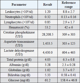
AbstractBackground: In this report, we describe the clinical, macro-, and microscopic findings of a newborn zebra victim of a fatal trauma and its possible causes are discussed in detail. Case Description: A plains zebra (Equus quagga) born in a Portuguese zoo was found in hypothermia and unable to get up. The animal was admitted to the hospital 36 hours later and, after the first hour of treatment and no attempts to get up, it began to convulse and died. At necropsy, subdural hematoma and atlantoaxial dislocation were the main findings. Conclusion: The inability to pinpoint the exact moment of the lesions’ onset determines the importance of a thorough surveillance of the periparturient period and parturition of captive animals. Furthermore, literature concerning zebras, their reproduction, and neonatal period is scarce, reinforcing the need to report these cases. Keywords: Fatal, Newborn, Trauma, Congenital, Zebra. IntroductionGenus Equus is composed of three zebra species, namely Grevy’s (E. grevyi), mountain (E. zebra) and plains (E. quagga) (Harley et al., 2009). Plains zebras are widespread across Africa, but in many countries are only found in protected areas and zoos (King and Moehlman, 2016). This species can be held successfully in captivity, when provided with a large open enclosure and access to shelter, shade, food, and water (Husher, 2010). They are seasonally polyestrous and the gestation period takes roughly 375 days. In the wild, foals are born throughout the year with peaks during the rainy months; however, in zoos, zebras have given birth in every month of the year, with clusters in spring and summer (Smuts, 1976; Nunez et al., 2011). The mechanics and labor stages of zebra’s parturition are very similar to those of domestic horses (Nunez et al., 2011). After birth, the foal must be able to get up and start walking and should be fed within 67 minutes after birth (Husher, 2010). The passive transfer of immunoglobulins through colostrum is essential for strengthening the foal’s immune system in the neonatal period (Rizzoni and Miyauchi, 2012). Sometimes, due to multifactorial neonatal pathologies, the foal hardly gets up and starts suckling, which can determine the foal’s life or death. In this report, we describe the clinical, macro-, and microscopic findings of a newborn zebra victim of both congenital and traumatic fatal cervical injuries. Case DetailsA female E. quagga was born in Maia Zoo in Porto, Portugal (41°14’.25" N, 8°37’47.2" W). After natural fertilization, the mother normally delivered the foal without any human assistance on a cold, rainy night of January 2021. The dam was multiparous, and shared with the male and another female, an outdoor space of about 600 m2 with two collection parks. In the zoo, there were only records of aggression of the male zebra toward other mature males and never toward the offspring. In addition, the zookeepers have never witnessed any kind of hostile behavior by the female zebras against newborns. In the morning, the newborn was found in hypothermia semi-submerged and lying in the mud, unable to get up. Commercial artificial colostrum was administered, and after heating, the foal was able to get up with human help for some instants and seemed to be recovering from the shock. Both mother and foal were then isolated in one of the collection parks. On the next day, the newborn’s clinical condition worsened and the animal was referred to the UP Clinical Equine Center where it was admitted 36 hours after birth in decubitus and shock, prostrate and listless. The clinical examination revealed hypothermia (35°C), tachycardia (>120 bpm), congestive mucous membranes with increased CRT, and absent intestinal motility in the four quadrants of the abdomen. A blood sample was collected, and the results are summarized in Table 1. The treatment started with IV fluid therapy: Gelaspan (40 mg/ml), Ringer’s Lactate (20 mg/kg in the first half hour and then 4 ml/kg/hour), and Glucose 5% (4 mg/kg/minute). Dexamethasone (0.05 mg/kg, IV) was also administered. Nasogastric intubation was performed and no gastric reflux was observed. After the first hour of treatment, the animal’s clinical condition improved but no attempts to get up were noted. From then on, the situation worsened, and the foal began to convulse. Diazepam was administered, initially via rectal and IV, and butorphanol (0.1 mg/kg, IV) as an analgesic, but both were apparently ineffective in controlling seizures. The baby zebra went into respiratory arrest and died. As the musculature of the cervical region was macroscopically deformed and presented increased stiffness, a radiographic examination was carried out before necropsy. Laterolateral and ventrodorsal radiographs of the cervical spine were carried out revealing atlantoaxial joint deviation, C2–C3 subluxation, C3 cranial vertebral body surface flattening, and multiple fracture lines in the cranial vertebral body of C3 (Fig. 1). The corpse was submitted for necropsy examination to the Institute of Biomedical Sciences Abel Salazar (ICBAS)-UP Veterinary Pathology lab (Fig. 2a). Table 1. Changes in hematological and biochemical parameters of a zebra’s blood sample. Reference values are in accordance with Husher (2010).
At necropsy, the tracheal lumen presented white foam. Edema and congestion of the larynx were observed. The lungs showed a general bright red color, more evident in the right lobe. The upper third of the esophageal mucosa exhibited a reddish color, suggesting congestion and inflammation. The left temporomandibular joint showed signs of subluxation associated with periocular tissue’s hematoma and hyphema of the left eye (Fig. 2c). The head presented several concussions and bruises with approximately 6.0 cm of diameter, located in the left supraorbital fossa and with sizes varying between 5.0 × 2.0 and 3.0 × 2.5 cm in the dorsal region of the nose (Fig. 2b and c). A subdural hematoma (Fig. 2d) and an atlantoaxial dislocation were also identified. Representative samples of lung, liver, spleen, kidney, heart, gastrointestinal (GI) tract, and central nervous system (CNS) were collected and fixed in 10% neutral buffered formalin for histopathological examination. Tissue samples were routinely processed and paraffin-embedded and 2 μm thick sections were routinely processed. Histologically, the lungs showed signs of pulmonary immaturity and moderate, acute, and multifocal interstitial pneumonia with the presence of mixed inflammatory infiltrate, composed of neutrophils, eosinophils, and some macrophages, associated with some bacillary Gram-positive bacteria. Areas of alveolar septal thickening and widespread congestion were observed. Microscopically, the cerebellum revealed diffuse multifocal edema between the pia mater and the cerebellar cortex. In the cerebrum, discrete edema of the leptomeninges and focal subarachnoid hemorrhage were also observed. In the remaining tissues, no relevant microscopic alterations were detected. DiscussionThe lack of information regarding the parturition and pre- and post-parturition periods precludes the determination of the pathological circumstances underlying these lesions, namely their causes and chronological sequence. Many common diseases of domestic horses have never been reported in zebras, probably due to incomplete documentation of zebras in captivity rather than a true absence of disease; therefore, one should be aware of the potential for any equine disease to develop in wild equids (Wiedner et al., 2012). According to the clinicopathological findings, some differential diagnoses shall be considered. Pathologies that affect equine neonates may have noninfectious, infectious, immunological, and traumatic causes (Rizzoni and Miyauchi, 2012). Hypoxic ischemic encephalopathy (HIE), known as dummy foal syndrome and as neonatal maladjustment syndrome, remains a systemic manifestation of perinatal asphyxia syndrome. HIE attempts to describe the suspected underlying pathophysiology of the syndrome, which is related with peripartum hypoxia. This can affect other organs such as GI, urinary, cardiovascular, respiratory, and endocrine systems. Affected foals are normal at birth but can develop signs of CNS abnormalities within a few hours, varying from mild depression with loss of the suck reflex to grand mal seizure activity (Wilkins, 2004). Although some clinical findings such as suckling difficulty and seizures may be consistent, some characteristic signs of this pathology were not detected in the presented clinical case (hyperexcitability, tremors, and protrusion of the tongue) (Toribio, 2019). Additionally, Wobbler’s syndrome or cervical vertebrae stenotic myelopathy is characterized by cervical instability and has been reported as a hereditary condition in captive zebras, showing signs of ataxia and motor incoordination (Wiedner et al., 2012). There are two main anatomical characteristics associated with this syndrome: static narrowing of the cervical vertebrae canal through which runs the spinal cord and dynamic compression of the spinal cord by the cervical vertebrae (Rossdales, 2013). Furthermore, Wobbler’s syndrome has been categorized into three main types depending on when clinical signs are seen in relation to the positioning of the neck (Rossdales, 2013).
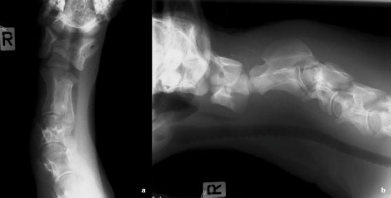
Fig. 1. (a) Ventrodorsal and (b) lateral left-right radiographs of the cranial cervical spine evidencing surrounding soft tissue edema, fracture of C3 cranial vertebral body (dislocated dorsally and with surface flattening), C2–C3 subluxation, and atlantoaxial joint deviation.
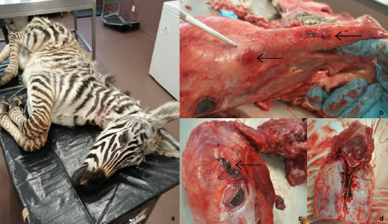
Fig. 2. (a) Zebra (Eqqus quagga) submitted to necropsy. (b) Concussive hemorrhagic lesions located in the right dorsal nasal region (arrows). (c) Hematoma in the left supraorbital fossa (arrow) and hyphema of the left eye. (d) Subdural hematoma. Neonatal septicemia is one of the main infectious causes of death in foals and can have a bacterial, viral, or fungal origin. Clinical signs depend on the type of pathogen involved and of the foal’s immune system condition, in which the failure of passive immunity transfer plays a major role. Early signs include depression, decreased sucking reflex, increased recumbency, fever, hypothermia, increased respiratory rate, injected mucous membranes, decreased capillary refill time (CRT), and tachycardia or bradycardia (Wilkins, 2004; Rizzoni and Miyauchi, 2012). This condition presupposes a generalized infection, for which no evidence was encountered in this zebra. Equine herpesvirus is another cause of death leading to abortion, respiratory disorders, and neurologic disease with ataxia. The clinical picture includes feverish states, nasal discharge, and petechiae, also not herein observed (Husher, 2010). Immunological causes comprise neonatal isoerythrolysis related with type II hypersensitivity reaction and blood incompatibility, which usually begins some hours after colostrum ingestion (Rizzoni and Miyauchi, 2012). To the best of our knowledge, this condition remains unreported in zebra or zebroid foals. In this case, the absence of anemia with a decrease in hematocrit and severe icterus (Wiedner et al., 2012), also makes this diagnosis less probable. Several traumatic causes must also be considered including dystocia, trauma at birth, or traumatic injuries in the postpartum period. Dystocia can lead to neurological conditions due to ischemia and tissue hypoxia; however, this birth was apparently normal, and it does not explain the macroscopic lesions found. Birth trauma occurs due to the pressure exerted on the foal during its passage through the birth canal. This is not usually a life-threatening condition, and it is more common in primiparous mares due to the tightness of the birth canal (Elks and Bidstrup, 2008). Thus, traumatic injuries in the postpartum period must be taken into account, which can be of unknown origin or a consequence of mother’s aggression. Although none of these causes can be completely ruled out, the combined imaging and necropsy findings are highly suggestive of the occurrence of mixed events, being very likely that both congenital malformations and traumatic events led to the death of this animal. Abnormal cervical vertebrae growth pattern, such as flattening of the cranial surface of C3 vertebral body, might have conducted to a narrowed spinal canal, or a progressive narrowing upon the movement of the neck and the vertebrae relative to each other, ultimately leading to spinal cord compression. Indeed, cases of Wobbler’s syndrome categorized as type 1 rarely occur but are usually present since birth, arising from the second and third cervical vertebrae where vertebral column is fixed in a flexed position at the site of the malformation of the cervical vertebrae. In horses, the onset of associated clinical signs is typically between 6 months and 3 years of age (Rossdales, 2013); however, the successive traumatic events must have precipitated the development of the clinical picture in this zebra. Although no aggressive maternal behavior was referred, there are reports of zebras’ infanticide where males fatally attack foals, being 45% of these directed to female foals (Pluháček and Bartoš, 2000). Some hours after birth, the foal and the mother were placed together in a park apart from the other animals. If there was any type of aggression until this isolation, it may have been caused by the male. At this time, zookeepers identified no suspected lesions; however, as restless and flighty animals (Husher, 2010), the mother could have caused the trauma by kicking the newborn as an attempt to lift her up or frightened by any external stimulus. Injuries in periocular tissues occur when there is a direct kick to the face, by a falling rock, from running into an obstacle, or from a fall (Gelatt, 2021). A more intense traumatic injury in the left part of the head, associated with the forces caused by the impact and with other consecutive traumatic events ended up aggravating an already fragile and unstable cervical area. All these lesions associated with edema and hemorrhage may have resulted in compression of the spinal cord, explaining the later neurological signs observed. The concussions distributed throughout the cranial cavity, the hyphema and the hematoma of the periocular tissues also suggest repeated traumatic injuries of lesser intensity or may be due to the convulsive condition. Additionally, equids often use the head and cervical muscles to lift themselves, so failed attempts to rise up can also be the cause. The inflammatory processes observed in the meninges can be explained as a consequence of the vascular impairment and compression of surrounding structures. Furthermore, delayed ingestion of colostrum may have resulted in a failure in the transfer of passive immunity that did not allow the newborn to react, and thus have been primary found fallen, in shock and hypothermic. Equids are also susceptible to pneumonia if they lie down for an extended period (Cook, 2011). The weakened immune system, associated with the muddy semi-submerged spot where the zebra was found, may have contributed to the bacterial pneumonia histologically encountered. In conclusion, the clinical, macro-, and microscopic findings are compatible with both congenital and traumatic cervical lesions that contributed together to the death of this newborn zebra. Wild animals are associated with various environments (wildlife and captivity) and express a variety of behaviors in specific conditions. A previous study refers trauma as the second leading cause of death in three different Italian zoos, which may occur because of fights with conspecifics (Scaglione et al., 2019). However, circumstances underlying trauma can be complex, and the causes of death can effectively be multifactorial, so the real context is rarely unveiled. Preventive medicine plays an important role to protect the health of captive animals and the information retained from postmortem examination can help future healthcare decisions in living animals (Scaglione et al., 2019). Even though similarities between captive zebras and domestic equids can be found, the literature concerning zebras, their reproduction, and neonatal period is still scarce, which reinforces the need to build knowledge and report cases regarding these animals. AcknowledgmentsThe authors would like to thank the support received by the Veterinary Medicine Master of the ICBAS, University of Porto. This article reports some of the research’s Master work of Gabriela Silva under the supervision of Irina Amorim. Conflict of interestThe authors declare no conflict of interest. Authors’ contributionsGFS and JEG wrote the manuscript. NA, LMA, and RC carried out the clinical evaluation and clinical care. CSB analyzed the radiographic examination. GFS, JEG, ACS, FF, and IA conducted the necropsy. FF carried out the HE staining. IA and LMA critically revised the manuscript for important intellectual content. All authors read and approved the final manuscript. ReferencesCook, C. 2011. Why does a broken leg mean the end for a horse? The Guardian. Available via: https://www.theguardian.com/sport/blog/2011/sep/23/claims-five-broken-leg-horse Elks, W. and Bidstrup, I. 2008. Birth trauma. Hoofbeats Magazine, pp: 68–61. Gelatt, K. 2021. Eye emergencies—special pet topics—veterinary manual. MSD Veterinary Manual. Available via: https://www.msdvetmanual.com/special-pet-topics/emergencies/eye-emergencies Harley, E., Knight, M., Lardner, C., Wooding, B. and Gregor, M. 2009. The Quagga project: progress over 20 years of selective breeding. South Afr. J. Wildl. Res. 39(2), 155–163. Husher, S. 2010. Husbandry guidelines for the plains Zebra Equus quagga (formerly Equus burchelli). Captive Animals Certificate III RUV 30204/1068, pp: 1–80. Available via: https://aszk.org.au/wp-content/uploads/2020/06/Plains-Zebra-Equus-quagga-Husher-S.2010.pdf King, S.R.B. and Moehlman, P.D. 2016. Equus quagga. The IUCN red list of threatened species 2016: e.T41013A45172424; 10.2305/IUCN.UK.2016-2.RLTS.T41013A45172424.en Nunez, C.M.V, Asa, C.S. and Rubenstein, D.I. 2011. Zebra reproduction. In Equine reproduction, 2nd ed. Eds., McKinnon A.O., Squires E.L., Vaala W.E. and Varner D.D. Wiley-Blackwell, pp: 2851–2865. Pluháček, J. and Bartoš, L. 2000. Male infanticide in captive plains zebra, Equus burchelli. Anim. Behav. 59(4), 689–694. Rizzoni, L. and Miyauchi, T. 2012. Principais doenças dos neonatos equinos [Main diseases of neonatal foals]. Acta Vet. Brasil. 6(1), 9–16. Rossdales, Veterinary Surgeons. 2013. Cervical vertebrae stenotic myelopathy (Wobblers Syndrome). Available via: www.rossdales.com (Accessed 22 November 2021). Scaglione, F., Biolatti, C., Pregel, P., Berio, E., Cannizzo, F., Biolatti, B. and Bollo, E. 2019. A survey on zoo mortality over a 12-year period in Italy. PeerJ, 7, 6198. Smuts, G.L. 1976. Reproduction in the zebra mare (Equus burchelli antiquorum) from the Kruger National Park. Koedoe 19, 89–132. Toribio, R.E. 2019. Equine neonatal encephalopathy: facts, evidence, and opinions. Vet. Clin. North Am. Equine Pract. 35(2), 363–378. Wiedner, E.B., Lindsay, W.A. and Isaza, R. 2012. Management of zebras and zebra hybrids (zebroids). Compend. Contin. Educ. Vet. 34(9), E4. Wilkins, P.A. 2004. Disorders of foals. Equine Int. Med. 2004, 1381–1440. | ||
| How to Cite this Article |
| Pubmed Style Silva GF, Gomes JE, Cunha R, Canadas-Sousa A, Faria F, Baptista C, Alvura N, . Fatal congenital and traumatic cervical spine injuries in a captive newborn plains zebra (Equus quagga). Open Vet J. 2022; 12(1): 75-79. doi:10.5455/OVJ.2022.v12.i1.9 Web Style Silva GF, Gomes JE, Cunha R, Canadas-Sousa A, Faria F, Baptista C, Alvura N, . Fatal congenital and traumatic cervical spine injuries in a captive newborn plains zebra (Equus quagga). https://www.openveterinaryjournal.com/?mno=131274 [Access: April 19, 2024]. doi:10.5455/OVJ.2022.v12.i1.9 AMA (American Medical Association) Style Silva GF, Gomes JE, Cunha R, Canadas-Sousa A, Faria F, Baptista C, Alvura N, . Fatal congenital and traumatic cervical spine injuries in a captive newborn plains zebra (Equus quagga). Open Vet J. 2022; 12(1): 75-79. doi:10.5455/OVJ.2022.v12.i1.9 Vancouver/ICMJE Style Silva GF, Gomes JE, Cunha R, Canadas-Sousa A, Faria F, Baptista C, Alvura N, . Fatal congenital and traumatic cervical spine injuries in a captive newborn plains zebra (Equus quagga). Open Vet J. (2022), [cited April 19, 2024]; 12(1): 75-79. doi:10.5455/OVJ.2022.v12.i1.9 Harvard Style Silva, G. F., Gomes, . J. E., Cunha, . R., Canadas-Sousa, . A., Faria, . F., Baptista, . C., Alvura, . N. & (2022) Fatal congenital and traumatic cervical spine injuries in a captive newborn plains zebra (Equus quagga). Open Vet J, 12 (1), 75-79. doi:10.5455/OVJ.2022.v12.i1.9 Turabian Style Silva, Gabriela Fernandes, José Eduardo Gomes, Raquel Cunha, Ana Canadas-Sousa, Fátima Faria, Cláudia Baptista, Nuno Alvura, and Luis Miguel Atayde and Irina Amorim. 2022. Fatal congenital and traumatic cervical spine injuries in a captive newborn plains zebra (Equus quagga). Open Veterinary Journal, 12 (1), 75-79. doi:10.5455/OVJ.2022.v12.i1.9 Chicago Style Silva, Gabriela Fernandes, José Eduardo Gomes, Raquel Cunha, Ana Canadas-Sousa, Fátima Faria, Cláudia Baptista, Nuno Alvura, and Luis Miguel Atayde and Irina Amorim. "Fatal congenital and traumatic cervical spine injuries in a captive newborn plains zebra (Equus quagga)." Open Veterinary Journal 12 (2022), 75-79. doi:10.5455/OVJ.2022.v12.i1.9 MLA (The Modern Language Association) Style Silva, Gabriela Fernandes, José Eduardo Gomes, Raquel Cunha, Ana Canadas-Sousa, Fátima Faria, Cláudia Baptista, Nuno Alvura, and Luis Miguel Atayde and Irina Amorim. "Fatal congenital and traumatic cervical spine injuries in a captive newborn plains zebra (Equus quagga)." Open Veterinary Journal 12.1 (2022), 75-79. Print. doi:10.5455/OVJ.2022.v12.i1.9 APA (American Psychological Association) Style Silva, G. F., Gomes, . J. E., Cunha, . R., Canadas-Sousa, . A., Faria, . F., Baptista, . C., Alvura, . N. & (2022) Fatal congenital and traumatic cervical spine injuries in a captive newborn plains zebra (Equus quagga). Open Veterinary Journal, 12 (1), 75-79. doi:10.5455/OVJ.2022.v12.i1.9 |

