


| Case Report | ||
Open Vet J. 2022; 12(4): 439-444 Open Veterinary Journal, (2022), Vol. 12(4): 439–444 Case Report Continuous hemilaminectomy of nine vertebrae can be performed safely in large breed dogs: A case report of a German Shepherd Dog with intervertebral disc extrusion and extensive extradural hemorrhageFelix Lackmann*, Sabine Schulze, and Peter BöttcherSmall Animal Clinic, Freie Universität Berlin, Berlin, Germany Submitted: 15/03/2022 Accepted: 19/06/2022 Published: 10/07/2022 *Corresponding Author: Felix Lackmann. Small Animal Clinic, Freie Universität Berlin, Berlin, Germany.Email: felix.lackmann [at] fu-berlin.de © 2022 Open Veterinary Journal
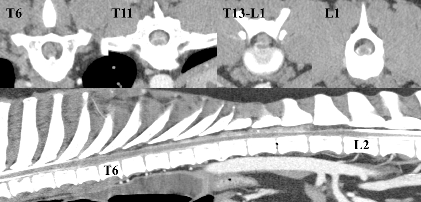
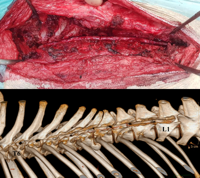
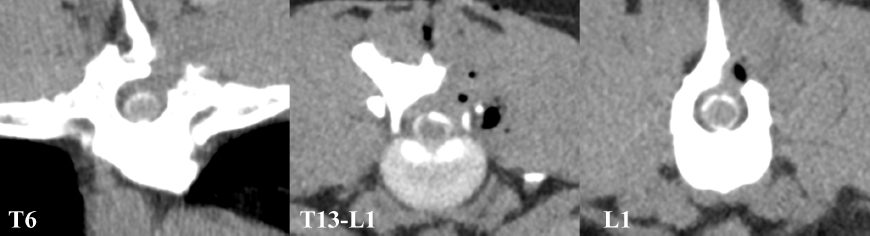
AbstractBackground: Extended, continuous hemilaminectomy has only been reported in small to medium-sized dogs so far. It remains unclear whether excessive continuous hemilaminectomy can also be performed safely in large breed dogs. Case Description: We describe the surgical treatment and clinical outcome of a 5-year-old German Shepherd Dog that presented with paraplegia and deep pain perception following a short episode of bilateral hind-limb lameness, secondary to jumping off of a car. Computed tomography-myelography revealed that the paraplegia originated from extensive extradural spinal cord compression (Th6-L1), due to intervertebral disc extrusion and associated epidural hemorrhage. The dog was treated with a continuous hemilaminectomy involving nine vertebrae (Th6-L1) and recovered completely with no remaining neurological deficits, within 6 months. Conclusion: The rapid, uncomplicated, and complete functional recovery in the presented case emphasizes the practicability of extensive, continuous hemilaminectomies, also in large breed dogs. Keywords: Dog, Epidural hemorrhage, Hemilaminectomy, Intervertebral disc extrusion, Paraplegia. IntroductionIntervertebral disc prolapse is a very common neurological disease in dogs (Goggin et al., 1970; Noyes et al., 2017). Herniated disc material can lead to spinal cord compression as well as extradural hemorrhage when the vertebral venous plexus is lacerated. In rare cases, bleeding from the lacerated venous plexus can result in extensive epidural hemorrhage, known as disc extrusion with extensive epidural hemorrhage (DEEH), contributing significantly to the extradural spinal cord compression (Olby et al., 2000; Tartarelli et al., 2005). So far, reported evidence on the significance of DEEH remains unclear. While Woelfel et al. (2021) reported DEEH in combination with loss of deep pain perception to be associated with a worse prognosis for return to function, Mateo et al. (2011) did not find significant differences when comparing DEEH to intervertebral disc extrusion (IVDE) without extensive epidural hemorrhage. Because Tartarelli et al. (2005) showed that decompression of DEEH over multiple vertebrae would result in the same prognosis as localized decompression of IVDE without extensive epidural hemorrhage, continuous hemilaminectomy might be considered standard care in the context of DEEH. However, the maximal extent of continuous hemilaminectomy that can be performed safely in dogs is unknown. Limiting factors might be the potentially associated increase of instability of the spinal column, the development of domino lesions, the formation of excessive scoliosis secondary to unilateral muscle dissection, as well as the increased risk of mid and long-term myelopathy, because of laminectomy membrane formation (Sharp, 2004; Janssen et al., 2011). In large breed dogs, continuous hemilaminectomy might be considered safe up to a length of seven vertebrae (Tartarelli et al., 2005). Successful and uncomplicated surgical decompression over a longer extent has only been documented in small and mid-sized dogs so far (Hirano et al., 2020; Nakamoto et al., 2021). This case report describes the uncomplicated, full neurological recovery of a German Shepherd Dog with paraplegia and intact deep pain perception, following continuous unilateral hemilaminectomy over the length of nine vertebrae, which we performed because of extensive thoracolumbar DEEH. Case DetailsHistory and clinical workupA 5-year-old, 28 kg, spayed male German Shepherd Dog was presented to the emergency service of the Small Animal Clinic of the Freie Universität Berlin for evaluation of acute, moderate bilateral hind limb lameness. The lameness occurred after the dog had jumped off of a car. On physical examination, only palpation of the cranial lumbar spine revealed pain. Otherwise, both the neurological as well as the orthopedic examination were unremarkable. A tentative diagnosis of mild intervertebral disc disease was made and the dog was treated symptomatically with nonsteroidal anti-inflammatory medication (Robenacoxib, Onsior 40 mg once daily, Elanco GmbH, Cuxhaven, Germany) and strict cage rest for at least 5 days. However, the dog was re-presented the same night with acute paraplegia with intact deep pain perception, accompanied by normal to increased spinal reflexes in both pelvic limbs. The cutaneous trunci reflex was absent caudal to the thoracolumbar junction. On palpation, the whole back of the dog appeared to be painful. The remaining neurological examination revealed no abnormalities. Based on the neurological findings, an upper motor neuron lesion localized to the Th3-L3 spinal cord segment was suspected. The exact localization of the maximal spinal cord lesion was assumed to be at the level of Th12-L1, at the level or slightly cranial to the cut-off of the cutaneous trunci reflex. Results of a complete blood count and blood chemistry were unremarkable, as well as thoracic and abdominal radiographs. Axial computed tomography (CT) with lumbar myelography (Phillips Brilliance TM CT 16, Phillips GmbH Market DACH Healthcare, Hamburg, Germany) under general anesthesia revealed dorsal to dorsolateral, mostly left-sided extradural spinal cord compression, extending from Th6 to L1 (Fig. 1). The degree of extradural spinal cord compression varied between 20% and 50% of the cross area of the spinal canal, being most pronounced at Th13-L1. The extradural compressive mass was mostly homogenous in appearance with a mean Hounsfield Unit (HU) of 54, slightly higher than the spinal cord (42 HU). At some locations, small hyperdense bodies within the mass were also visible. At the intervertebral space Th13-L1, a vacuum phenomenon was also noted. No further abnormalities were detected. Based on CT-myelography and the results of clinical history and physical examination, DEEH was suspected, but the most likely differential diagnosis at this point included, among others, malignancy and coagulopathy. Because mucosal bleeding time, as well as full coagulation profile, revealed no abnormalities, coagulopathy was excluded from the list of differential diagnoses. Surgical techniqueThe dog was positioned in sternal recumbence and decompressive surgery was performed to gain access to the vertebral canal by left-sided mini-hemilaminectomy at Th13-L1. A pasty, bright red substance protruded from the spinal canal, once the lamina was penetrated. Based on the cytological examination, the substance was classified to be hemorrhage and cartilage without any sign of malignancy, resulting in the final diagnosis of DEEH. The mini-hemilaminectomy was subsequently extended towards cranial and caudal, up to the point, where normal epidural fat and a non-compressed spinal cord were present. Using suction and gentle probing allowed for complete evacuation of epidural hemorrhage mixed with extruded disc material from within the spinal canal, reaching from Th6 to L1 (Fig. 2). Before routine wound closure, morphine (Morphinsulfat-GRY, 0.1 mg/kg, TEVA GmbH, Ulm, Germany) was dropped onto the exposed dura. No fat graft or the like was applied. Postoperative CT confirmed the complete resolution of extradural spinal cord compression along the entire extent of the performed hemilaminectomy (Fig. 3). In total, anesthesia time was 7 hours and surgery took 5 hours. HistologyMultiple samples of peri-spinal tissue at the levels Th6-Th7, Th10-Th11, and Th13-L1 were sent for histopathologic examination, and were defined to be fibrocartilage material with acute hemorrhage in all locations, further strengthening the clinical diagnosis of acute DEEH.
Fig. 1. Preoperative CT myelographic images. Note the extent of dorsal to dorsolateral left-sided extradural compression, starting at Th6 and reaching until L1. Spinal compression was most severe at the Th13-L1 level, reaching about 50%.
Fig. 2. Left-sided intraoperative photograph as well as post-operative 3D volume rendering of the spinal column (cranial to the left). Notice the continuous hemilaminectomy extending from Th6 to L1 and the fully decompressed spinal cord along the same extent. On the 3D rendering, remaining contrast media of the pre-operative myelography mimics remaining compressive radio-opaque material within the spinal canal. Postoperative carePostoperatively, the dog received intravenous infusion of balanced electrolyte solution (Sterofundin, B. Braun, 2 ml/kg/h, Tuttlingen, Germany) and fentanyl (Fentadon, 1 µg/kg/hour, Dechra GmbH, Aulendorf, Germany) for 5 days, along with robenacoxib once daily, orally (Onsior, 40 mg, Elanco GmbH, Cuxhaven, Germany). Physiotherapy was initiated immediately the day after surgery. Bladder management consisted of permanent urinary catheterization. First signs of regaining pelvic limb motor function could be observed 6 days postoperatively. Two days later, the dog could walk with minimal support and showed independent urination. On day 11, the dog was discharged with ambulatory paraparesis and without signs of pain. The owners continued physiotherapy for approximately 3 months. Follow-upA follow-up examination four and a half weeks after discharge showed a mild ambulatory paraparesis with slightly decreased proprioception and normal spinal cord reflexes in the pelvic limbs. Palpation of the spinal column revealed no pain and wound healing had been uneventful. While the left latissimus dorsi muscle showed some mild atrophy extending from Th6 to Th9, no signs of scoliosis were present. At 6, 11, and 36 months postoperatively, the dog continued to be without any sign of pain and did not show any neurological deficits. Latissimus dorsi muscle atrophy had disappeared already at the 6-month re-check. DiscussionThis is the first report of a large breed dog with extensive spinal cord compression following DEEH, which had been treated with an extensive hemilaminectomy over nine vertebrae. Other accepted treatment options than surgical decompression along the complete length of the DEEH, are local decompression at the site of the herniated disk (local hemilaminectomy) or conservative therapy (Tartarelli et al., 2005; Langerhuus and Miles, 2017; Hirano et al., 2020; Nakamoto et al., 2021; Woelfel et al., 2021). While no data exists on the conservative treatment of dogs with DEEH, non-surgical management of IVDE alone can result in a good outcome (Langerhuus and Miles, 2017), but surgical decompression is usually considered the treatment of choice for non-ambulatory patients (Moore et al., 2016; Langerhuus and Miles, 2017; Jeffery et al., 2018). In the present case, conservative treatment had been rejected, because of the acute onset and progressing neurological deterioration, as well as the degree of spinal cord compression reaching up to 50% of the spinal canal’s cross-section, judged on CT-myelography. A local hemilaminectomy did not appear to be the appropriate approach either, because the wide extent of substantial extradural spinal cord compression, demonstrated on CT-myelography, was felt to be only resolvable by a more extensive decompressing technique. A hypothesis, which was proven right during hemilaminectomy, as the extent of the hemilaminectomy was based on the appearance of normal epidural fat as well as visibly normal/non-compressed spinal cord within the cranial and caudal extent of the hemilaminectomy. Woelfel et al. (2021) and Tartarelli et al. (2005) found a positive clinical correlation between the outcome after IVDE and DEEH with a more extensive surgical decompression. Similarly, Nakamoto et al. (2021) and Hirano et al. (2020) showed that extensive hemilaminectomy combined with durotomy in small breed dogs resulted in a good clinical outcome.
Fig. 3. Postoperative transverse CT images, corresponding to the locations in Figure 1. The spinal cord is no longer displaced or compressed by extradural material. Potential disadvantages of extensive hemilaminectomy in comparison to local hemilaminectomy may be, among others, (i) prolonged anesthesia as well as surgical time, (ii) a potential risk of inducing spinal column instability, probably propagating further IVDD, (iii) spinal column deformation due to extensive muscle dissection as well as (iv) the increased risk of laminectomy membrane formation because of the extensive exposure of the spinal cord. Prolonged anesthesia increased the risk of surgical site infections (Yap et al., 2015), which is not specific to spinal surgery. However, Fenn et al. (2020) reported a negative association between the duration of surgery and outcome in dogs with surgically treated acute severe spinal cord injury caused by thoracolumbar IVDE without deep pain perception. Considering those, extended hemilaminectomy probably bears an inherent risk, because of the increased anesthetic as well as surgical time that is necessary. Strict adherence to aseptic principles, repeated intraoperative intravenous application of antibiotics, in our case cephazolin starting 30 minutes before surgery and repetition every 90 minutes until wound closure, as well as meticulous anesthetic monitoring, including blood pressure measurement, to prevent operation-related hypoperfusion of the spinal cord. Spine instability after extensive hemilaminectomy is probably the type of potential complication many surgeons would be most concerned about. When considering the three-column concept of spinal fractures which divides the spine into three horizontal sections, the dorsal, middle, and ventral column (Shores, 1992; Jeffery, 2010), a hemilaminectomy, whether performed locally or in a continuous way, will only affect the middle segment. According to Jeffery et al. (2018), instability of the vertebral column will only result if at least two columns are affected. Even bilateral hemilaminectomy, as well as bilateral pediculectomy, have both been successfully performed in dogs without major postoperative complications (Swaim and Vandevelde, 1977; Muir et al., 1995; Necas, 1999), which is by the three-column concept. However, when adding a fenestration to a standard hemilaminectomy, vertebral instability is significantly increased which, once again, is following the three-column concept, because fenestration results in damage to the ventral column, too. Whether excessive continuous hemilaminectomy can truly be compared to vertebral fracture-luxation and therefore be interpreted in the context of the three-column concept, remains to be investigated. Nevertheless, the work by Corse et al. (2003) who looked at instability after continuous hemilaminectomy of up to four vertebrae in an ex vivo model, suggests that continuous hemilaminectomy does not behave differently than localized vertebral surgery, and therefore will not result in significant instability, regardless of the number of vertebrae involved. A formation of excessive scoliosis secondary to unilateral muscle dissection after extensive hemilaminectomy has been mentioned as another potential complication of extended hemilaminectomy (Sharp, 2004; Janssen et al., 2011). However, to the best of our knowledge, no peer-reviewed paper has ever mentioned scoliosis to be associated with hemilaminectomy, which is echoed by the current case. We speculate that unless the nerve roots are transected, muscle elevation alone would probably not result in excessive asymmetrical muscle pull along the lateral spinal column. Nevertheless, some form of muscle trauma/denervation probably occurs, as indicated by muscular atrophy, which is resolved completely within 6 months. Being a case report, conclusions should be drawn with caution and we acknowledge that the observed positive outcome may also have been attributed to the very high compliance of the dog owners, who meticulously took care of the physio-therapeutically support postoperatively, which is known to have positive effects on rehabilitation of dogs with IVDD (Gandini et al., 2003; Millis and Ciuperca, 2015; Hodgson et al., 2017; Woelfel et al., 2021). ConclusionThis case report illustrates the option and feasibility of extensive hemilaminectomy over nine vertebral bodies. The uncomplicated recovery observed in our case, suggests that even in large breed dogs, extensive continuous hemilaminectomy can be performed safely. Furthermore, we question if nine vertebrae should be taken as the maximal extent for continuous hemilaminectomy, because of the reported low complication rates following continuous hemilaminectomy in dogs overall, as well as the three-column concept, suggest that there is no absolute limit. AcknowledgmentsThe authors thank Luca Bertzbach for writing assistance and language editing. Conflict of interestThe authors declare that they have no conflicts of interest. ReferencesCorse, M.R., Renberg, W.C. and Friis, E.A. 2003. In vitro evaluation of biomechanical effects of multiple hemilaminectomies on the canine lumbar vertebral column. Am. J. Vet. Res. 64, 1139–1145. Fenn, J., Ru, H., Jeffery, N.D., Moore, S., Tipold, A., Soebbeler, F.J., Wang-Leandro, A., Mariani, C.L., Early, P.J., Munana, K.R. and Olby, N.J. 2020. Association between anesthesia duration and outcome in dogs with surgically treated acute severe spinal cord injury caused by thoracolumbar intervertebral disk herniation. J. Vet. Intern. Med. 34, 1507–1513. Gandini, G., Cizinauskas, S., Lang, J., Fatzer, R. and Jaggy, A. 2003. Fibrocartilaginous embolism in 75 dogs: clinical findings and factors influencing the recovery rate. J. Small Anim. Pract. 44, 76–80. Goggin, J.E., Li, A.S. and Franti, C.E. 1970. Canine intervertebral disk disease: characterization by age, sex, breed, and anatomic site of involvement. Am. J. Vet. Res. 31, 1687–1692. Hirano, R., Asahina, R., Hirano, T., Hyakkoku, A., Miura, R., Kunihiro, T. and Nakamoto, Y. 2020. Outcomes of extensive hemilaminectomy with durotomy on dogs with presumptive progressive myelomalacia: a retrospective study on 34 cases. BMC Vet. Res. 16, 476. Hodgson, M.M., Bevan, J.M., Evans, R.B. and Johnson, T.I. 2017. Influence of in-house rehabilitation on the postoperative outcome of dogs with intervertebral disk herniation. Vet. Surg. 46, 566–573. Janssen, M.M., de Wilde, R.F., Kouwenhoven, J.W. and Castelein, R.M. 2011. Experimental animal models in scoliosis research: a review of the literature. Spine J. 11, 347–358. Jeffery, N.D. 2010. Vertebral fracture and luxation in small animals. Vet. Clin. North Am. Small Anim. Pract. 40, 809–828. Jeffery, N.D., Harcourt-Brown, T.R., Barker, A.K. and Levine, J.M. 2018. Choices and decisions in decompressive surgery for thoracolumbar intervertebral disk herniation. Vet. Clin. North Am. Small Anim. Pract. 48, 169–186. Langerhuus, L. and Miles, J. 2017. Proportion recovery and times to ambulation for non-ambulatory dogs with thoracolumbar disc extrusions treated with hemilaminectomy or conservative treatment: a systematic review and meta-analysis of case-series studies. Vet. J. 220, 7–16. Mateo, I., Lorenzo, V., Foradada, L. and Munoz, A. 2011. Clinical, pathologic, and magnetic resonance imaging characteristics of canine disc extrusion accompanied by epidural hemorrhage or inflammation. Vet. Radiol. Ultrasound 52, 17–24. Millis, D.L. and Ciuperca, I.A. 2015. Evidence for canine rehabilitation and physical therapy. Vet. Clin. North Am. Small Anim. Pract. 45, 1–27. Moore, S.A., Early, P.J. and Hettlich, B.F. 2016. Practice patterns in the management of acute intervertebral disc herniation in dogs. J. Small Anim. Pract. 57, 409–415. Muir, P., Johnson, K.A., Manley, P.A. and Dueland, R.T. 1995. Comparison of hemilaminectomy and dorsal laminectomy for thoracolumbar intervertebral disc extrusion in dachshunds. J. Small Anim. Pract. 36, 360–367. Nakamoto, Y., Uemura, T., Hasegawa, H., Nakamoto, M. and Ozawa, T. 2021. Outcomes of dogs with progressive myelomalacia treated with hemilaminectomy or with extensive hemilaminectomy and durotomy. Vet. Surg. 50, 81–88. Necas, A. 1999. Clinical aspects of surgical treatment of thoracolumbar disc disease in dogs. A retrospective study of 300 cases. Acta Vet. Brno. 68, 121–130. Noyes, J.A., Thomovsky, S.A., Chen, A.V., Owen, T.J., Fransson, B.A., Carbonneau, K.J. and Matthew, S.M. 2017. Magnetic resonance imaging versus computed tomography to plan hemilaminectomies in chondrodystrophic dogs with intervertebral disc extrusion. Vet. Surg. 46, 1025–1031. Olby, N.J., Munana, K.R., Sharp, N.J. and Thrall, D.E. 2000. The computed tomographic appearance of acute thoracolumbar intervertebral disc herniations in dogs. Vet. Radiol. Ultrasound 41, 396–402. Sharp, N.J.H. 2004. Small animal spinal disorders: diagnosis and surgery. Maryland, MI: Mosby Ltd. Shores, A. 1992. Spinal trauma. Pathophysiology and management of traumatic spinal injuries. Vet. Clin. North Am. Small Anim. Pract. 22, 859–888. Swaim, S.F. and Vandevelde, M. 1977. Clinical and histologic evaluation of bilateral hemilaminectomy and deep dorsal laminectomy for extensive spinal cord decompression in the dog. J. Am. Vet. Med. Assoc. 170, 407–413. Tartarelli, C.L., Baroni, M. and Borghi, M. 2005. Thoracolumbar disc extrusion associated with extensive epidural haemorrhage: a retrospective study of 23 dogs. J. Small Anim. Pract. 46, 485–490. Woelfel, C.W., Robertson, J.B., Mariani, C.L., Munana, K.R., Early, P.J. and Olby, N.J. 2021. Outcomes and prognostic indicators in 59 paraplegic medium to large breed dogs with extensive epidural hemorrhage secondary to thoracolumbar disc extrusion. Vet. Surg. 50, 527–536. Yap, F.W., Calvo, I., Smith, K.D. and Parkin, T. 2015. Perioperative risk factors for surgical site infection in tibial tuberosity advancement: 224 stifles. Vet. Comp. Orthop. Traumatol. 28, 199–206. | ||
| How to Cite this Article |
| Pubmed Style Lackmann F, . Continuous hemilaminectomy of nine vertebrae can be performed safely in large breed dogs: a case report of a German Shepherd Dog with intervertebral disc extrusion and extensive extradural hemorrhage.. Open Vet J. 2022; 12(4): 439-444. doi:10.5455/OVJ.2022.v12.i4.4 Web Style Lackmann F, . Continuous hemilaminectomy of nine vertebrae can be performed safely in large breed dogs: a case report of a German Shepherd Dog with intervertebral disc extrusion and extensive extradural hemorrhage.. https://www.openveterinaryjournal.com/?mno=101824 [Access: April 19, 2024]. doi:10.5455/OVJ.2022.v12.i4.4 AMA (American Medical Association) Style Lackmann F, . Continuous hemilaminectomy of nine vertebrae can be performed safely in large breed dogs: a case report of a German Shepherd Dog with intervertebral disc extrusion and extensive extradural hemorrhage.. Open Vet J. 2022; 12(4): 439-444. doi:10.5455/OVJ.2022.v12.i4.4 Vancouver/ICMJE Style Lackmann F, . Continuous hemilaminectomy of nine vertebrae can be performed safely in large breed dogs: a case report of a German Shepherd Dog with intervertebral disc extrusion and extensive extradural hemorrhage.. Open Vet J. (2022), [cited April 19, 2024]; 12(4): 439-444. doi:10.5455/OVJ.2022.v12.i4.4 Harvard Style Lackmann, F. & (2022) Continuous hemilaminectomy of nine vertebrae can be performed safely in large breed dogs: a case report of a German Shepherd Dog with intervertebral disc extrusion and extensive extradural hemorrhage.. Open Vet J, 12 (4), 439-444. doi:10.5455/OVJ.2022.v12.i4.4 Turabian Style Lackmann, Felix, and . 2022. Continuous hemilaminectomy of nine vertebrae can be performed safely in large breed dogs: a case report of a German Shepherd Dog with intervertebral disc extrusion and extensive extradural hemorrhage.. Open Veterinary Journal, 12 (4), 439-444. doi:10.5455/OVJ.2022.v12.i4.4 Chicago Style Lackmann, Felix, and . "Continuous hemilaminectomy of nine vertebrae can be performed safely in large breed dogs: a case report of a German Shepherd Dog with intervertebral disc extrusion and extensive extradural hemorrhage.." Open Veterinary Journal 12 (2022), 439-444. doi:10.5455/OVJ.2022.v12.i4.4 MLA (The Modern Language Association) Style Lackmann, Felix, and . "Continuous hemilaminectomy of nine vertebrae can be performed safely in large breed dogs: a case report of a German Shepherd Dog with intervertebral disc extrusion and extensive extradural hemorrhage.." Open Veterinary Journal 12.4 (2022), 439-444. Print. doi:10.5455/OVJ.2022.v12.i4.4 APA (American Psychological Association) Style Lackmann, F. & (2022) Continuous hemilaminectomy of nine vertebrae can be performed safely in large breed dogs: a case report of a German Shepherd Dog with intervertebral disc extrusion and extensive extradural hemorrhage.. Open Veterinary Journal, 12 (4), 439-444. doi:10.5455/OVJ.2022.v12.i4.4 |

